

 PDF
PDF ePub
ePub Citation
 Print
Print


INTRODUCTION
A cutaneous horn (cornu cutaneum) is a relatively rare epidermal tumor that generally appears as a conical projection of hyperkeratotic epidermis. It is named after horns of other animal species because of its gross resemblance. In dermatology, "horn" is the clinical term for a circumscribed, conical, markedly hyperkeratotic lesion in which the height of the keratotic mass amounts to at least half of its largest diameter1. Cutaneous horns may arise from any part of the body, and 30% arise from the face and scalp2. They are thought to result from underlying benign, premalignant or malignant pathologic lesion.
We report here on a case of a cutaneous horn originating from keratoacanthoma in a 76-year-old Korean woman.
CASE REPORT
A 76-year-old female patient presented with a club-shaped fungating mass growing from her right temple area. She had recognized a small erythematous exophytic mass 3 years previously, and the mass remained unchanged until 3 months prior to this visit. Since then it had grown rapidly, but there was no associated pain, pruritus or bleeding.
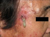
On physical examination, she had a 3×3 cm area of an erythematous patch with a central 1×1 cm sized hypertrophic nodule on her right temple. A yellowish-white colored cylindrical horn with a base diameter of 0.7 cm and a height of 2.7 cm arose from the central nodule (Fig. 1). The patient had no other specific findings.
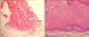
A central crater-like keratin plug surround by epidermis was found on the histologic findings. The lesion was well demarcated from the dermis and it has an eosinophilic glassy appearance as a consequence of keratinization. Exceedingly thick hyperkeratosis and parakeratosis were observed above the epidermis. An infiltration of inflammatory cells was seen in the dermis (Fig. 2, 3).
Based on the clinical and pathologic findings, we diagnosed this lesion as a cutaneous horn originating from keratoacanthoma, and the lesion was totally excised. Up to the present date, she is in good physical health without recurrence.
DISCUSSION
Cutaneous horn is a clinical term denoting distinctive, highly confined, protruding firm projections on the skin surface. Many old medical texts in the Middle Ages described cutaneous horn as a marker of a witch3. Cutaneous horns are of various shapes and they range from 2~60 mm in size. The color is usually skin-colored or erythematous and it may present as white when finely scaled. Cutaneous horn can be related to many underlying diseases, including actinic keratosis, verruca vulgaris, seborrheic keratosis, trichilemmoma, squamous cell carcinoma and melanoma4,5. On rare occasion it can be accompanied with keratoacanthoma. One such case has been previously reported in Korea6. Generally, one-half of cutaneous horns arise from benign lesions and 16% to 20% arise from definitely malignant lesions, but most of them arise from actinic keratosis2,5. In Korea, about 80% of such cases arise from benign lesions, and mostly from viral warts. Fifteen percent of the cases arise from premalignant lesion and 5% arise from malignant lesion7. Cutaneous horns rising from premalignant or malignant lesions signify the importance of a thorough histological examination.
The pathogenesis of cutaneous horn is not yet clear. It is thought continuous stimulus may affect the formation of a cutaneous horn. Old age and abundant blood vessels at the base are also associated with cutaneous horn4,8.
The diagnosis of cutaneous horn can be clinically settled when the height of the keratotic mass amounts to at least half of its largest diameter1.
The treatment and prognosis of cutaneous horn entirely depend on its base lesion, and for the most part cutaneous horn is totally excised for cosmetic reasons.
Keratoacanthoma is a relative common epithelial tumor. It grows rapidly and reaches a maximum size around 10 to 12 weeks after which it forms a crateriform ulcer9. It generally originates as a solitary lesion and it usually forms on the face, forearm and dorsum of the hand, which are the most sun-exposed areas. Demographically, it occurs 2 to 3 times more often in men and its incidence peaks at the ages of 50 to 69 years old10.
This lesion starts with rapidly enlarging firm erythematous papules with smooth surfaces. In the mature stage, the central keratotic core is formed and then falls off naturally after which a crateriform ulcer occurs. The crateriform ulcer disappears spontaneously leaving a flat hypopigmented scar. Pathologically, the mature lesion exhibits epithelial proliferation with atypical keratinocytes and mitoses, along with a central keratotic plug.
Keratoacanthoma usually heals spontaneously leaving a scar, but rapidly growing lesions can cause wide destruction of tissue. For example, a case was presented in which a giant keratoacanthoma destroyed a patient's entire nose11. Since there can be such consequences, complete surgical excision is recommended for most cases. Imiquimod has been reported to be effective in some cases12.
Our patient had a typically shaped cutaneous horn and on the histologic findings, the base of the horn showed the characteristic histologic features of keratoacanthoma. Clinically, most case of keratoacanthoma regresses spontaneously within one year. But like our patient, a few cases have been reported to remain without regression even one year after occurrence13-15. Such cases in which the base is keratoacanthoma are rare, and only 1 such case was previously reported in Korea by Hwang et al.6. So, we report here on an unusual case of a cutaneous horn that originated from the keratoacanthoma of a 76-year-old female patient.

 XML Download