

 PDF
PDF ePub
ePub Citation
 Print
Print


I. Introduction
In modern society, the number of people who become blind due to the removal of visual organs is rapidly increasing due to trauma to the eye, tumors, or diseases. Recently, a type of artificial implant has been designed to restore the shape of the eye to enable normal daily activities and social function. These eye implants involve an artificial eye. They are used to repair cosmetic problems or restore the natural appearance of patients. Currently, there is no objective and standardized technique, and the process can be expensive and time consuming. Therefore, many people with visual impairment can experience social discomfort because they do not wear an artificial eye due to economic costs, or because they wear low-quality artificial eyes or artificial eyes that are not suitable for them. To reduce the time and cost of the manufacturing process, studies on artificial eye fabrication using advanced equipment, such as 3D printers have been actively conducted. In 2013, the fripp design company in the UK worked with Manchester Metropolitan University to successfully produce artificial eyes of color with 3D printing, reducing both the time and money required for the fabrication process. Design work on the shape, color, and blood vessel distribution is required to make an artificial eye using this automated equipment. Therefore, there is a need to develop a standardization model before automated artificial eyes can be produced on a wider scale. Currently, most artificial eyes are made by hand, but standardization of iris color is essential to make artificial eyes using advanced equipment, such as 3D printers.
Since the iris is the part of the eye that is colored, visual judgment is an important step in making an artificial eye. The iris consists of layers, including the anterior border layer, stroma, sphincter and dilator muscle fibers, and the posterior pigment epithelium. The anterior layer and stroma are the most important parts in terms of the color of the eye [1]. Wielgus and Sarna [2] showed that the color of the iris results from the amount of melanin and that brown eyes have 40% more melanin than other eye colors. The iris has several structural layers, and the iris color and pattern are generated according to the shape of the pigment and the structures of each layer [3]. Attempts to classify iris colors date back to 1843 in the work by Petrequin [4]. Thereafter, a genetic approach was used to increase the number of iris color categories from 3 to 8 [56]. The first attempt to standardize artificial eyes by classifying iris colors was made by Rudolf Martin [7], who made 16 glass eyes. However, lightness, chroma, hue, and color attributes were not defined, so there were limitations in the use of the original iris color classification as a diagnostic kit. In a previous study in the Netherlands, 24 iris colors were classified using 67 eye images [8]. In addition, two skilled separators studied iris photography to develop a classification method based on 5 iris colors [9]. The 5 colors of the iris are blue, gray, green, light brown, and brown. Therefore, this 5 color system cannot be applied to the classification of the iris color of Koreans, whose eyes are homogeneously colored brown. Research has been conducted in the United States to digitize and analyze iris color information obtained from photographs [10]. In previous studies [1112], we investigated the distribution of iris colors by comparing the color of a sample with the color of the standard color table developed by Kim et al. [11], who investigated the relationship between iris coloration and skin color by analyzing photographs of patients with skin-related diseases. Park [13] introduced an iridescent coloration table for the manufacture of artificial eye using 9.03YR, 2.06Y, and lightness 3.11, based on iris colorimetry of Koreans [14]. It is known that iris formation tissues change through a random process for the first year or two after birth so that each person has a unique iris pattern, while the iris pattern is maintained after these early years in most individuals. The purpose of this study was to classify the color and pattern of the iris in Koreans using a colorimetric photographic technique based on the measurement of iris color using digital image analysis to obtain iris images of Korean eyes.
II. Methods
1. Subjects
The data collection period was from July 2015 to February 2016. Of the 145 patients who visited the Department of Ophthalmology, Yonsei University Hospital during this 8-month period, iris color analysis was valid in 100 samples, and iris pattern analysis was valid in 131 samples. The study was approved by the Institutional Review Board (IRB) of Yonsei University Hospital (No. IRB-2015-3045-001).
2. Method
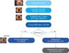
The experimental process is shown in Figure 1. The digital color values were first extracted from the digital image of each eye. The representative color combination was then extracted, and the representative colors were statistically grouped.
1) Extracting digital color data from the actual image
(1) Photographic environment and camera settings
Because eye color can be affected by ambient light, eyes were digitally photographed in the darkest possible environment to minimize the effects of light. The shooting distance was about 10 cm from the front, and a microscope (Slit Lamp BQ-900; Haag-Streit AG, Koeniz, Switzerland) was used for the lighting. The digital camera was equipped with 18 million effective pixels of the Advanced Photo System type-C (APS-C) and a complementary metal-oxide semiconductor (CMOS) image sensor. The subjects were repeatedly photographed because it was difficult to focus. We analyzed 100 iris pictures of patients at Department of Ophthalmology, Yonsei University College of Medicine. The size of each image was 2456 × 2304, and the file type was JPG.
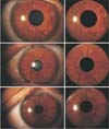
(2) Masking image
The iris area was extracted from the whole image by masking the other areas, including the pupil and sclera (Figure 2). All of the other parts except the iris were treated with black to prevent the inclusion of colors outside the iris in the clustering process.
(3) Digital analysis
The color extraction operation was performed assuming that the RGB digital values of the images photographed in the same environment were all standard RGB (sRGB). Although there are as many colors as there are digital values in an image, usually few color attributes, such as lightness, hue, or chroma are perceived by our eyes. Because of the texture of the iris plane as well as shading due to the iris pattern, the iris cannot be defined as having a single color (Figure 3). The hue will be constant because the color of the iris is determined by the amount of color. Therefore, the minimum number of hues of clustered colors is determined by reducing the number of clusters from the hundreds of colors in an image. In most images, the hue was constant with minimal variation when clustered with 10 colors.
2) Color analysis
(1) Representative color
The image of the iris extracted from each photograph was subjected to simplification by clustering of the RGB color values of each pixel with ten colors using the K-mean process with Forgy's algorithm [15]. The K-mean algorithm is a method to cluster color by minimizing the scatter of the distance within each cluster. These ten RGB colors were converted into CIELAB data (Table 1). CIELAB L* is the lightness, and a* and b* indicate hue and chroma, respectively. Here, L* represents lightness in the range of 0 to 100, and +a* represents red, −a* represents green, +b* represents yellow, and −b* represents blue. Valid color data was screened by a lightness value under 50 (0–100). This procedure was used to remove the specular aspect caused by the spherical shape of the eye, which produces high lightness values. The number of valid colors for each eye varied from three to a maximum of seven. The C* (chroma) and hab* (hue) values were calculated for these colors, and the average hue (ave_h), standard deviation (stdev), maximum chroma (maxC), maximum lightness (maxL), median chroma (medianC), and median lightness (medianL) were also calculated. Among the selected colors, the highest value of C* (chroma) was selected as the representative color of the iris. This is because the iris is composed of various colors in a complex way, but when the eye, which is a convex shape, is recognized along with the eye, it is recognized as having the color with the highest chroma.
(2) Statistical analysis
Statistical analysis was performed with the possibility that the iris L*, a*, and b* obtained from the previous procedure were related to each other. The correlation of age, iris, and CIE L*a*b* values was analyzed by the Pearson's correlation coefficient in all patients, and each correlation of the CIE L*a*b* values of the iris was examined. We also compared the CIE L*a*b* and RGB values of each patient by gender and age using the Student t-test and the median score. Excel and SPSS version 21.0 (IBM, Armonk, NY, USA) were used as statistical programs.
3) Pattern analysis
The pattern data extracted from each photograph were grouped by pattern analysis based on a literature review [13] and the opinions of design experts.
III. Results
1. Iris Image Analysis Result
1) General characteristics of the subjects
The subjects were 24 males (24.0%) and 76 females (76.0%). By age group, 3 subjects (3.0%) were 0–9 years old, 8 (8.0%) were 10–19 years old, 18 (18.0%) were 20–29 years old, and 13 (13.0%) were 30–39 years old (Table 2).
2) Differences in color value between males and females
The mean ± standard deviation for the value of L* for the color value was 21.8 ± 6.5 for males and 21.6 ± 6.02 for females (p = 0.878). The value of a* was 22.4 ± 5.6 for males and 22.2 ± 5.2 for females. The mean value of b* was 28.9 ± 7.3 for men and 28.6 ± 6.6 for women (p = 0.844). The value of C* was 36.6 ± 9.0 for men and 36.2 ± 8.2 for women (p = 0.844). There were no statistically significant differences in these values between the two groups (Table 3).
3) Differences in color index value by age group
There was no significant difference in the mean and standard deviation of the color values by age for the values of L* (p = 0.206) and hab* (p = 0.335), while the values of a* (p = 0.0332), b* (p = 0.047), and C* (p = 0.036) were significantly different by age group (Table 4).
4) Representative iris color
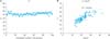
Figure 4A shows a plot of 100 hue values of the highest chroma among 10 extracted colors per eye. As indicated in Figure 4A, all 100 participants had a similar hue value. Figure 4B shows a graph plotting the lightness (L*) value of the representative color and the max value of chroma. The relationship between the max value and the lightness value of the chroma is relatively high, with a correlation coefficient of 0.68. This indicates that the chroma of the 10 colors of one eye was correctly extracted by the representative colors. Therefore, the representative color was determined to be the representative color of the total 100 samples clustered into 10 colors.
The representative color for each eye is plotted on the a*–b* plane and the C*–L* plane (Figure 5). The color of the iris exists in the positive plane of a*–b* and is distributed in the value between red and yellow. It should be noted that the range of values of hue angle (hab*) is small (45 to 55; range within 10). This indicates that the hue does not vary, but the chroma does vary, implying that the coloration of Koreans' irises appears to vary in a similar hue rather than different hues.
5) Iris color
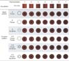
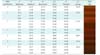
In the iris color distribution analysis, 100 irises were classified into 7 representative colors. The color difference (ΔE*ab) was used to reduce the color distribution to seven colors. A total of 4,950 color differences were calculated by forming groups of similar colors by sorting of 100 samples. Seven coloring groups were formed by grouping irises with minimal color difference. The average hue value in each group was calculated, and the corresponding representative color was selected from 2 to 3 colors in one group. As mentioned above, because the eyes are not in a single plane, various chroma and lightness values are grouped in the same color. The lightness, a*–b*, hue angle, and chroma values for the 7 representative colors are described in Table 5.
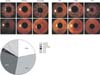
6) Iris pattern
For analysis of the iris pattern, 145 pieces of photographic data were used. A total of 131 patterns were classified into three basic patterns and three complex patterns. Among the basic patterns, the crown pattern appears shows sharp cogwheel shapes, whereas the flower pattern shows round petal-like shapes. The sunshine pattern comprises radially configured straight lines. A complex pattern is a pattern in which two of the three basic patterns are found together. The distribution of the sample data for these six patterns is shown in Figure 6. The three basic patterns of crown, flower, and sunshine accounted for 81% of the iris patterns, and the remaining 19% had complex patterns. The total representative color and pattern chart is shown in Figure 7.
IV. Discussion
This study aimed to classify the Korean iris colors extracted from digital photographs and apply them to the development of artificial eyes. Even with a digital photograph, it is difficult to judge the color of a human eye using a photograph alone. First, accurate photography of the specimen is necessary. It is recommended that the highlight be photographed in the pupil because it is not recognized as a color when caused by the reflection of the sclera, but it is recognized as an additional color in a photograph. Because the iris color of Koreans is generally dark, the iris color classification interval cannot be explained by genetics or disease, as has been determined by previous studies [1116]. Additional research is needed because the process of verifying iris color will be more reliable if it is explained in relation to genetic diseases. Although a previous study [16] found that age and gender have a significant effect on iris color, the lightness and color of the iris are significantly correlated with age [11]. This indicates that the lightness and color of the iris depend on other permanent factors or the race of individuals rather than differences, as reported by Edwards et al. [17], and that CIELAB values of the iris are simply due to age. In a study by Kim et al. [11], a higher L* value of the iris was correlated with a higher a*b* value of the iris, indicating the iris melanin constitution of color, especially since it depends on the hue. In addition, iris coloration is not affected by age and gender. When images are collected for color iris analysis, such as slit lamp images, the color and pattern of the iris image give light to the eyes, which can be displayed slightly differently than in a real eye. Future research is needed to study color differences visible on the standard model and the visual results regarding coloration and pattern.
In conclusion, we classified Korean iris colors and patterns for application in the fabrication of artificial eyes. We constructed a method to automatically extract colors from only photographs of subjects and classified them into seven color groups as light and dark brown based on previous iris color research. Unlike the eyes of individuals from other racial backgrounds, the Korean iris patterns corresponding to brown colors were classified into 6 categories.


 XML Download