

 PDF
PDF ePub
ePub Citation
 Print
Print


A 69-year-old woman visited the emergency room for aggravating dyspnea. She has suffered from bronchial asthma for the previous one year and varicose veins for decades. Her blood pressure was 142/82 mmHg, heart rate 108/min, and respiration rate 22/min. Her body mass index was 40.7 kg/m2.
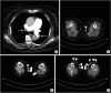
Chest radiograph was unremarkable, and electrocardiogram showed sinus tachycardia with T-wave inversion at leads V1-4. Laboratory study revealed elevated D-dimer of 11.90 mg/L (reference, < 0.49). Transthoracic echocardiography demonstrated right ventricular dysfunction with elevated pulmonary artery systolic pressure of 63 mmHg. Pulmonary embolism (PE) computed tomography showed filling defects in both main pulmonary, lobar, and segmental arteries and the right popliteal vein, indicating venous thromboembolism (VTE) (Figure 1, arrows). In addition, massive thrombosis in severe varicose veins was noted in bilateral great saphenous veins (GSVs) and muscle perforators (Figure 1, arrowheads). Endovenous laser ablation for varicose veins of both GSVs was performed after 6-months of anticoagulation therapy.
Superficial vein thrombosis (SVT) is estimated to be twice more prevalent than deep vein thrombosis (DVT) and PE, and DVT is a comorbidity in 18% and PE in 7% of patients with SVT.1) Varicose veins have been reported in 32%-100% of SVT patients, and their presence in SVT has been suggested to be a negative risk factor for concomitant VTE.1)2)3)4) However, as in this case, DVT with submassive PE can develop in a setting of long-standing varicose veins with SVT, so their clinical significance should not be neglected. Immediate assessment for concurrent VTE is required if clinically suspected.
 XML Download