Journal List > J Cardiovasc Imaging > v.26(4) > 1109822

Kazmi, Khanna, Soni, and Kapoor: Asymptomatic Multiple Lobulated Giant Left Ventricular Pseudoaneurysms Following Double Valve Replacement
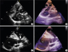
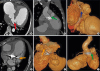
A 34-year-old male underwent double valve replacement with mechanical mitral and aortic prosthesis for rheumatic heart disease-severe mitral stenosis and moderate aortic stenosis and was asymptomatic at 1-year follow-up. On echocardiogram, a large aneurysmal sac arising from the left ventricle (LV) originating infero-posteriorly, passing anteriorly and compressing the right atrium and right ventricle was delineated (Figure 1A, B; Movie 1, 2). There was to-and-fro color flow into the cavity through a narrow neck (~20 mm) suggesting a large pseudoaneurysm (PSA) (Figure 1C, D; Movie 3). Normally functioning mitral (mean gradient 3.0 mmHg) and aortic (mean gradient 10 mmHg) prosthesis were noted. CT angiogram confirmed a giant lobulated PSA (13 × 12 × 10 cm) from the postero-inferior LV, coursing across AV groove and extending inferolaterally compressing the right hemi-diaphragm and superior surface of liver (Figure 2A, B, C). Though the superior part of sac had myocardial covering, the lower extension was covered with pericardium only. Two additional small PSAs were noted, one from superolateral LV wall (Figure 2D, E) and another from juxtavalvular aortic area (Figure 2B, F). The patient was advised urgent aneurysmectomy which he refused and was lost to follow-up.
LV PSAs can develop following valve replacement due to inadvertent intraoperative LV free wall invasion,1) mitral annular disruption, oversized prosthesis2) or myocardial erosion by prosthetic valve struts. Although prone to rupture,3) it may not be unusual to detect incidental asymptomatic PSAs.4) A multi-modality imaging using 2D, 3D echocardiography and CT helps in accurately delineating the surgical anatomy.

Figures and Tables

Figure 1

Transthoracic echocardiography. (A) Apical 4 chamber view with tilt revealed a PSA arising from LV and compressing RA and RV anteriorly and (B) the anatomy was better delineated using 3D imaging. (C, D) Modified parasternal short axis view localizes the origin of the aneurysm from the base of LV through a narrow neck along with delineation of its size and inferior extensions. LV: left ventricle, PSA: pseudoaneurysm, RA: right atrium, RV: right ventricle.

jcvi-26-250-g001
Figure 2

CT angiogram. (A) A large lobulated pseudoaneurysm (PSA) with narrow neck (red arrow) is seen arising from the base of the left ventricle wall, near the posterior atrioventricular groove. (B) This is further extending along the posterior atrioventricular groove and indenting on the dome of diaphragm anteriorly. (C) Volume rendered image is clearly depicting the neck (blue arrow) of aneurysm. Axial oblique coronary CT angiogram (D) and volume rendered image (E) show another small lobulated PSA with narrow neck (orange arrow), arising from the lateral wall of the left ventricle, near mitral valve. Coronal oblique coronary CT angiogram (B) and volume rendered image (F) show the third small lobulated PSA (green arrows in B and F), juxta aortic annulus.

jcvi-26-250-g002

Notes

Conflict of Interest The authors have no financial conflicts of interest.

References

1. Roberts WC, Morrow AG. Causes of early postoperative death following cardiac valve replacement. Clinico-pathologic correlations in 64 patients studied at necropsy. J Thorac Cardiovasc Surg. 1967; 54:422–437.
2. Waller BF, Taliercio CP, Clark M, Pless JE. Rupture of the left ventricular free wall following mitral valve replacement for mitral stenosis: a cause of complete (fatal) or contained (false aneurysm) cardiac rupture. Clin Cardiol. 1991; 14:341–345.
crossref
3. Frances C, Romero A, Grady D. Left ventricular pseudoaneurysm. J Am Coll Cardiol. 1998; 32:557–561.
crossref
4. Hulten EA, Blankstein R. Pseudoaneurysms of the heart. Circulation. 2012; 125:1920–1925.
crossref

SUPPLEMENTARY MATERIALS

Movie 1

Movie 2

Movie 3

TOOLS
Similar articles