Journal List > J Cardiovasc Ultrasound > v.16(3) > 1018101

Nagueh: Tissue Doppler Imaging for the Assessment of Left Ventricular Diastolic Function

Introduction

Echocardiography plays an essential role in the evaluation of left ventricular (LV) diastolic function. Over the past decade, the diagnostic armamentarium was fortified by tissue Doppler imaging (TDI). The technique is based on recording the high amplitude, low frequency signals generated by tissue motion.1) Myocardial velocities can be measured in a longitudinal and a radial direction, and can be used to compute regional displacement and deformation. TDI has been successfully applied to study segmental LV systolic and diastolic function, since myocardial velocities are strongly related to interstitial fibrosis,2) beta-adrenergic receptor density,2) and regional cytokine expression.3) For examining global LV diastolic function, mitral annulus velocities are recorded by Pulsed-wave Doppler and the velocities can be measured on line from the spectral display (Fig. 1).

Relation of Early Diastolic Velocity to LV Relaxation and Preload

A number of observational studies4)5) have shown an inverse relation between age and the mitral annulus early diastolic velocity (e'). This observation is similar to other Doppler parameters which are age dependent, as the E/A (peak early filling velocity to peak late filling velocity) ratio in mitral inflow, and S/D (peak systolic to peak diastolic) ratio in pulmonary venous flow. In animal studies,6)7) e' related well to the time constant of LV relaxation (τ) and - dP/dt. Likewise, human studies8-10) have shown a significant inverse correlation between e' and τ.
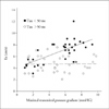
The relation between e' and preload is more complicated. When LV relaxation is normal,6)7) preload is directly related to e'. On the other hand, preload has a minimal effect on e' with impaired LV relaxation (Fig. 2). These animal observations were also noted in humans when saline and nitroglycerin infusion8) had no effect on septal e' velocity in cardiac patients, whereas mean pulmonary capillary wedge pressure was directly related to e' in normal subjects.11) In summary, it is valid to use e' as a surrogate marker of LV relaxation in cardiac patients where LV relaxation is impaired, but not in normal individuals.

Relation of E' to Regional Function

Lateral e' velocity is usually higher than septal e' velocity.4) This difference can be exaggerated or reversed depending on the presence and extent of cardiac pathology. For example, this difference is exaggerated in patients with hypertrophic cardiomyopathy,12) septal infarction,13) or those with primary pulmonary hypertension.14) However, in patients with lateral wall infraction, septal e' velocity can be much higher than the lateral e' velocity.
An animal study15) showed that in the setting of myocardial ischemia, e' is dependent not only on global LV systolic and diastolic function, but also on regional thickening. Accordingly, it is imperative to use the average of septal and lateral e' velocities when drawing conclusions on LV diastolic function in patients with regional dysfunction.

Relation of E' to Cycle Length

Few studies have addressed this important question. We have noted in an animal model that e' is affected heart rate.15) Heart rate affected the transmitral pressure gradient (a higher heart rate reduces the transmitral pressure gradient), which in turn led to a reduced e'. Therefore, caution should be exercised in using e' to draw conclusions on LV relaxation in normal subjects with sinus tachycardia. On the other, e' relates significantly to τ in patients with cardiac disease, whether in regular tachycardia,16) or atrial fibrillation.17)

Doppler Assessment of LV Filling Pressures

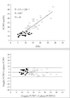
The ratio of mitral E velocity to annular e' velocity corrects for the effect of LV relaxation on mitral E velocity, and can be used to estimate LV filling pressures (Fig. 3). This method has been validated by several laboratories and used for patients with normal and depressed EF,4)10)18) sinus tachycardia,16)18) atrial fibrillation,17) pulmonary hypertension,14) and hypertrophic cardiomyopathy.12) It is usually not critical to use TDI in predicting LV filling pressures in patients with depressed EF. However, in patients with normal EF, TDI is the most reliable method for the assessment of LV relaxation and filling pressures.13)19) Importantly, 2 studies have shown the lateral velocity13)19) to be more accurate in that regard. However, if the septal velocity is used, an E/e' ratio <8 favors the presence of normal filling pressures, whereas a ratio >15 is usually associated with increased LV diastolic pressures.10) If the average of septal and lateral e' is used, a ratio <8 identifies patients with normal filling pressure, whereas a ratio >13 identifies those with increased filling pressures.13) When the ratio falls between these cutoffs, other echocardiographic measurements are needed. These include mitral inflow velocities, pulmonary venous flow velocities, pulmonary artery pressures, and LA volume index.

Comparison with BNP

BNP is currently used to determine the underlying etiology of dyspnea, as cardiac patients usually have an increased BNP level. The comparative performance of BNP and Doppler echocardiography was recently reported. BNP had a significant positive correlation with mean wedge pressure, but with a wide scatter.20) It also failed to detect changes in filling pressures with therapy. On the other hand, TDI (E/e' ratio) was more accurate in identifying patients with increased filling pressures, and tracked well the changes in mean wedge pressure with therapy.20)

Time Intervals

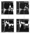
We observed in an animal model a decrease in the peak e' velocity and a delay in its onset with myocardial ischemia.15) Furthermore, e' is delayed (Fig. 4) such that it occurs after the onset of mitral E velocity.6)7) The time interval: TE-e' relates well with invasive indices of LV relaxation, and has been used for the estimation of LV filling pressures (in conjunction with end systolic pressure and isovolumetric relaxation time) in normal individuals, in patients with depressed EF, those with normal EF, and patients with mitral valve disease.21)22) The highest accuracy was obtained when the average time interval was computed from septal, lateral, anterior, and inferior TD signals.21) Recently, it was successfully used to identify patients with increased LV filling pressures23) when the E/e' fell in the indeterminate range of 8-15.

Role of TDI in Constrictive Pericarditis

Most patients with pericardial constriction have a normal or increased e' velocity (>7 cm/s). In this disease, septal e' velocity is usually higher than lateral e' velocity, and the E/e' ratio is inversely related to LV filling pressures.24) Recent studies have also noted that the presence of a reduced annular systolic velocity and a prolonged TE-e' are of value in borderline cases25) as they identify patients with primarily myocardial (as opposed to pericardial) disease.

Late Diastolic Velocity (a')

This velocity is affected by left atrial dP/dt and LV end diastolic pressure. Reduced left contractility leads to a decreased a' velocity. Likewise, an increase in LV end diastolic pressure (after-load) also leads to a decrease in a'.6) In patients with early diastolic dysfunction (impaired relaxation filling pattern), e'/a' ratio is reduced. With progressive diastolic dysfunction, a' velocity decreases, and e'/a' ratio is ≥1.8) Recently, e'/a' ratio was reported to identify well patients with diastolic heart failure as determined by high fidelity LV pressure/volume measurements.19)

Prognostic Power of Myocardial Diastolic Velocities

Mitral annulus e' velocity, a' velocity, and E/e' ratio have been shown to provide important prognostic information in several patient groups. This includes patients with heart failure,26-29) acute myocardial infarction,30) chronic renal failure,31) non-valvular atrial fibrillation,32) mitral regurgitation,33) cardiomyopathy,34)35) general community,36) and hypertension.37) In many of the above studies, TDI provided prognostic information that was incremental to clinical data, and conventional echocardiographic measurements.

Conclusion

Echocardiography plays an essential role in the evaluation of LV diastolic function in cardiac patients. The technique provides important diagnostic and prognostic information in this population as discussed in the above paragraphs.

Figures and Tables

Fig. 1
Tissue Doppler signals from the lateral side of the mitral annulus. S: systolic velocity during ejection, Ea: early diastolic mitral annulus velocity, Aa: late diastolic mitral annulus velocity.
jcu-16-76-g001
Fig. 2
Relation between maximal transmitral pressure gradient and e' velocity in the presence of normal or enhanced LV relaxation (solid circles and continuous line), and impaired LV relaxation (open circles and interrupted line).6)
jcu-16-76-g002
Fig. 3
Correlation between E/e' ratio and mean pulmonary capillary wedge pressure (PCWP) in 60 patients with cardiac disease.4)
jcu-16-76-g003
Fig. 4
Mitral inflow and TD signals at baseline and after circumflex occlusion from one of the canine experiments.21) Notice the reduced and delayed e' after circumflex occlusion.
jcu-16-76-g004

References

1. Waggoner AD, Bierig SM. Tissue Doppler imaging: a useful echocardiographic method for the cardiac sonographrer to assess systolic and diastolic left ventricular function. J Am Soc Echocardiogr. 2001. 14:1143–1152.
crossref
2. Shan K, Bick RJ, Poindexter BJ, Shimoni S, Letsou GV, Reardon MJ, Howell JF, Zoghbi WA, Nagueh SF. Relation of tissue Doppler derived myocardial velocities to myocardial structure and beta-adrenergic receptor density in humans. J Am Coll Cardiol. 2000. 36:891–896.
crossref
3. Kalra DK, Ramchandani M, Zhu X, Lawrie G, Reardon MJ, Mann DL, Zoghbi WA, Nagueh SF. Relation of tissue Doppler-derived myocardial velocities to serum levels and myocardial gene expression of tumor necrosis factor-alpha and inducible nitric oxide synthase in patients with ischemic cardiomyopathy having coronary artery bypass grafting. Am J Cardiol. 2002. 90:708–712.
crossref
4. Nagueh SF, Middleton KJ, Kopelen HA, Zoghbi WA, Quinones MA. Doppler tissue imaging: a non-invasive technique for evaluation of left ventricular relaxation and estimation of filling pressures. J Am Coll Cardiol. 1997. 30:1527–1533.
crossref
5. De Sutter J, De Backer J, Van de Veire N, Velghe A, De Buyzere M, Gillebert TC. Effects of age, gender, and left ventricular mass on septal mitral annulus velocity (E') and the ratio of transmitral early peak velocity to E' (E/E'). Am J Cardiol. 2005. 95:1020–1023.
crossref
6. Nagueh SF, Sun H, Kopelen HA, Middleton KJ, Khoury DS. Hemodynamic determinants of mitral annulus diastolic velocities by tissue Doppler. J Am Coll Cardiol. 2001. 37:278–285.
crossref
7. Hasegawa H, Little WC, Ohno M, Brucks S, Morimoto A, Cheng HJ, Cheng CP. Diastolic mitral annular velocity during the development of heart failure. J Am Coll Cardiol. 2003. 41:1590–1597.
crossref
8. Sohn DW, Chai IH, Lee DJ, Kim HC, Kim HS, Oh BH, Lee MM, Park YB, Choi YS, Seo JD, Lee YW. Assessment of mitral annulus velocity by Doppler tissue imaging in evaluation of left ventricular diastolic function. J Am Coll Cardiol. 1997. 30:474–480.
crossref
9. Oki T, Tabata T, Yamada H, Wakatsuki T, Shinohara H, Nishikado A, Luchi A, Fukuda N, Susumo I. Clinical application of pulsed tissue Doppler imaging for assessing abnormal left ventricular relaxation. Am J Cardiol. 1997. 79:921–928.
crossref
10. Ommen SR, Nishimura RA, Appleton CP, Miller FA, Oh JK, Relfield MM, Tajik AJ. Clinical utility of Doppler echocardiography and tissue Doppler imaging in the estimation of left ventricular filling pressures: a comparative simultaneous Doppler-catheterization study. Circulation. 2000. 102:1788–1794.
crossref
11. Firstenberg MS, Levine BD, Garcia MJ, Greenberg NL, Cardon L, Morehead AJ, Zuckerman J, Thomas JD. Relationship of echocardiographic indices to pulmonary capillary wedge pressures in healthy volunteers. J Am Coll Cardiol. 2000. 36:1664–1669.
crossref
12. Nagueh SF, Lakkis NM, Middleton KJ, Spencer WH 3rd, Zoghbi WA, Quinones MA. Doppler estimation of left ventricular filling pressures in patients with hypertrophic cardiomyopathy. Circulation. 1999. 99:254–261.
crossref
13. Rivas-Gotz C, Manolios M, Thohan V, Nagueh SF. Impact of left ventricular ejection fraction on estimation of left ventricular filling pressures using tissue Doppler and flow propagation velocity. Am J Cardiol. 2003. 91:780–784.
crossref
14. Ruan Q, Nagueh SF. Clinical application of tissue Doppler imaging in patients with idiopathic pulmonary hypertension. Chest. 2007. 131:395–401.
crossref
15. Nagueh SF, Rao L, Soto J, Middleton KJ, Khoury DS. Haemodynamic insights into the effects of ischaemia and cycle length on tissue Doppler-derived mitral annulus diastolic velocities. Clin Sci (Lond). 2004. 106:147–154.
crossref
16. Sohn DW, Kim YJ, Kim HC, Chun HG, Park YB, Choi YS. Evaluation of left ventricular diastolic function when mitral E and A waves are completely fused: role of assessing mitral annulus velocity. J Am Soc Echocardiogr. 1999. 12:203–208.
crossref
17. Sohn DW, Song JM, Zo JH, Chai IH, Kim HS, Chun HG, Kim HC. Mitral annulus velocity in the evaluation of left ventricular diastolic function in atrial fibrillation. J Am Soc Echocardiogr. 1999. 12:927–931.
crossref
18. Nagueh SF, Mikati I, Kopelen HA, Middleton KJ, Quinones MA, Zoghbi WA. Doppler estimation of left ventricular filling pressure in sinus tachycardia. A new application of tissue Doppler imaging. Circulation. 1998. 98:1644–1650.
crossref
19. Kasner M, Westermann D, Steendijk P, Gaub R, Wilkenshoff U, Weitmann K, Hoffmann W, Poller W, Schultheiss HP, Pauschinger M, Tschope C. Utility of Doppler echocardiography and tissue Doppler imaging in the estimation of diastolic function in heart failure with normal ejection fraction: a comparative Doppler-conductance catheterization study. Circulation. 2007. 11:637–647.
20. Dokainish H, Zoghbi WA, Lakkis NM, Al-Bakshy F, Dhir M, Quinones MA, Nagueh SF. Optimal noninvasive assessment of left ventricular filling pressures: a comparison of tissue Doppler echocardiography and B-type natriuretic peptide in patients with pulmonary artery catheters. Circulation. 2004. 109:2432–2439.
crossref
21. Rivas-Gotz C, Khoury DS, Manolios M, Rao L, Kopelen HA, Nagueh SF. Time interval between onset of mitral inflow and onset of early diastolic velocity by tissue Doppler: a novel index of left ventricular relaxation: experimental studies and clinical application. J Am Coll Cardiol. 2003. 42:1463–1470.
crossref
22. Diwan A, McCulloch M, Lawrie GM, Reardon MJ, Nagueh SF. Doppler estimation of left ventricular filling pressures in patients with mitral valve disease. Circulation. 2005. 111:3281–3289.
crossref
23. Min PK, Ha JW, Jung JH, Choi EY, Choi D, Rim SJ, Jang Y, Shim WH, Cho SY, Chung N. Incremental value of measuring the time difference between onset of mitral inflow and onset of early diastolic mitral annulus velocity for the evaluation of left ventricular diastolic pressures in patients with normal systolic function and an indeterminate E/E'. Am J Cardiol. 2007. 100:326–330.
crossref
24. Ha JW, Oh JK, Ling LH, Nishimura RA, Seward JB, Tajik AJ. Annulus paradoxus: transmitral flow velocity to mitral annular velocity ratio is inversely proportional to pulmonary capillary wedge pressure in patients with constrictive pericarditis. Circulation. 2001. 104:976–978.
25. Choi EY, Ha JW, Kim JM, Ahn JA, Seo HS, Lee JH, Rim SJ, Chung N. Incremental value of combining systolic mitral annular velocity and time difference between mitral inflow and diastolic mitral annular velocity to early diastolic annular velocity for differentiating constrictive pericarditis from restrictive cardiomyopathy. J Am Soc Echocardiogr. 2007. 20:738–743.
crossref
26. Wang M, Yip G, Yu CM, Zhang Q, Zhang Y, Tse D, Kong SL, Sanderson JE. Independent and incremental prognostic value of early mitral annulus velocity in patients with impaired left ventricular systolic function. J Am Coll Cardiol. 2005. 45:272–277.
crossref
27. Dokainish H, Zoghbi WA, Lakkis NM, Ambriz E, Patel R, Quinones MA, Nagueh SF. Incremental predictive power of B-type natriuretic peptide and tissue Doppler echocardiography in the prognosis of patients with congestive heart failure. J Am Coll Cardiol. 2005. 45:1223–1226.
crossref
28. Yamamoto T, Oki T, Yamada H, Tanaka H, Ishimoto T, Wakatsuki T, Tabata T, Ito S. Prognostic value of the atrial systolic mitral annular motion velocity in patients with left ventricular systolic dysfunction. J Am Soc Echocardiogr. 2003. 16:333–339.
crossref
29. Troughton RW, Prior DL, Frampton CM, Nash PJ, Pereira JJ, Martin M, Fogarty A, Morehead AJ, Starling RC, Young JB, Thomas JD, Lauer MS, Klein AL. Usefulness of tissue Doppler and color M-mode indexes of left ventricular diastolic function in predicting outcomes in systolic left ventricular heart failure (from the ADEPT study). Am J Cardiol. 2005. 96:257–262.
crossref
30. Hillis GS, Moller JE, Pellikka PA, Gersh BJ, Wright RS, Ommen SR, Reeder GS, Oh JK. Noninvasive estimation of left ventricular filling pressure by E/E' is a powerful predictor of survival after acute myocardial infarction. J Am Coll Cardiol. 2004. 43:360–367.
crossref
31. Sharma R, Pellerin D, Gaze DC, Mehta RL, Gregson H, Streather CP, Collinson PO, Brecker SJ. Mitral peak Doppler E-wave to peak mitral annulus velocity ratio is an accurate estimate of left ventricular filling pressure and predicts mortality in end-stage renal disease. J Am Soc Echocardiogr. 2006. 19:266–273.
crossref
32. Okura H, Takada Y, Kubo T, Iwata K, Mizoguchi S, Taguchi H, Toda I, Yoshikawa J, Yoshida K. Tissue Doppler-derived index of left ventricular filling pressure, E/E', predicts survival of patients with non-valvular atrial fibrillation. Heart. 2006. 92:1248–1252.
crossref
33. Bruch C, Klem I, Breithardt G, Wichter T, Gradaus R. Diagnostic usefulness and prognostic implications of the mitral E/E' ratio in patients with heart failure and severe secondary mitral regurgitation. Am J Cardiol. 2007. 100:860–865.
crossref
34. McMahon CJ, Nagueh SF, Pignatelli RH, Denfield SW, Dreyer WJ, Price JF, Clunie S, Bezold LI, Hays AL, Towbin JA, Eidem BW. Characterization of left ventricular diastolic function by tissue Doppler imaging and clinical status in children with hypertrophic cardiomyopathy. Circulation. 2004. 109:1756–1762.
crossref
35. McMahon CJ, Nagueh SF, Eapen RS, Dreyer WJ, Finkelshtyn I, Cao X, Eidem BW, Bezold LI, Denfield SW, Towbin JA, Pignatelli RH. Echocardiographic predictors of adverse clinical events in children with dilated cardiomyopathy: a prospective clinical study. Heart. 2004. 90:908–915.
crossref
36. Redfield MM, Jacobsen SJ, Burnett JC Jr, Mahoney DW, Bailey KR, Rodeheffer RJ. Burden of systolic and diastolic ventricular dysfunction in the community: appreciating the scope of the heart failure epidemic. JAMA. 2003. 289:194–202.
crossref
37. Wang M, Yip GW, Wang AY, Zhang Y, Ho PY, Tse MK, Yu CM, Sanderson JE. Tissue Doppler imaging provides incremental prognostic value in patients with systemic hypertension and left ventricular hypertrophy. J Hypertens. 2005. 23:183–191.
crossref
TOOLS
Similar articles