

 PDF
PDF Citation
 Print
Print


Abstract
Objectives
To document the first known case of posterior migration of a herniated disc in a lumbar flexion-distraction injury.
Summary of Literature Review
Lumbar disc herniation is sometimes confused with epidural hematoma, especially when the disc migrates posterior to the thecal sac. There has been no report of posterior migration of a herniated disc after a lumbar flexion-distraction injury.
Materials and Methods
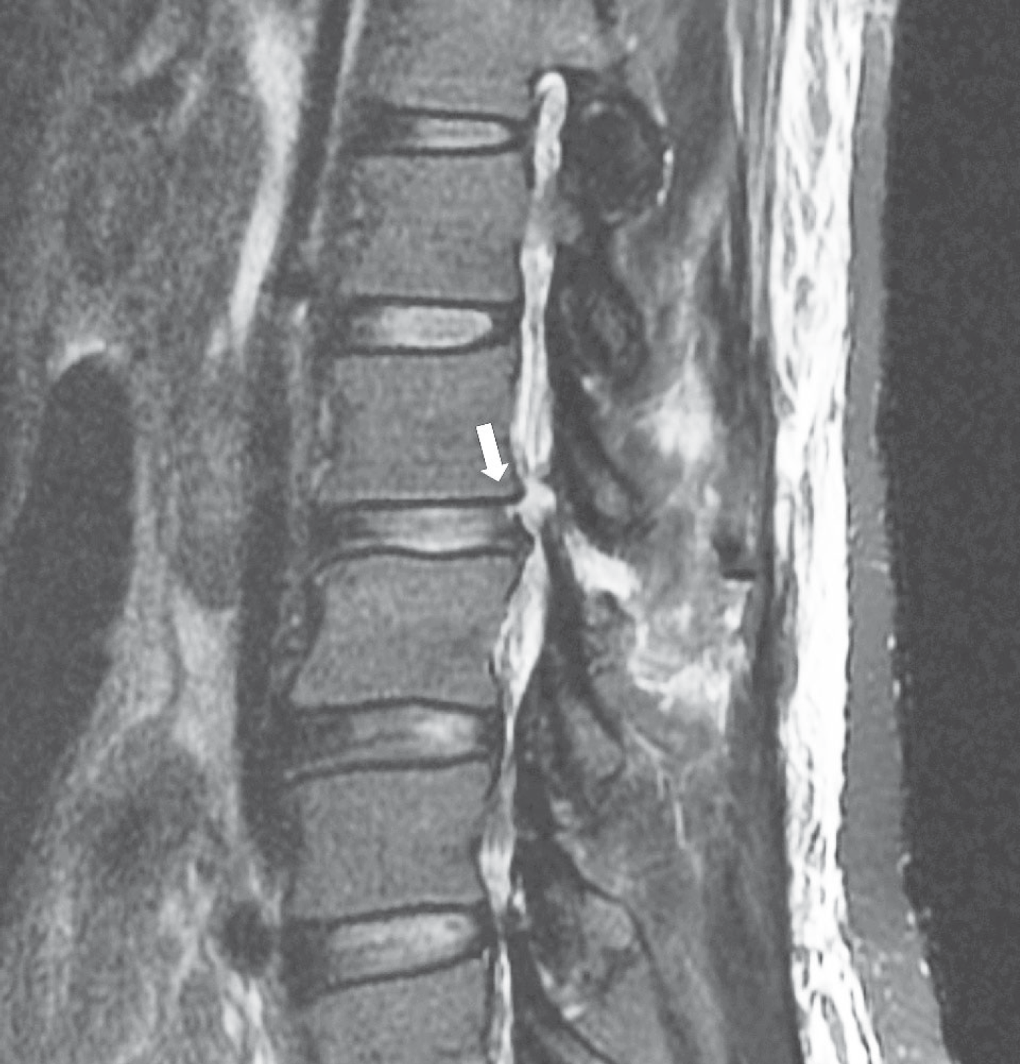
A 47-year-old woman with no pertinent medical history was diagnosed with a flexion-distraction injury of the L2–L3 vertebrae after a motor vehicle accident. The patient had no neurological deficit initially. Magnetic resonance imaging (MRI) showed a space-occupying lesion with T2 hyperintensity and T1 isointensity on the dorsal side of the thecal sac at L2–L3. After posterior lumbar fixation and fusion, progressive leg weakness occurred 1 week postoperatively.
Results
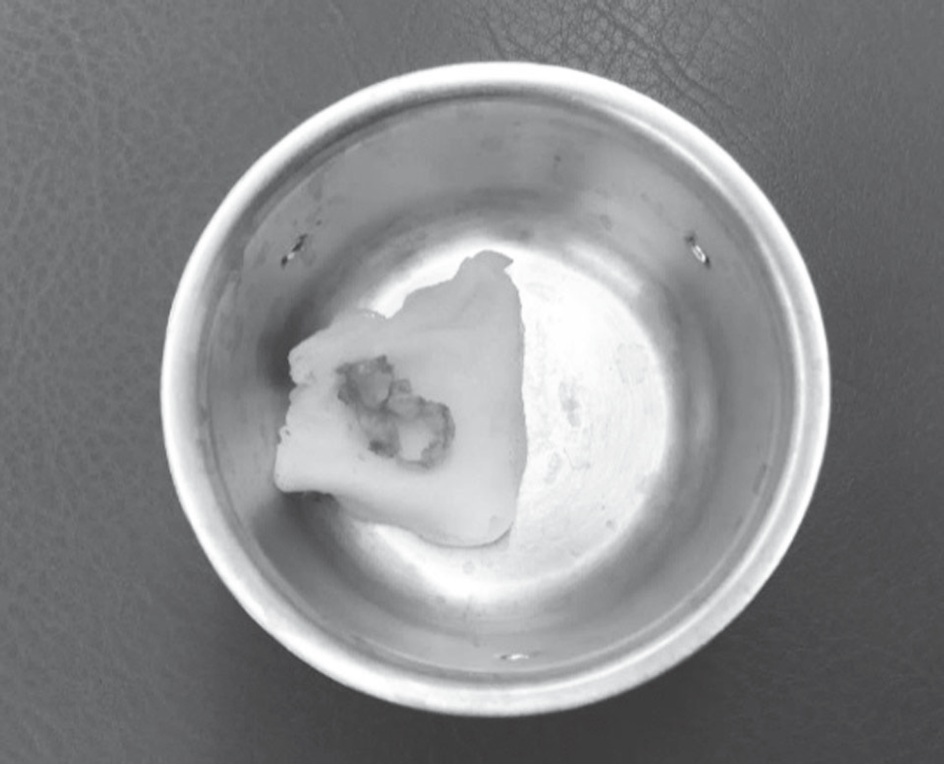
A second operation revealed no evidence of epidural hematoma, but a sequestrated disc. Decompression and sequestrectomy were performed, and the patient's neurological status had recovered fully at 4 months postoperatively.
Conclusions
This case highlights the potential for posterior migration of a herniated disc with flexion-distraction injuries of the thoracolumbar spine. Discontinuity of the posterior annulus fibrosus on MRI may aid the distinction of posterior migration of a herniated disc from epidural hematoma. Because posterior migration of a herniated disc is associated with progressive neurological deficits, surgeons must consider decompression surgery when such herniation is suspected, even in the absence of neurological symptoms.
REFERENCES
1. Kim JH, Kim SH, Lee SK, et al. Traumatic lumbar disc herniation mimicking epidural hematoma: A case report and literature review. Medicine. 2019 May; 98(18):E15438. DOI: 10.1097/MD.0000000000015438.
2. Roaf R. A study of the mechanics of spinal injuries. The Journal of Bone and Joint Surgery. British volume. 1960 Nov; 42:810–23. DOI: 10.1302/0301-620X.42B4.810.
3. Jain N, Crouser N, Yu E. Lumbar intervertebral disc herniation masquerading as an epidural hematoma: A case report and review of the literature. JBJS case connector. 2018 Jul-Sep; 8(3):E59. DOI: 10.2106/JBJS.CC.17.00300.
4. Dorsay T, Helms C. Mr imaging of epidural hematoma in the lumbar spine. Skeletal radiology. 2002 Dec; 31(12):677–85. DOI: 10.1007/s00256-002-0584-y.

5. Weinstein J, Lurie J, Tosteson T, et al. Surgical vs nonop-erative treatment for lumbar disk herniation: The spine patient outcomes research trial (sport) observational cohort. Jama. 2006 Nov; 296(20):2451–9. DOI: 10.1001/jama.296.20.2451k.
6. Tamburrelli F, Meluzio M, Masci G, et al. Etiopathogenesis of traumatic spinal epidural hematoma. Neurospine. 2018 Mar; 15(1):101–7. DOI: 10.14245/ns.1834938.469.pp.
7. Akhaddar A, El-Asri A, Boucetta M. Posterior epidural migration of a lumbar disc fragment: A series of 6 cases: A review. Journal of Neurosurgery: Spine. 2011 Jul; 15(1):117–28. DOI: 10.3171/2011.3.SPINE10832.
8. Elsharkawy A, Hagemann A, Klassen P. Posterior epidural migration of herniated lumbar disc fragment: A literature review. Neurosurgical review. 2019 Jan. 1-13. DOI: 10.1007/s10143-018-01065-1.
9. Lewis S, Amritanand R. Traumatic disc herniation following flexion-distraction injury of the thoracolumbar spine: A rare presentation. Journal of Trauma & Treatment. 2016 Jan; 5:293. DOI: 10.4172/2167-1222.1000293.
10. Finkelstein J, Wai E, Jackson S, et al. Single-level fixation of flexion distraction injuries. Clinical Spine Surgery. 2003 Jun; 16(3):236–42.
Fig. 1.
Anteroposterior (A) and lateral (B) radiographs of the lumbar spine at the initial assessment show a compression fracture of L3 (arrow) and increased distance between the L2 and L3 spinous processes (bidi-rectional arrow).
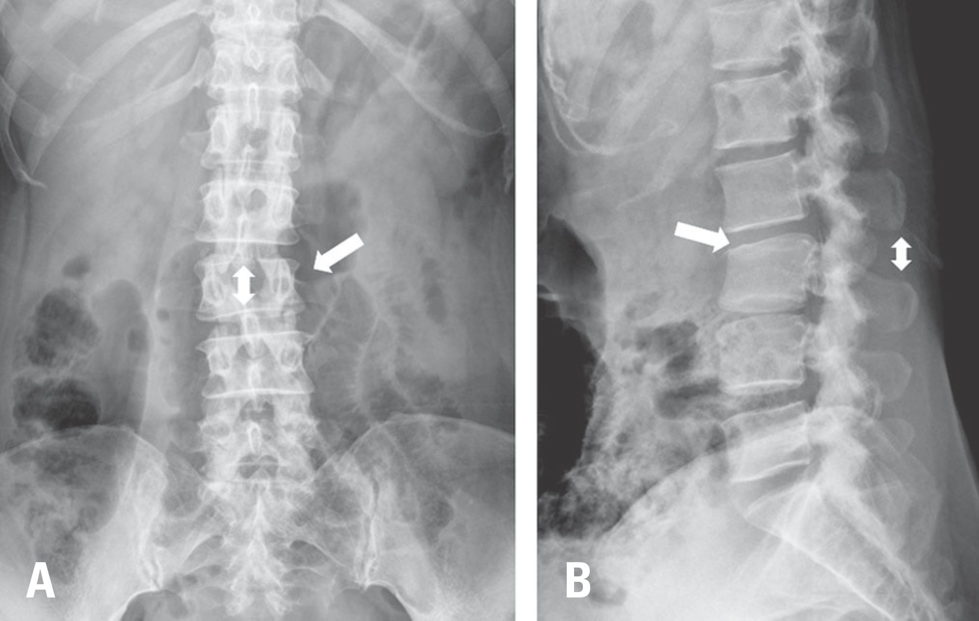
Fig. 2.
T2-weighted sagittal (A) and axial (B) magnetic resonance images of the lumbar spine at the initial assessment show a hyperintense lesion (white arrow). T1-weighted sagittal (C) and axial (D) images show an isointense lesion (gray arrow).
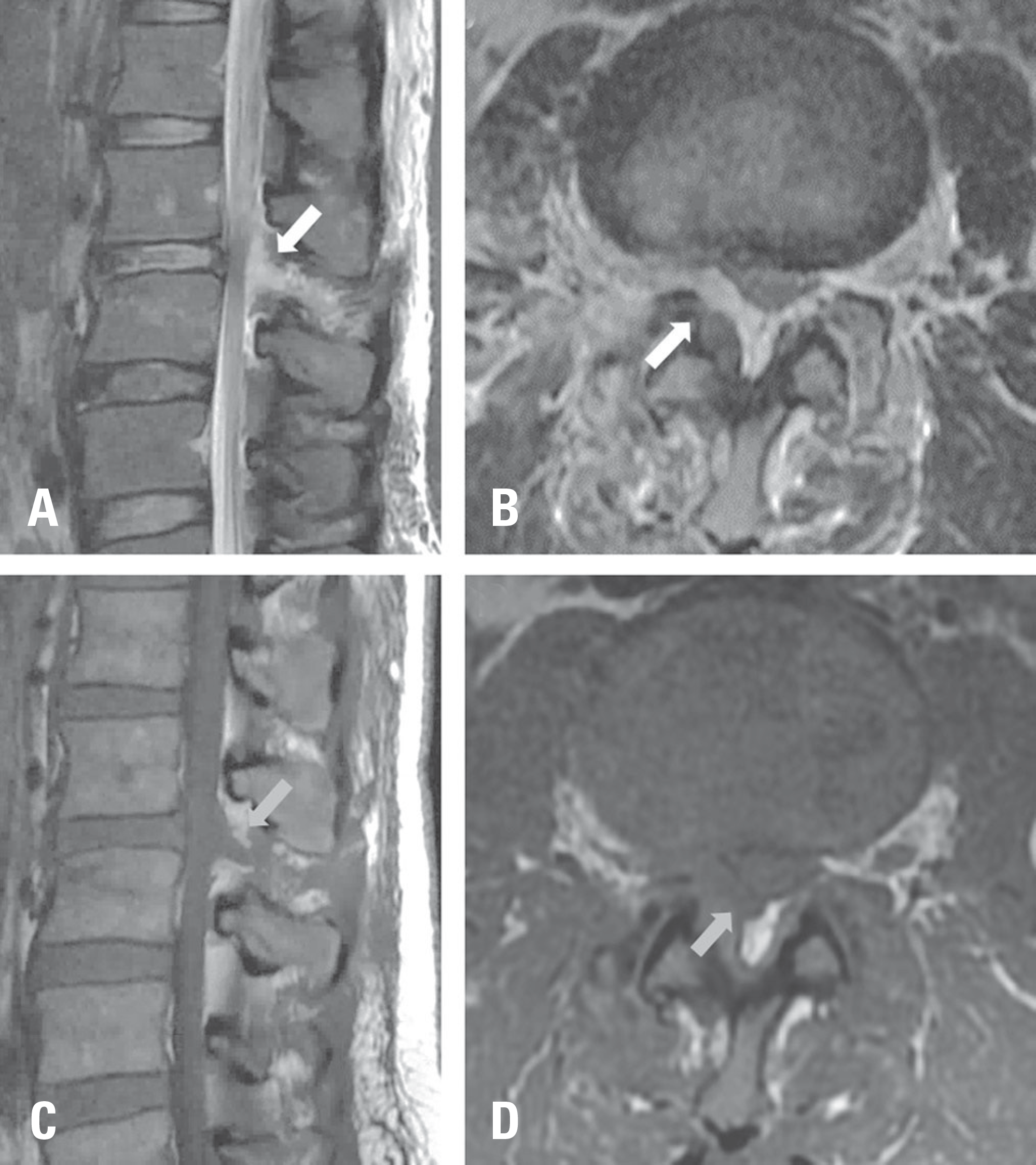
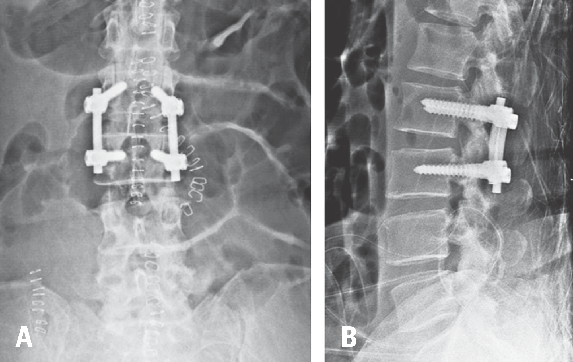
Fig. 3.
Anteroposterior (A) and lateral (B) radiographs of the lumbar spine after the first operation show single-level fusion with instrumentation and the correction of spinal alignment.
 XML Download