

 PDF
PDF ePub
ePub Citation
 Print
Print


Abstract
Objective
We report a case of treated failure spinal construct and pseudarthrosis in a patient with Parkinson's disease.
Summary of Literature Review
There have been no reports about revision surgery due to failure and pseuarthrosis of degenerative lumbar spine disease in patients with Parkinson's disease.
Materials and Methods
A 55-year-old female who had been diagnosed with Parkinson's disease 4 years ago presented with back pain and radiating pain on both legs. Radiographic assessment showed spinal stenosis from L2 to L5 combined with degenerative spondylolisthesis at L3-4. Posterior decompression, instrumentation, and posterolateral fusion were performed and her symptoms improved.
Results
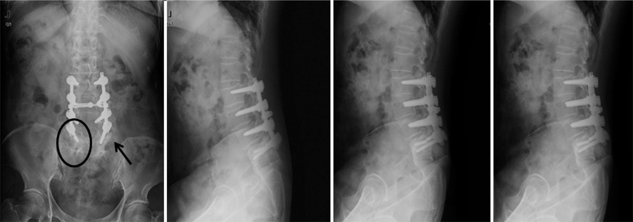
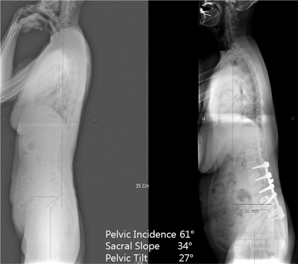
Two years after the operation, she complained of severe back pain without injury. A simple X-ray showed the pull out of bilateral L5 screws, and revision surgery was performed. Three years after the revision, she underwent re-reoperation due to metal failure. The breakage of a unilateral pedicle screw at L5 was found, and her fusion level was extended to S1 with a posterior lumbar interbody fusion with cages and alar screws. Finally, she has not shown any further failure but, a sagittal imbalance and aggravation of pelvic incidence due to Parkinson's disease have been detected.
REFERENCES
1. Adams RD, Victor M Principles of Neruology 2nd ed. New York: McGraw-Hill;1981. 807.
2. Seo WK, Koh SB, Kim BJ, Yu SW, Park MH, Park KW, Lee DH. Prevalence of Parkinson's disease in Korea. J Clin Neurosci. 2007; 14:1155–7.

3. Yoon HK, Kim BK, Shin DE, Song SJ, Park HK, Chang JH. Bipolar hemiarthroplasty of displaced femoral neck fracu-tres ion Parkinsonism patients. J of Korean Fracture Society. 2005; 18:126–30.
4. Hoehn MM, Yahr MD. Parkinsonim: onset, progression, and mortality. Neurology. 1967; 17:427–42.
5. Babat LB, McLain RF, Bingaman W, Kalfas I, Young P, Rufo-Smith C. Spinal surgery in patients with Parkinson’s disease: contruct failure and progressive deformity. Spine (Phila Pa 1976). 2004; 29:2006–12.
6. Laroche M, Delisle M, Aziza R, Laqarrique J, Mazieres B. Is camptrocormia a primary muscular disease? Spine (Phila Pa 1976). 1995; 20:1011–6.
7. Gau YL, Lonstein JE, Winter RB, Koop S, Denis F. Luque-Galveston procedure for correction and stabilization of neuromuscular scoliosis and pelvic obliquity: a review of 68 patients. J Spinal Disord. 1991; 4:399–410.
8. Bell DF, Moseley CF, Koresca J. Unit rod segmental spinal instrumentation in the management of patients with progressive mneuromuscular spinal deformity. Spine (Phila Pa 1976). 1989; 14:1301–7.
9. Banta JV, Drummond DS, Ferguson RL. The treatment of neuromuscular scoliosis. Instruct Course Lect. 1999; 64:551–62.
10. Swank SM, Cohen DS, Brown JC. Spine fusion in cerebral palsy with L-rod segmental spinal instrumentation. A comparison of single and two-stage combined approach with Zilke instrumentation. Spine (Phila Pa 1976). 1989; 14:750–9.
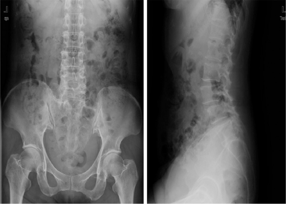
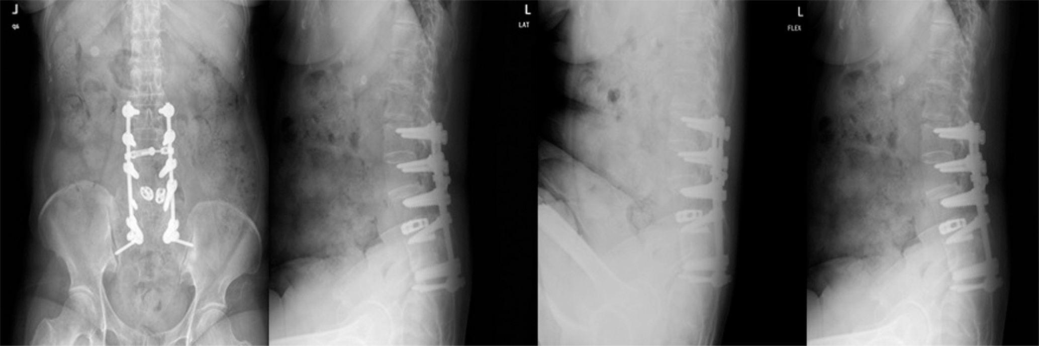
Fig. 3.
X-rays show halo (arrows) around bilateral L5 screws and loosening of L5 screws (circles) were found on dynamic study after revision.
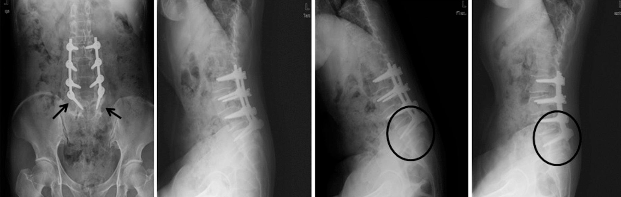
Fig. 4.
2 year after revision, screw breakage of left L5 (arrow) was found and halo (circles) was found around right L5 pedicle screw.
 XML Download