

 PDF
PDF ePub
ePub Citation
 Print
Print


Abstract
Objectives
In patients with fracture-dislocation of the lumbar spine with neurologic deficit and hemodynamic instability, minimally invasive surgery made/produced good clinical results. So the authors have reported the results with literature review
Summary of Literature Review
In patients with unstable lumbar spine fracture-dislocation, early surgical treatment has been preferred due to its many advantages of anatomical reduction, nerve decompression, recovery of nerve function, and early rehabilitation, etc. But for patients with unstable lumbar spine fracture-dislocation and who are hemodynamically unstable, the surgical treatment is generally delayed, so there are many cases that cannot fulfill the expectation of neurologic recovery.
Materials and Methods
In patients with unstable lumbar 2-3 spine fracture-dislocation and who are hemodynamically unstable, applying the concept of stage operation, postural reduction and minimal invasive percutaneous pedicle screw fixation were conducted as soon as possible. Then after recover of general condition, decompression and posterior fusion were conducted as a second stage operation.
Results
After the first stage operation, motor grade was improved from 3 to 4 below the L3 spine level in postoperative physical examination. The second stage operation was conducted two weeks later and neurologic symptom was more improved after the second stage operation.
Conclusions
In patients with lumbar spine fracture-dislocation having hemodynamic instability and neurologic deficit, early minimally invasive fixation for reducing complications of open reduction and internal fixation may contribute to improving general conditions and recovery of neurologic deficits.
REFERENCES
1. Hsieh CT, Chen GJ. Complete fracture-dislocation of the thoracolumbar spine without paraplegia. Am J Emerg Med. 2008; 26:E5–7.

2. Bedbrook GM. Treatment of toracolumbar dislocation and fracture with paraplegia. Clin Orthop Relat Res. 1975; 112:27–43.
3. Roberts JB, Curtiss PH Jr. Stability of the Thoracic and Lumbar Spine in Traumatic Paraplegia following Fracture of Fracture-Dislocation. J Bone Joint Surg Am. 1970; 52:1115–30.
4. Holdsworth FW, Hardy A. Early Treatment of Paraplegia from Fractures of the Thoracolumbar Spine. J Bone Joint Surg Br. 1953; 35:540–50.
5. Dickson JH, Harrington PR, Erwin WD. Results of reduction and stabilization of severely fractured thoracic and lumbar spine. J Bone Joint Surg Am. 1978; 60:799–805.
6. Jacobs RR, Asden MA, Snider RK. Thoracolumbar spinal injuries. A comparative study of recumbent and operative treatment in 100 patients. Spine (Phila Pa 1976). 1980; 5:463–77.
Fig. 1.
Initial simple Lumbar spine x-ray and computed tomography scan and MRI T2 sagittal image demonstrate fracture-dislocation of L2 through L3 with facet fracture and severe canal compromise
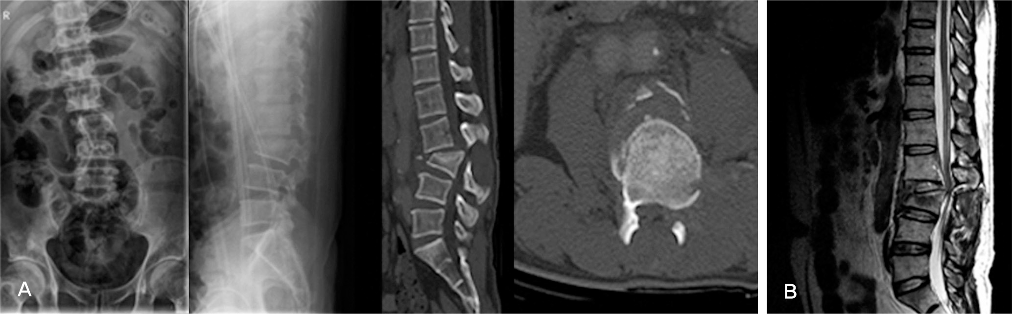
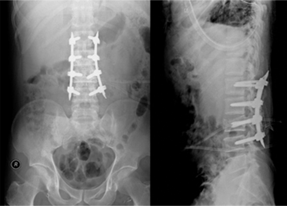
Fig. 2.
First staged operation was done a method of postural reduction and minimally invasive percutaneous pedicle screw fixation

Fig. 3.
(A) Post operative(two weeks after first stage operation) reconstruction CT demonstrate correction of sagittal balance with facet joint reduction (B) postoperative(two weeks after first stage operation) MRI sagittal and axial image demonstrate canal encroachment of herniated disc with soft tissue
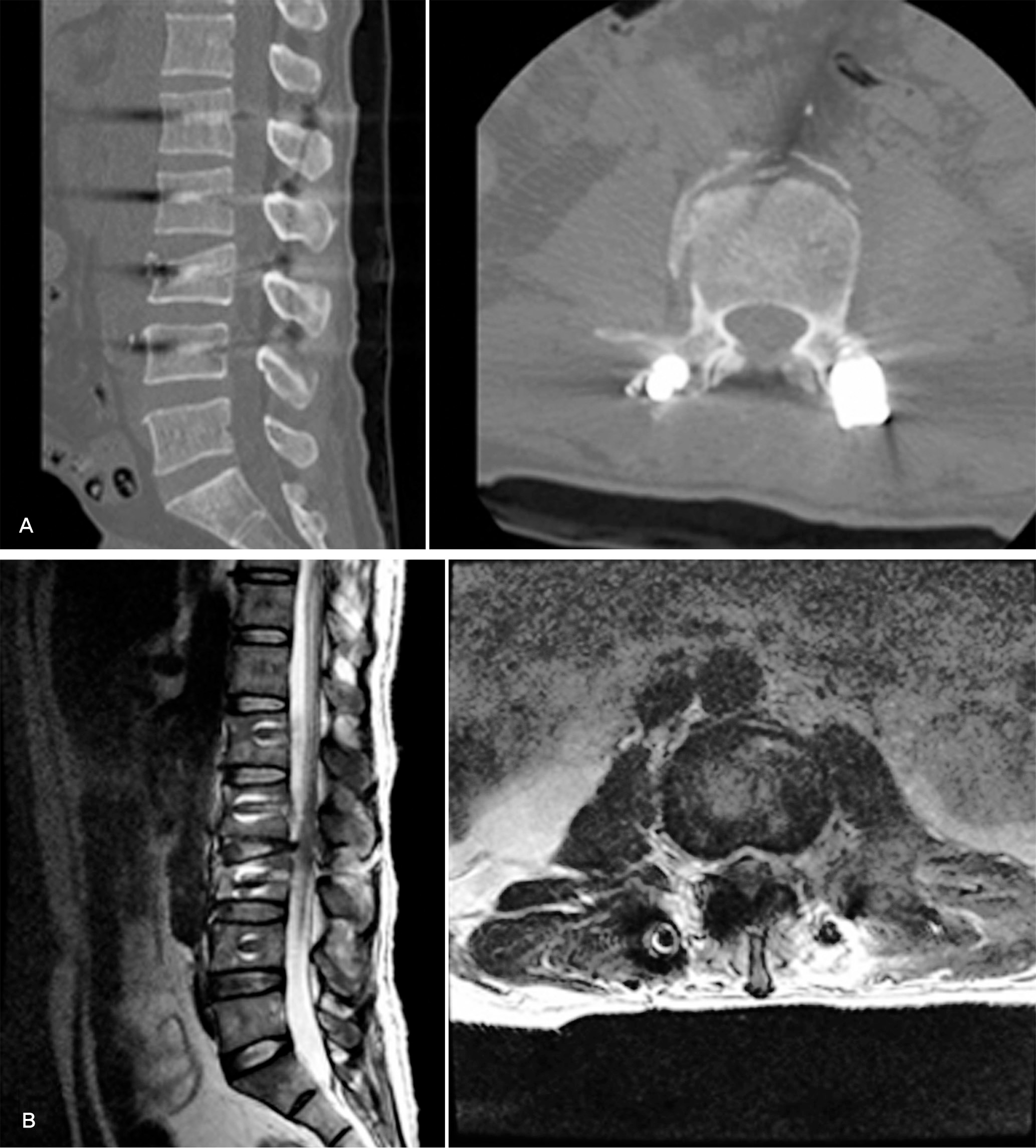
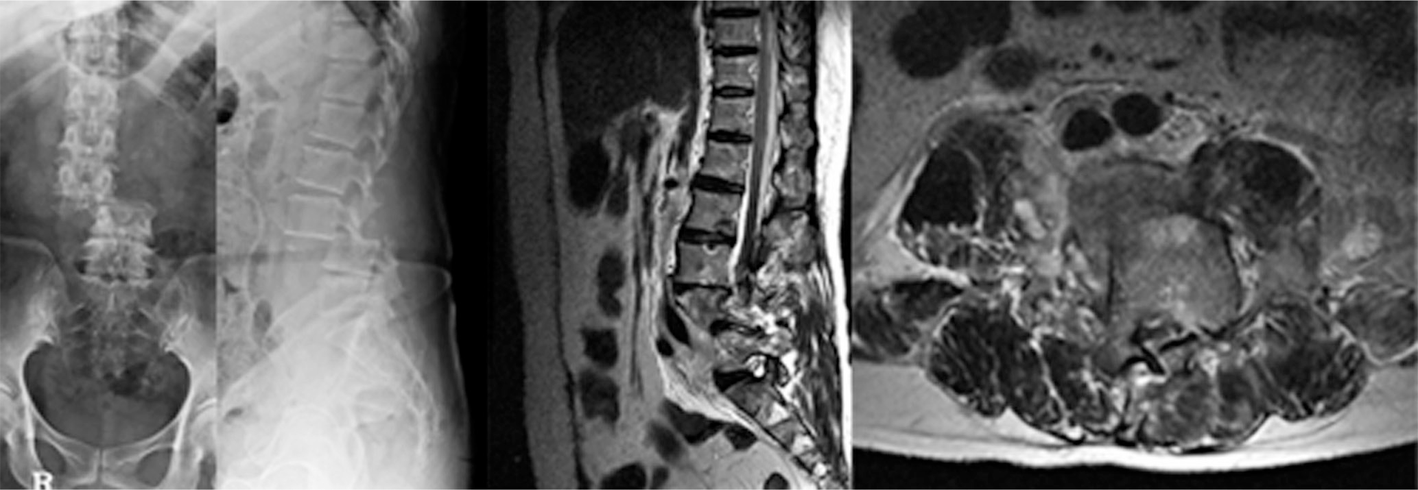
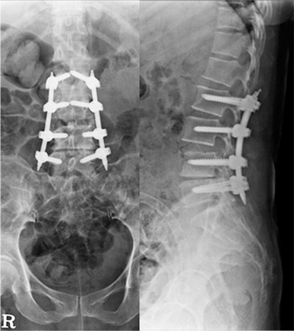
Fig. 4.
Second staged operation was done a method of partial laminectomy, discectomy and posterior fusion
 XML Download