

 PDF
PDF ePub
ePub Citation
 Print
Print


Abstract
Objectives
This study analyzed the clinical and radiographic results of the posterior lumbar interbody fusion performed on patients 50–65 and >7-years-of-age suffering from degenerative lumbar disease.
Literature Review Summary
Several studies on posterior lumbar interbody fusion performed on patients aged about 65 years reported insignificant age-related differences in the spinal-fusion results.
Materials and Methods
The records of 121 patients with degenerative lumbar disease treated with posterior lumbar interbody fusion between 2004 and 2010 were assessed. The patients’ clinical results, visual analogue scale (VAS) scores, Oswestry disability index (ODI) values, and complications before and after the surgery were compared. The radiographic results and changes in the fusion segmental angle before and after the surgery as well as in the height of the posterior intervertebral disc were also compared.
Results
In group A, comprising 44 patients >70-years-of-age, followup duration and number of comorbidities were 73.3 years and 17.8 months, respectively. In group B, comprising 77 patients 50–65-years-of-age, followup duration and number of comorbidities were 58.8 years and 31.8 months, respectively. In both groups, the average VAS scores and ODI values decreased. The incidence rate of vertebra-related postoperative complications was 13.6% in group A and 9.0% in group B. The incidence rate of general complications unrelated to the vertebra, was 18.1% in group A and 9.0% in group B.
REFERENCES
1. Okuda S, Oda T, Miyauchi A, Haku T, Yamamoto T, Iwasaki M. Surgical outcomes of posterior lumbar interbody fusion in elderly patients. J Bone Joint Surg Am. 2006; 88:2714–20.

2. Carreon LY, Puno RM, Dimar JR 2nd, Glassman SD, Johnson JR. Perioperative complications of posterior lumbar decompression and arthrodesis in older adults. J Bone Joint Surg Am. 2003; 85:2089–92.
3. Jo DJ, Jun JK, Kim KT, Kim SM. Lumbar Interbody Fusion Outcomes in Degenerative Lumbar Disease: Comparison of Results between Patients Over and Under 65 Years of Age. J Korean Neurosurg Soc. 2010; 48:412–8.
4. McAfee PC, Boden SD, Brantigan JW, et al. Symposium: a critical discrepancy-a criteria of successful arthrodesis following interbody spinal fusions. Spine (Phila Pa 1976). 2001; 26:320–34.
5. Deyo RA, Ciol MA, Cherkin DC, Loeser JD, Bigos SJ. Lumbar spinal fusion. A cohort study of complications, reoperations, and resource use in the Medicare population. Spine (Phila Pa 1976). 1993; 18:1463–70.
6. Best NM, Sasso RC. Outpatient lumbar spine decompression in 233 patients 65 years of age or older. Spine (Phila Pa 1976). 2007; 32:1135–9.
7. Kim EH, Kim HT. En bloc partial laminectomy and posterior lumbar interbody fusion in foraminal spinal stenosis. Asian Spine J. 2009; 3:66–72.
8. Tan JS, Singh S, Zhu QA, Dvorak MF, Fisher CG, Oxland TR. The effect of cement augmentation and extension of posterior instrumentation on stabilization and adjacent level effects in the elderly spine. Spine (Phila Pa 1976). 2008; 33:2728–40.
9. Hu SS. Internal fixation in the osteoporotic spine. Spine (Phila Pa 1976). 1997; 22(24 Suppl):43–8.
10. Wittenberg RH, Lee KS, Shea M, White AA 3rd, Hayes WC. Effect of screw diameter, insertion technique, and bone cement augmentation of pedicular screw fixation strength. Clin Orthop Relat Res. 1993. 278–87.
11. Sarzier JS, Evans AJ, Cahill DW. Increased pedicle screw pullout strength with vertebroplasty augmentation in osteoporotic spines. J Neurosurg. 2002; 96(3 Suppl):309–12.
Figures and Tables%
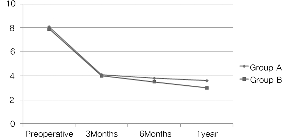
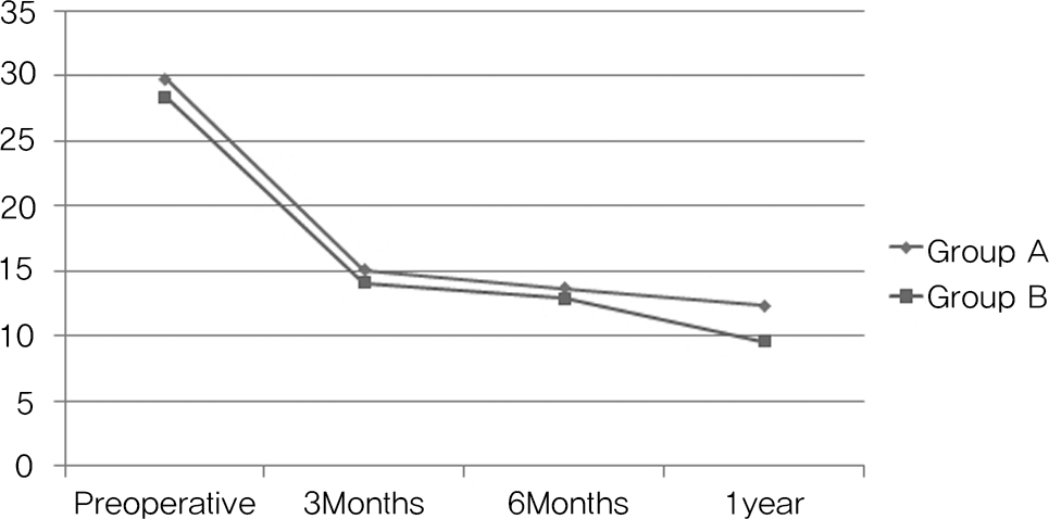
Fig. 3.
Course of the fusion segmental angle. A trend of postoperative increase, followed by gradual decrease was observed in both groups.
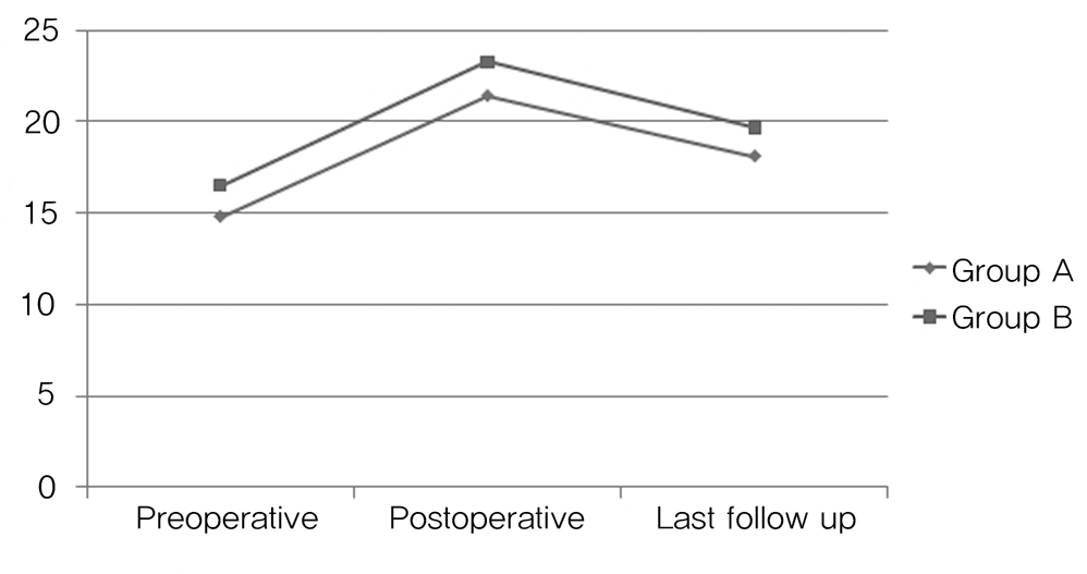
Fig. 4.
Course of the height of the posterior intervertebral disc. A trend of postoperative increase, followed by gradual decrease was observed in both groups.
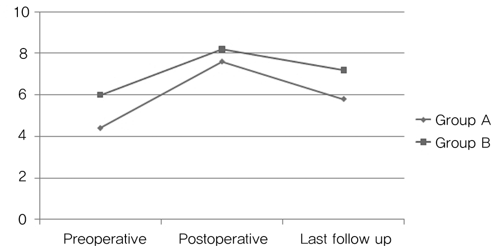
Table 1.
Characteristics of the patients in groups A and B
 XML Download