Abstract
Objectives
To compare the radiological and clinical results of the unipedicular and bipedicular approach of kyphoplasty for osteoporotic vertebral compression fractures.
Summary of Literature Review
A unipedicular rather than a bipedicular technique has been suggested to decrease the risks associated with surgical procedures.
Materials and Methods
Between July 2005 and May 2006, 136 vertebrae of 97 patients, who underwent kyphoplasty for osteoporotic vertebral compression fractures, were analyzed. Group 1, with the bipedicular approach, consisted of 86 vertebrae of 67 patients with a mean age of 72.2 years. Group 2, with unipedicular approach, consisted of 50 vertebrae of 30 patients with mean age of 73.4 years. The plain radiographs, MRI and surgical records were reviewed.
Results
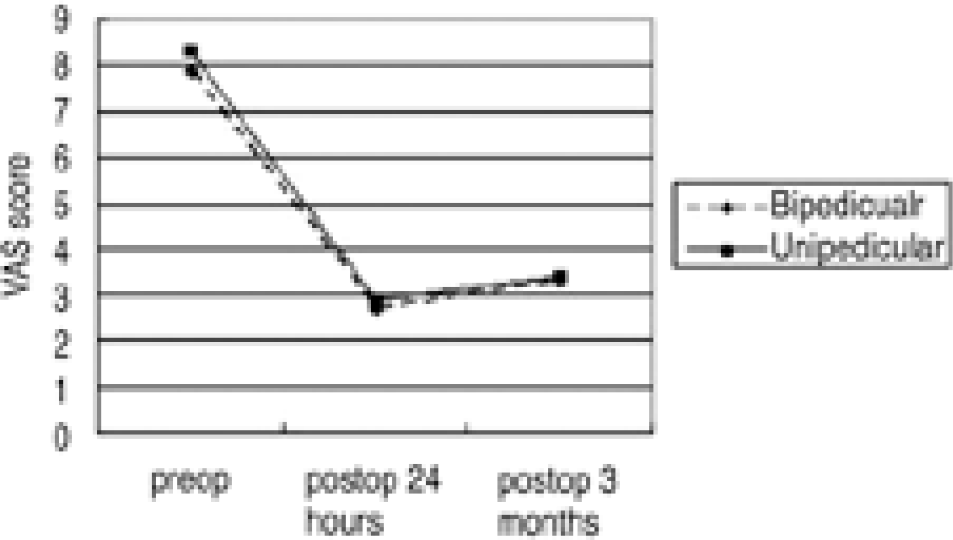
The mean operation time of the single vertebral body in group 2 was statistically lower than in group 1(p<0.05). There was more disruption of the medial wall of the pedicle in group 2 than in group 1(p<0.05). In the aspect of the volume of cement injected in the thoracolumbar junctional vertebrae, group 2 used significantly less cement than group 1(p<0.05). There were no significant differences in the cement leakage, vertebral height restoration, kyphotic deformity correction, admission time and VAS scores between groups 1 and 2(p>0.05).
Conclusion
There were no significant differences in clinical satisfaction and radiological results between the unipedicular and bipedicular kyphoplasty. The advantage of a unipedicular approach is the shorter procedure time than the bipedicular approach. This is particularly useful in multilevel compression fractures. The rate of the unipedicular approach in upper and mid thoracic spine is higher because of the higher convergence of the pedicle and the lower volume of vertebral body despite the disadvan-tages of instrument insertion through the medial pedicle wall.
REFERENCES
1). Hide IG, Gangi A. Percutaneous vertebroplasty: history, technique and current perspectives. Clin Radiol. 2004; 59:461–467.

2). Steinmann J, Tingey CT, Cruz G, Dai Q. Biomechanical comparison of unipedicular versus bipedicular kyphoplasty. Spine. 2005; 30:201–205.

3). Boszczyk B, Biershneider M, Hauck S, Beisse R, Potul-ski M, Jaksche H. Transcostovertebarl kyphoplasty of the mid and high thoracic spine. Eur Spine J. 2005; 14:992–999.
4). Tohmeh AG, Mathis JM, Fenton DC, et al. Biomechanical efficacy of unipedicular versus bipedicular vertebroplasty for the management of osteoporotic compression fractures, Spine. 1999; 24:1772–1776.
5). Kim AK, Jensen ME, Dion JE, Schweickert PA, Kauf-mann TJ, Kallmes DF. Unilateral transpedicular percutaneous vertebroplasty: Initial experience. Radiology. 2002; 222:737–741.

6). Mathis JM, Barr JD, Belkoff SM, Barr MS, Jensen ME, Deramond H. Percutaneous Vertebroplasty: A Developing Standard of Care for Vertebral Compression Fractures. Am J Neuroradiol. 2001; 22:373–381.
7). Belkoff SM, Mathis JM, Jasper LE, Deramond H. The biomechanics of vertebroplasty. The effect of cement volume on mechanical behavior. Spine. 2001; 26:1537–1541.
8). Liebschner MA, Rosenberg WS, Keaveny TM. Effects of bone cement volume and distribution on vertebral stiff-ness after vertebroplasty. Spine. 2001; 26:1547–1554.

9). Boszczyk BM, Biershneider M, Panzer S, et al. Fluoroscopic radiation exposure of the kyphoplasty patient. Eur Spine J. 2006; 15:347–355.

10). Manson NA, Phillips FM. Minimally invasive techniques for the treatment of osteoporotic vertebral fractures. Instr Course Lect. 2007; 56:273–285.

11). Garfin S, Lin G, Lieberman I, et al. Retrospective analysis of the outcomes of balloon kyphoplasty to treat vertebral compression fracture refractory to medical management. Eur Spine J. 2001; 10(suppl 1):S7.
Fig. 1.
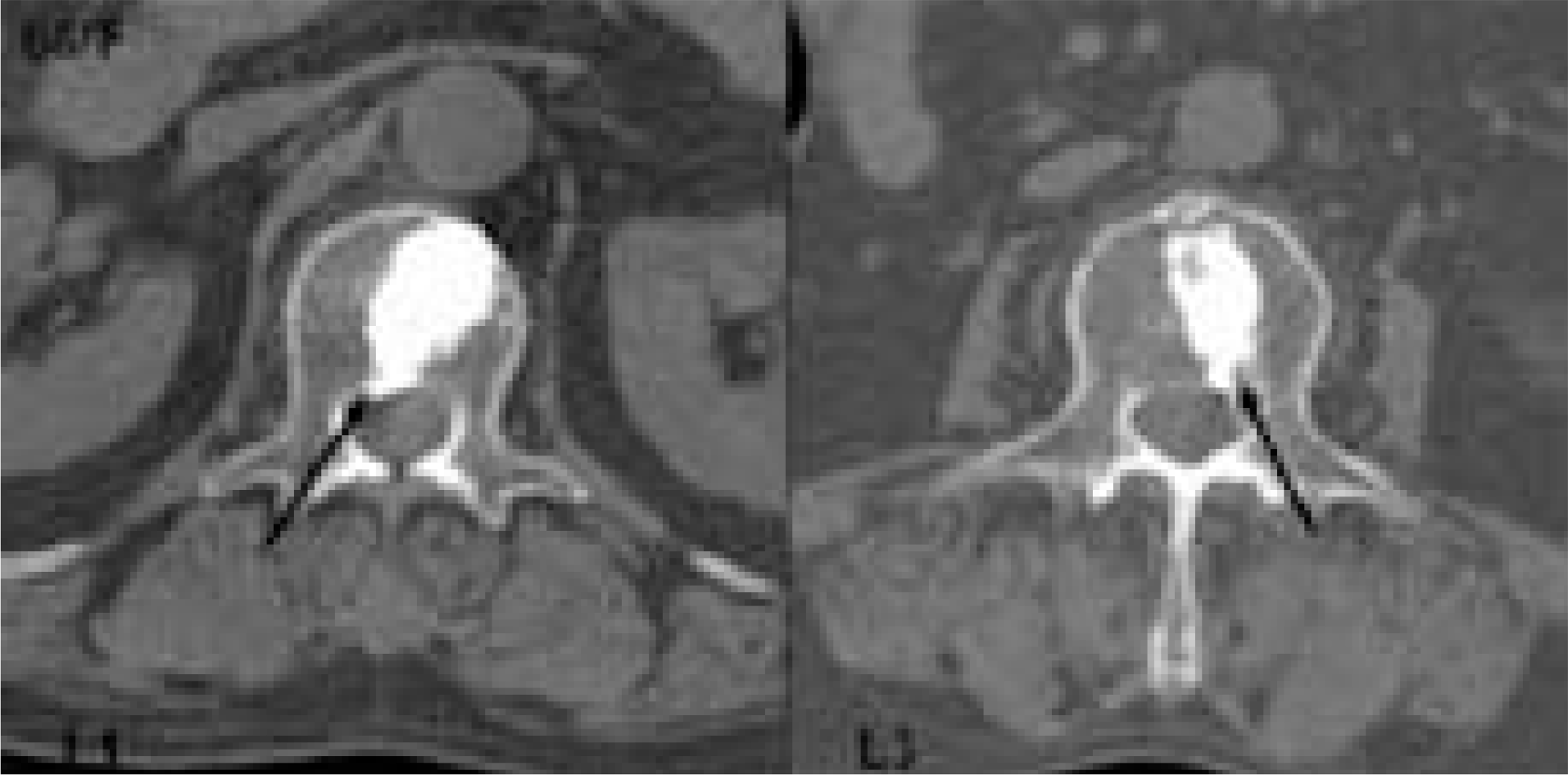
Preoperative CT scans. The solid arrow presented planned trajectory, the dotted arrow presented pedicle convergence. (A) T10, (B) T11, (C) T12.


 PDF
PDF ePub
ePub Citation
 Print
Print


 XML Download