

 PDF
PDF ePub
ePub Citation
 Print
Print


Abstract
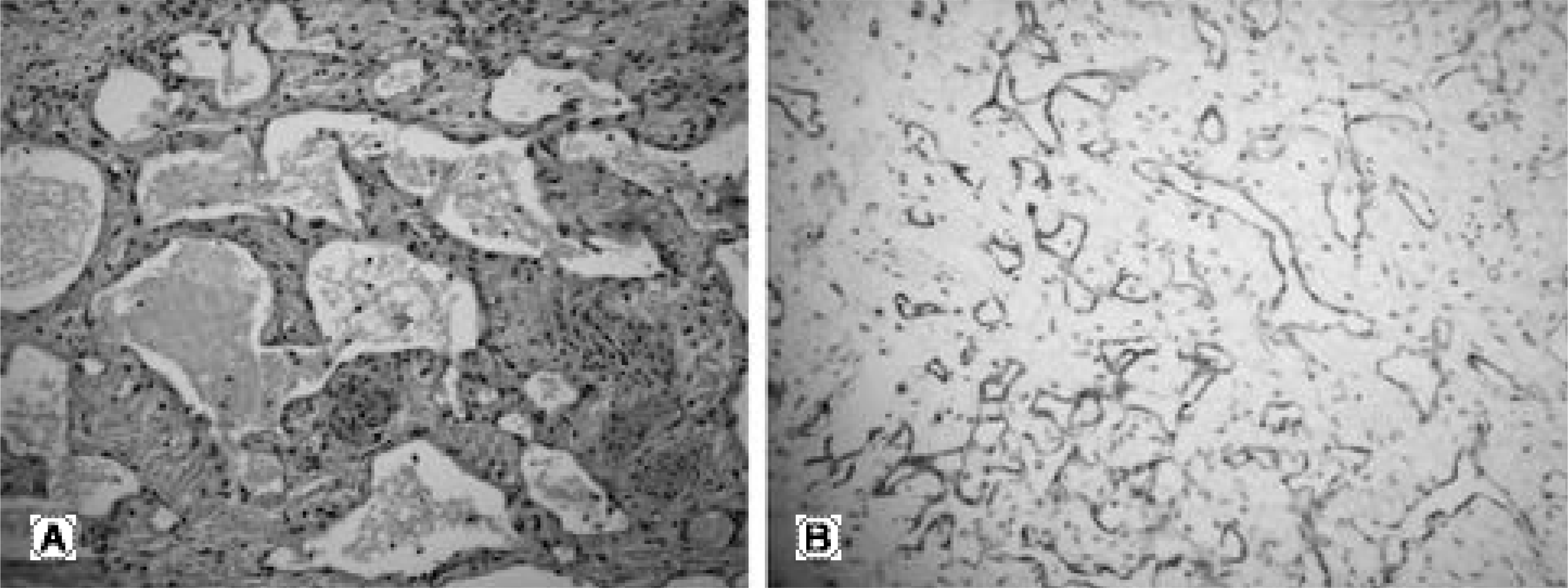
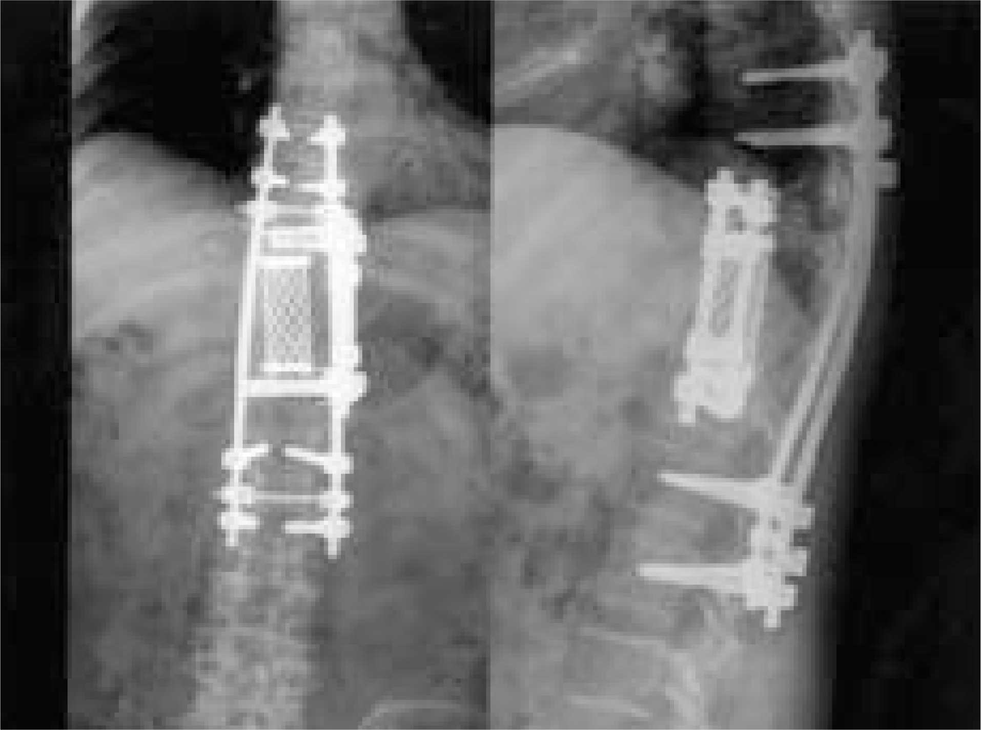
Gorham's disease is a rare condition of unknown etiology that is characterized by progressive osteolysis. A 48 year-old woman had a burst fracture at T10, which was treated by pedicle screw instrumentation at another hospital. She was transferred due to progressive paraparesis, which was not observed initially. A n MRI demonstrated severe cord compression at the T10 level. Under the assumption that the patient had a highly vascular metastatic tumor, an anterior decompression with instrumentation was performed. However, neurologic symptoms and bone destruction worsened after six weeks postoperatively. A repeat decompression was performed through the posterior route and long- level pedicle screw instrumentation was applied. A fter the second operation, Gorham's disease was confirmed histologically. Care must be taken not to overlook a pathologic fracture caused by a spinal tumor as a simple fracture, especially an osteoporotic one.
REFERENCES
1). Gorham LW, Wright AW, Shultz HH, Maxon FC Jr. Disappearing bones. A rare form of massive osteolysis. Am J Med. 1954; 17:674–682.

2). Heffez L, Doku HC, Carter BL, Feeney JE. Perspective on massive osteolysis. Report of a case and review of the literature. Oral Surg Oral Med Oral Pathol. 1983; 55:331–343.
3). Heyden G, Kindblom LG, Nielsen M. Di sapp earing bone disease. J bone Joint Surg Am. 1977; 59:57–61.
4). Jeong BY, Lee CK, Chang BS and Lee DH. Gorham's disease of the spine. J Kor Orthop Assoc. 2002; 37:443–6.
5). Johnson PM, McClure JG. Observations on massive osteolysis. A review of the literature and report of a case. Radiology. 1958; 71:28–41.
6). Drewry GR, Sutterlin CE, Martinez CR, Brantley SG. Gorham disease of the spine. Spine. 1994; 19:2213–2222.
7). Joseph J, Bartal E. Disappearing bone disease. A case report and review of the literature. J Ped Orthop. 1987; 7:584–588.
8). Pazzalia UE, Andrini L, Bonato M, Leutner M. Pathology of disappearing bone disease. A case report with immunohistochemical study. Int Orthop. 1997; 21:303–307.
9). Choma ND, Biscotti CV, Bauer TW, Metha AC, Licata AA. Gorham's syndrome. A case report and review of the literature. Am J Med. 1987; 83:1151–1156.
10). Campbell J, Almond HGA, Johnson R. Massive osteolysis of the humerus with spontaneous recovery. Report of a case. J Bone Joint Surg. Br. 1975; 57:238–240.
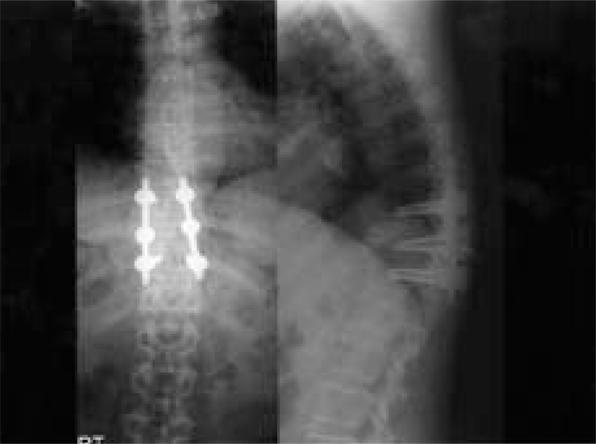
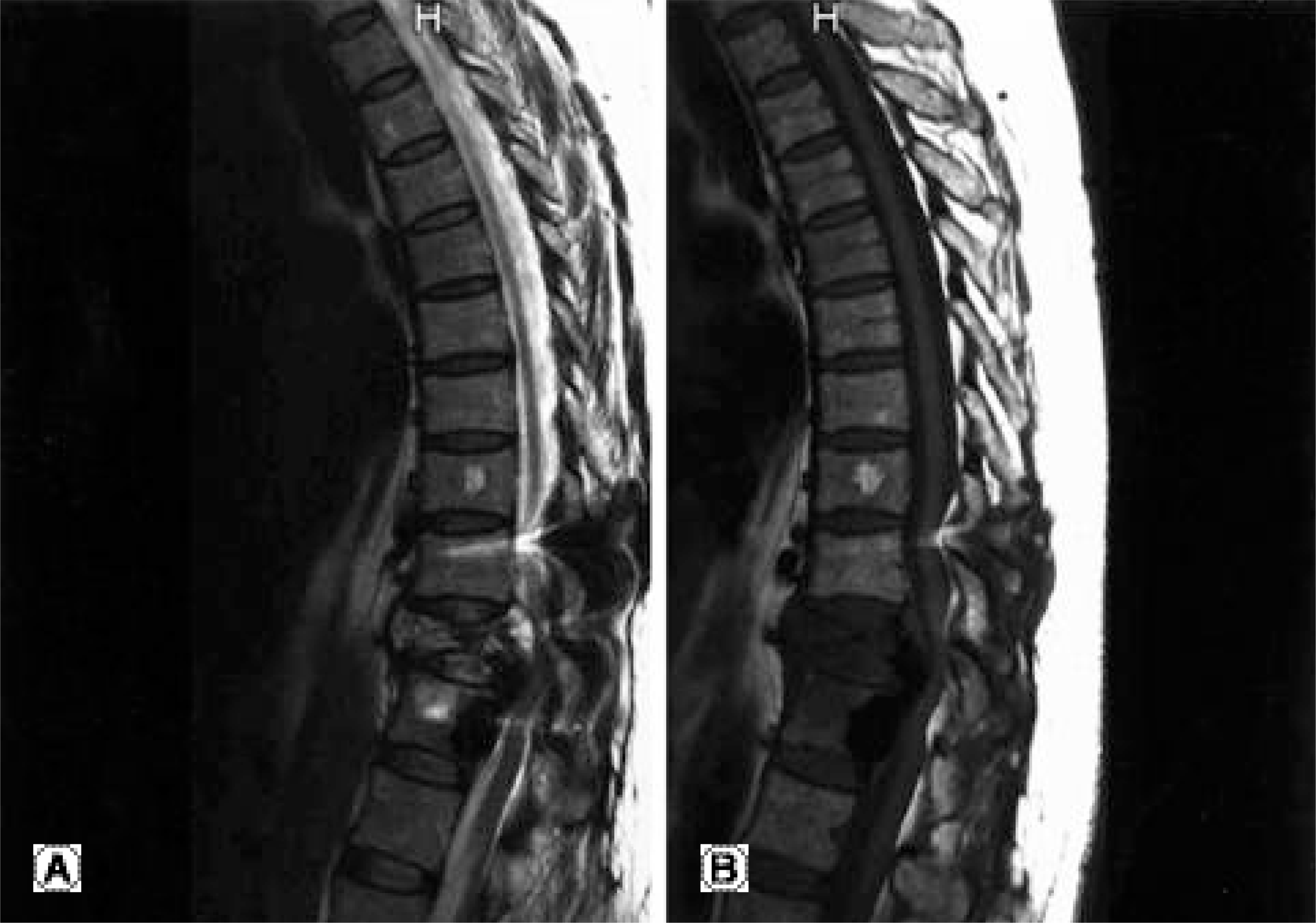
Fig. 2.
Preoperative MRI. (A) Sagittal T2-weighted image showing heterogeneous high signal intensities in T10 (B) Sagittal T1-weighted image showing low signal intensities in T10 vertebral body with spinal cord compression.
 XML Download