

 PDF
PDF ePub
ePub Citation
 Print
Print


Abstract
Objectives
To measure the lateral mass of cervical spine using MRI for lateral mass screw fixation and find out the ideal entry point and insertion angle and length of lateral mass screws.
Summary of Literature Review
Two methods of screw placement are in common use. The original technique, described by Ray-mond Roy-Camille, places the screw in a more or less straight sagittal direction and angling the screw laterally 10 to 20 degrees. Margerl technique involves placing the screw parallel to the facet joint and angling the screw laterally 25 to 30 degrees.
Materials and Methods
Axial MR images of the cervical spine parallel to the facet joints were obtained from C3 to C6 of 10 patients. The mean age of the patients were 48.0 years. The patients consisted of 6 male and 4 female patients. Ideal entry points, insertion angle and length of the lateral mass for lateral mass screw fixation were measured on the axial MR images using PACS digital measuring instrument.
Results
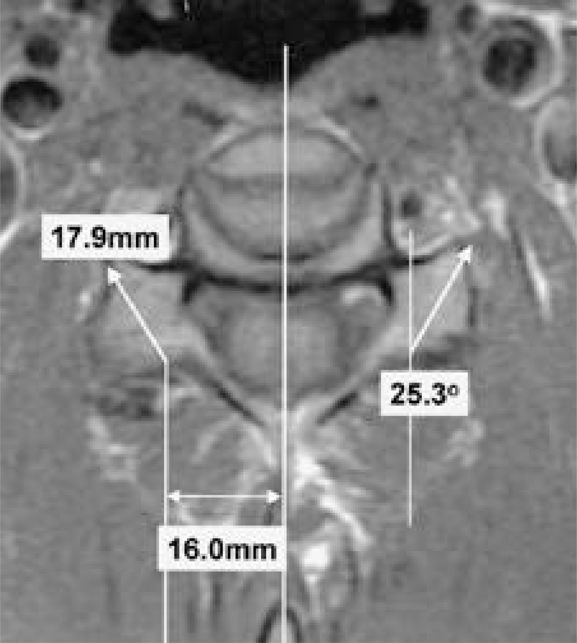
Ideal entry point of a lateral mass screw was center of lateral mass in sagittal plane, 16mm lateral to the midline of the cervical spine, ideal direction of the lateral mass screw was parallel to the facet joint and angling the screw laterally 25.3 degrees, and ideal length of lateral mass screw was 17.9mm.
REFERENCES
1). Anderson PA, Henley MB, Grady MS, Montesano PX, Winn HR. Posterior cervical arthrodesis with AO reconstruction plates and bone graft. Spine. 16(S):S72–9. 1991.

2). Coe JD, Warden KE, Sutterlin CE, McAfee PC. Biomechanical evaluation of cervical spinal stabilization methods in a human cadaveric model. Spine. 14(10):1122–31. 1989.
3). Cooper PR, Cohen A, Rosiello A, Koslow M. Posterior stabilization of cervical spine fractures and sublux -ations using plates and screws. Neurosurgery. 23:300–6. 1988.
4). Ebraheim NA, An HS, Jackson WT, Brow JA. Internal fixation of the unstable cervical spine using posteior Roy-Camille plates: preliminary report. J Orthop Trauma. 3:23–8. 1989.
5). Errico T, Uhl R, Cooper P, Casar R, McHenry T. Pullout strength comparison of two methods of orienting screw insertion in the lateral masses. J Spinal Dis. 7:429–38. 1992.
6). Gill K, Paschal S, Corin J, Asham R, Bucholz R. Posterior plating of the cervical spine: a biomechanical comparison of different posterior fusion technique. Spine. 13:813–6. 1988.
7). Graham AW, Swank ML, Kinard RE, Lowery GL, Dials BE. Posterior cervical arthrodesis and stabilization with a lateral mass plate: clinical and computed tomo -graphic evaluation of lateral mass screw placement and associated complications. Spine. 21:323–8. 1996.
8). Grob D, Magerl F. Dorsal spondylodesis of the cervical spine using a hooked plate. Orthopade. 16:55–61. 1987.
9). Lowery GL, Swank ML, McDonough RF. Surgical revision for failed anterior cervical fusions: articular pil -lar plating or anterior revision? Spine. 20:2436–42. 1995.
10). Magerl F. Posterior fusion using hook plates. Bulletin 61, Paoli, PA: Synthes.
11). Magerl F, Grob D. Stable dorsal fusion of the cervical spine(C2-T1) using hook plates. Kehr P, Weidner A, editors. Cervical spine. Vienna: Springer-Verlag;p. 217–21. 1987.
12). Montesano PX, Jauch E, Anderson PA, Benson DR, Hanson B. Biomechanics of cervical spine internal fixation. Spine. 16(suppl.):S10–6. 1991.
13). Roy-Camille RR, Sailant G, Mazel C. Internal fixation of the unstable cervical spine by posterior osteosynthesis with plate and screws. Cervical Spine Research Society, The cervical spine. 2nd ed.Philadelphia: JB Lippincott;p. 390–404. 1989.
14). Savini R, Parisini P, Cevellati S. The surgical treatment of late instability of flexion-rotation injuries in the lower cervical spine. Spine. 12:178–82. 1987.
15). Swank ML, Sutterlin CE III, Bossons CR, Dials BE. Rigid internal fixation with lateral mass plates in multi -level anterior and posterior reconstruction of the cervical spine. Spine. 22:274–82. 1997.
Figures and Tables%
Fig. 1.
Axial MR images of the cervical spine parallel to the facet joints were obtained from C3 to C6.

Fig. 2.
Ideal entry points, insertion angle and length of the lateral mass for lateral mass screw fixation were measured on the axial MR images using PACS digital measuring instr ument.
 XML Download