

 PDF
PDF ePub
ePub Citation
 Print
Print


Abstract
An accurate assessment of injuries to the spinal column and the neural tissues will facilitate the management of patients with injuries to the thoracic and lumbar spine. Routine radiological investigations are essential, but newer techniques are now available that define the extent of injuries in exquisite detail, providing a better understanding of not only the bony injuries, but also the extent of the soft tissue lesion, including the nervous system. The referring physician and the radiologist have many imaging techniques available for the diagnosis of the extent of thoracolumbar spine fracture. These include plain film radiography, computed tomography(CT), conventional polydirectional tomography, bone scan, magnetic resonance image(MRI), and myelography. These techniques are used alone or in combination to arrive at the correct diagnosis. It behooves the examining physician to be extremely thorough in identifying additional lession, not only for medicolegal reasons, but also to ensure that other potentially unstable lesions are not overlloked, since this could lead to neurological compromise if unsuspected. We describe the integrated use of multiple imaging techniques.
REFERENCES
1). An HS, Andreshak TG, Nguyen C, et al. Can we distin -guish between benign versus malignant compression fractures of the spine by magnetic resonance imaging? Spine. 20:1776–1782. 1995.
2). Atlas SW, Regenbogen V, Rogers LF, Kim KS. The radiographic characterisation of burst fractures of the spine. AJR. 147:575–582. 1986.
3). Berg EE. The sternal-rib complex: A possible fourth column in thoracic spine fractures. Spine. 18:1916–1919. 1993.
4). Bondurant FJ, Cotler HB, Kulkarni MV, et al. A cute spinal cord injury: A study using physical examination and magnetic resonance imaging. Spine. 15:161. 1990.
5). Brandser EA, el-Khoury GY. Imaging of Thoracic and Lumbar Spine Trauma. Radiol Clin North Am. 35:533–577. 1997.
6). Brian JM, Russel SV, Bradford LC, Frank JE. Diagnosis of subtle thoracolumbar burst fractures, Spine. 18:2282–2285. 1993.
7). Bridwell KH, DeWald RL. The Textbook of Spinal Surgery. 2nd ed. Phialdelphia: Lippincott-Raven Publishers;p. 1246. 1997.
8). Brightman RP, Milller CA, Rea GL, et al. Magn e tic resonance imaging of trauma to the thoracic and lumbar spine. Spine. 17:541. 1992.
9). Cammisa FP, Eismont FJ, Green BA. Dural laceration occuring with burst fractures associated laminar fractures. J Bone Joint Surg. 71-A:1044–1052. 1989.
10). Carl AL, Matsumoto M, Whalen JT. Anterior Dural Laceration Caused by Thoracolumbar and Lumbar Burst Fractures. J Spin Disord. 13:399–403. 2000.

11). Chung YK, Kim SW, Kim BS, et al. Radiographic comparison of stable burst fractures with compression fractures in thoracolumbar spine. J of korean spine surgery. 6:415–421. 1999.
12). Collier BD, Fogelman I, Rosenthall L. S kelet al Nuclear Medicine. Missouri: Mosby-Year Book Inc.;:224-259. 1996.
13). Daffner RH. Imaging of Vertebral Trauma. Philadelphia: Lippincott-Raven Publisher;s: 209-222. 1996.
14). Daffner RH, Deeb ZL, Goldberg AL, et al. The radiologic assessment of post-traumatic vertebral stability. Skeletal Radiol. 19:103. 1990.
15). Daffner RH, Deeb ZL, Rothfus WE. The posterior vertebral body line: Importance in detection of burst fractures. Am J Roentgenol. 148:93–96. 1987.
16). Denis F, Burkus JK. Diagnosis and treatment of cauda equina entrapment in the vertical lamina fracture of lumbar burst fractures. Spine. 16:433–439. 1991.
17). Domenicucci M, Preite R, Ramieri A, Osti MF, Ciap-petta P, Delfini R. Three-Dimensional Computed Tomographic Imaging in the Diagnosis of Vertebral Column Trauma: Experience Based on 21 Patients and Review of the Literature. J Trauma. 42:254–259. 1997.
18). El-Khoury GY, Moore TE, Kathol MH. Radiology of the thoracic spine. Clin Neurosurg. 38:261–295. 1992.
19). El-Khoury GY, Whitten CG. Trauma to the upper thoracic spine: Anatomy, biomechanics, and unique imaging features. Am J Roentgenol. 160:95–102. 1993.
20). Eubanks BA, Cann CE, Brandt-zawadaki M. CT measurement of the diameter of spinal and other bony canals: effects of section angle and thickness. Radiology. 157:243–246. 1985.
21). Ferguson RL, Allen DI. A mechanistic classification of thoracolumbar spine fractures. Clin Orthop. 189:77–88. 1984.
22). Flanders AE. Thoracolumbar Trauma Imaging Overview. Inst Course Lecture. 48:429–431. 1999.
23). Gehweiler JA, Daffner RH, Osborne RL. Relevant signs of stable and unstable thoracolumbar vertebral column trauma. Skeletal Radiol. 7:179–183. 1981.
24). Hirsh LF, Duarte L, Wolfson EH. Thoracic spinal cord injury without spine fracture in an adult: case report and literature review. Surg Neurol. 40:35–38. 1993.
25). Holdsworth FW. Fracture, dislocations and fracture-dis -locations of the spine. J Bone Joint Surg. 52-A:1534–1551. 1970.
26). Kanal E, Shellock FG. Polices, guidelines, and recommendations for MR imaging safety and patient management. J Magn Reson Imaging. 2:247–248. 1992.
27). Kaye JJ, Nance EP. Thoracic and lumbar spine trauma. Radiol Clin North Am. 28:361–377. 1990.
28). Keats TE. Atlas of Normal Roentgen Variants That May Simulate Disease. 6th ed. Missouri: Mosby-Year Book Inc;p. 239–293. 1996.
29). Keenen TL, Antony J, Benson DR. Dural tears associated with lumbar burst fractures. J Orthop Trauma. 4:243–245. 1990.
30). Kosling S, Dietrich K, Steinecke R, Kloppel R, Schulz HG. Diagnostic value of 3D CT surface reconstruction in spinal fractures, Eur Radiol. 7:61–64. 1997.
31). Kothari P, Freeman B, Grevitt M, Kerslake R. Injury to the spinal cord without radiological abnormality (SCIWORA) in adult. J Bone Joint Surg. 82-B:1034–1037. 2000.
32). Kram HB, Appel PL, Wohlmuth DA, et al. Diagnosis of traumatic thoracic aortic rupture: A 10 years retrospective analysis. Ann Thorac Surg. 47:282–286. 1989.
33). Kulkami M, McArdle CB, Kopanicky D. Acute spinal injury: MR imaging at 1.5T. Radiology. 164:837–843. 1987.
34). Lee HW, Kim HS, Kim DJ, Suk KS, Park JO, Kim NH. Reliability of Magnetic Resonance Imaging in Thoracolumbar Spinal Fractures. Spine. 25:2079–2084. 2000.
35). McAfee PC, Yuan HA. Computed tomography in spondylolisthesis. Clin Orthop. 166:62–71. 1982.
36). Miller CA, Hunt WE. Variation of impaction fracture of lumbar spine. J Neurosurg. 56:603. 1982.
37). Nykamp PW, Levy JM, Christensen F, Dunn R, Hubbard J. Computed tomography for a bursting fracture of the lumbar spine. J Bone Joint Surg. 60-A:1108–1109. 1978.
38). O'Callaghan JP, Ullrich CG, Yuan HA, et al. CT of facet dislocation in flexion injuries of the thoracolumbar spine; The “naked” facet. Am J Neuroradiol. 1:97–102. 1980.
39). Pang D, Wilberger JE. Spinal cord injury without radiographic abnormalities in children. J Neurosurg. 57:114–29. 1982.
40). Ramon S, Dominguez R. Ramirez, et al. Clinical and magnetic resonance Image correction in acute spinal cord injury. Spinal Cord. 35:664. 1997.
41). Resnick D, Niwayama G, Guerra J, et al. Spinal vacuum phenomenon: Anatomical study and review. Radiology. 139:341–348. 1981.
42). Saifuddin A, Noordeen H, Taylor BA, et al. The role of imaging in the diagnosis and manegement of thoracolumbar burst fractures: Current concept and a review of the literature. Skeletal Radiol. 25:603–613. 1996.
43). Shin BJ, Kwon H, Kim JB, et al. The Clinical Significance of Unilateral Multiple Transverse Process Fractures in Lumbar Spine, J Korean Spine Surg. 3:195–201. 1996.
44). Shin BJ, Yun TK, Kang HS, Lee JC, Kim KJ, Kim YI. Intraspinal gas with lumberjack injury. J of Korean Society of Spine Surgery. 8(1):81–85. 2001.
45). Silvestro C, Francaviglia N, Bragazzi R, et al. On the predictive value of radiological signs for the presence of dural lacerations related to fractures of the lower thoracic or lumbar spine. J Spinal Disord. 4:49–53. 1991.
46). Takada M, Wu CY, Lang TF, Genant HK. Vertebral Fracture Assessment using the Lateral Scoutview of Computed Tomography in Comparison with Radiographs. Osteoporosis Int. 8:197–203. 1998.
47). Young PC, Petersilage CA. MR imaging of the traumatized lumbar spine. Spine. 25:2079–2084. 1999.
48). Zanca P, Lodmell EA. Fracture of the spinous processes: “New” sign for the recognition of fractures of cervical and upper dorsal spinous processes. Radiology. 56:427–428. 1951.
Figures and Tables%
Fig. 1.
Wide gap between spinous processes in the AP(flexion-distraction) (A) and lateral(dislocation) (B) views that means posterior column injury.

Fig. 2.
Sagittal reconstruction view is useful in the evaluation of the canal encroachment, preoperatively (A) and canal clearance postoperatively (B).
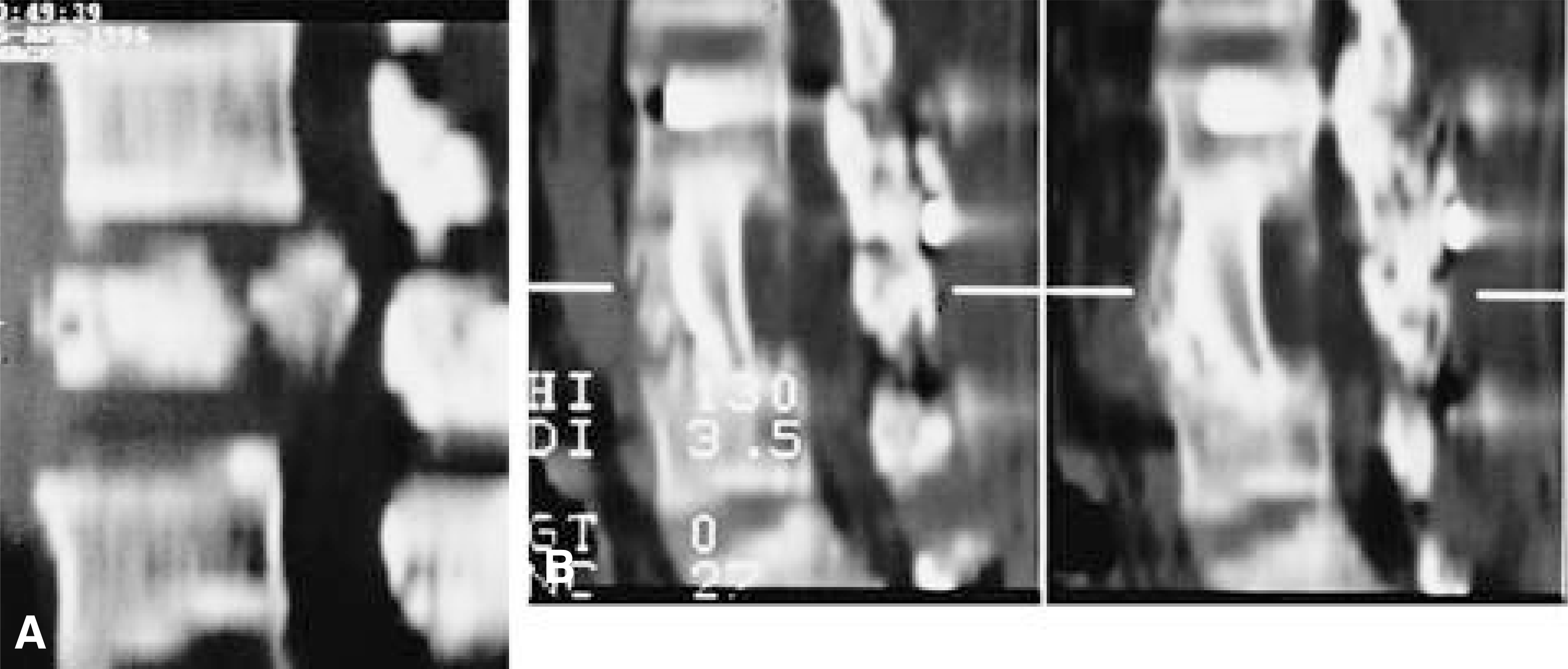
Fig. 3.
3D reconstruction view is useful only in the malalignment (A) but subtraction technique can demonstrate canal encroachment (B).
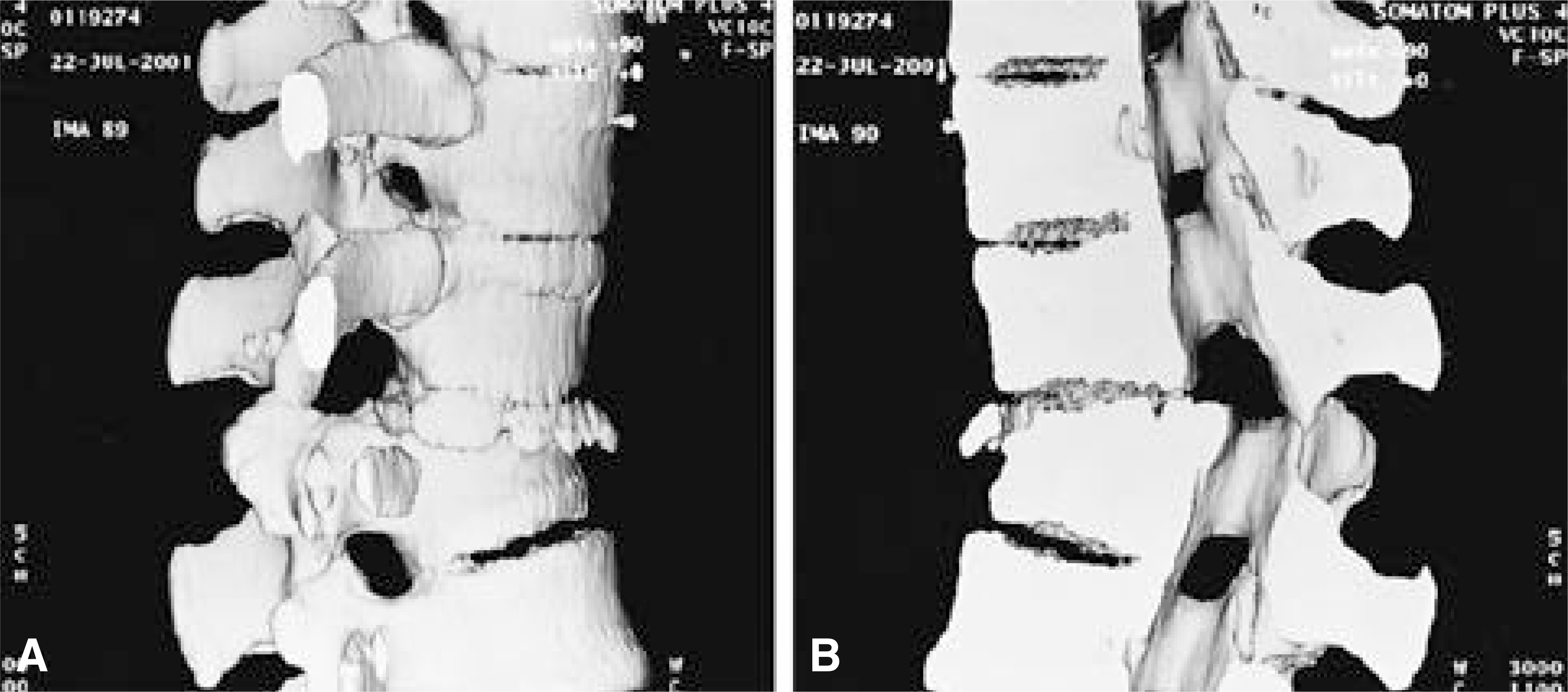
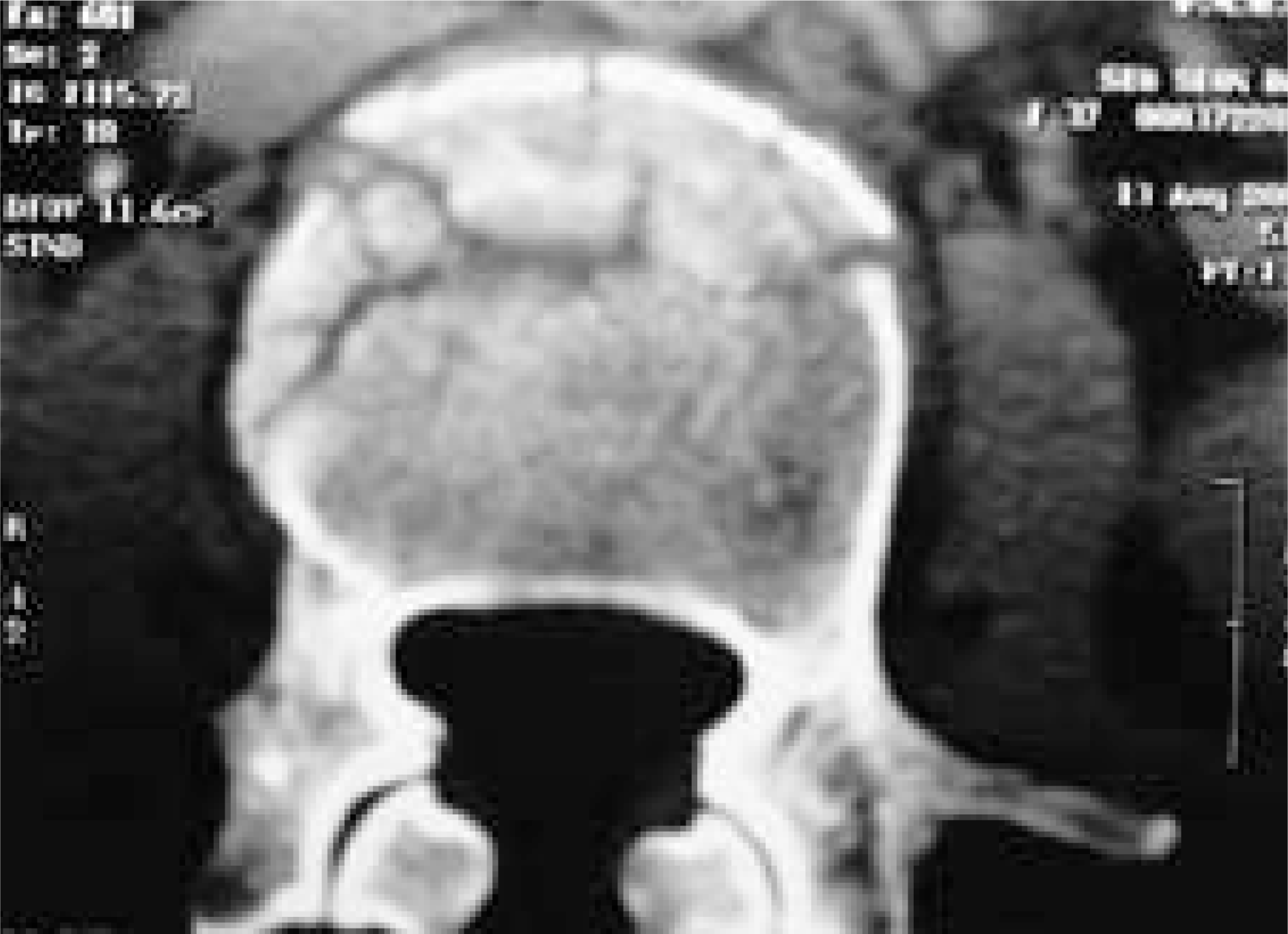
Fig. 5.
Several patterns of retropulsed fragments in the CT. A. single central. B. sagittal split. C. comminution. D. asym-metric. E. reverse cortical sign(arrows) that means rupture of the posterior longitudinal ligament.

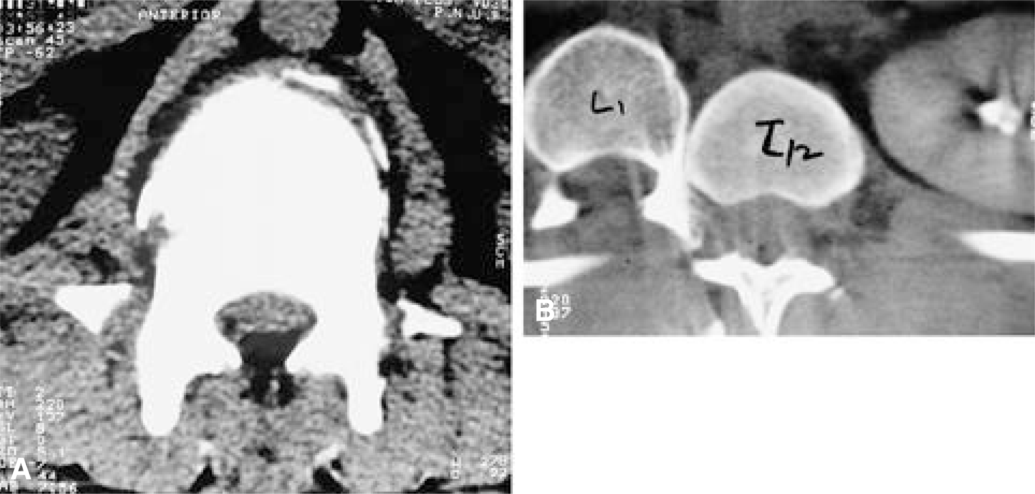
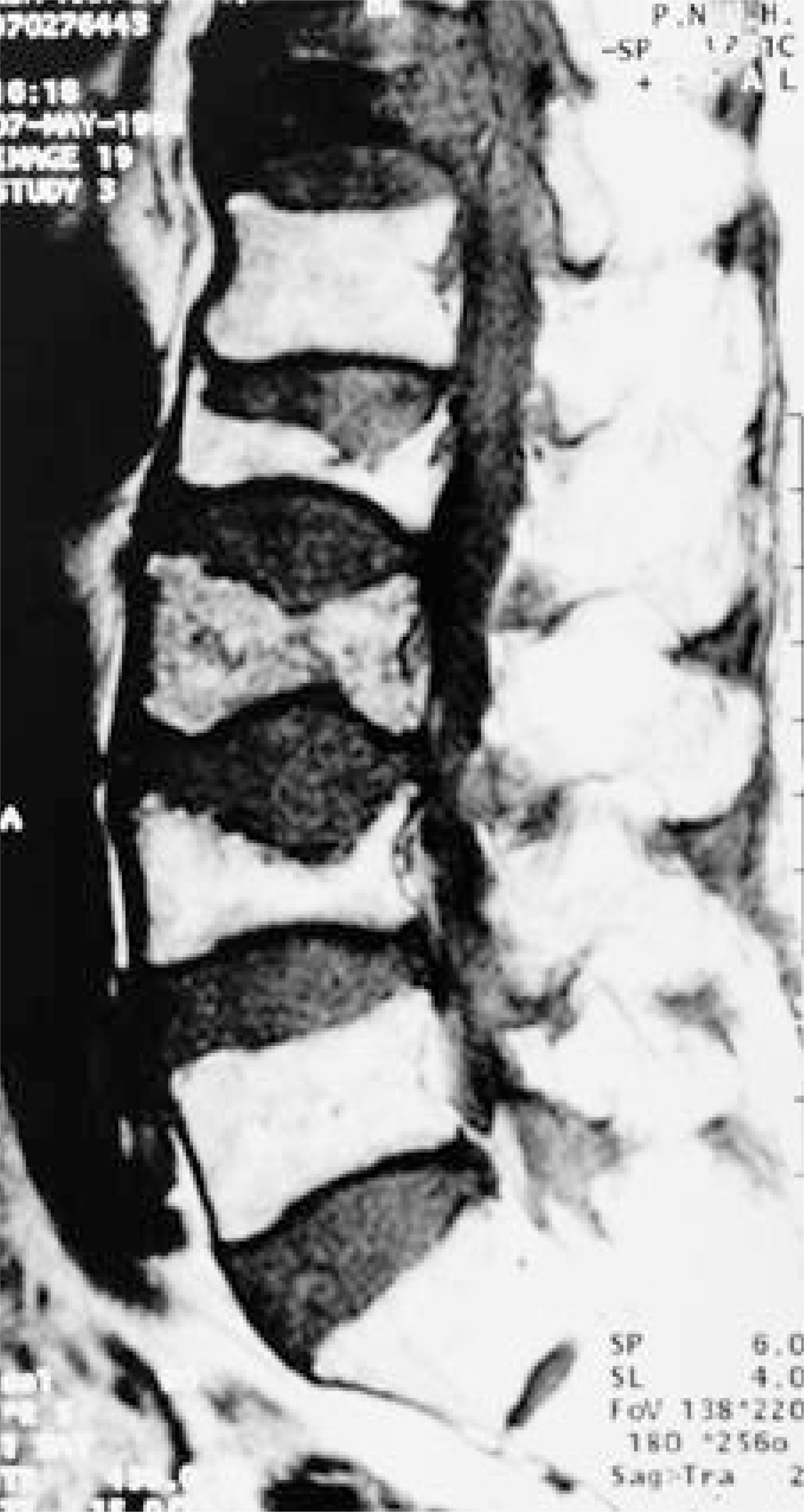
Fig. 8.
MR is superior to any other imaging modalities in evaluation of the posterior ligament complex injury(black arrow). And also is the only method of direct visualization of the nerve injury(white arrow).
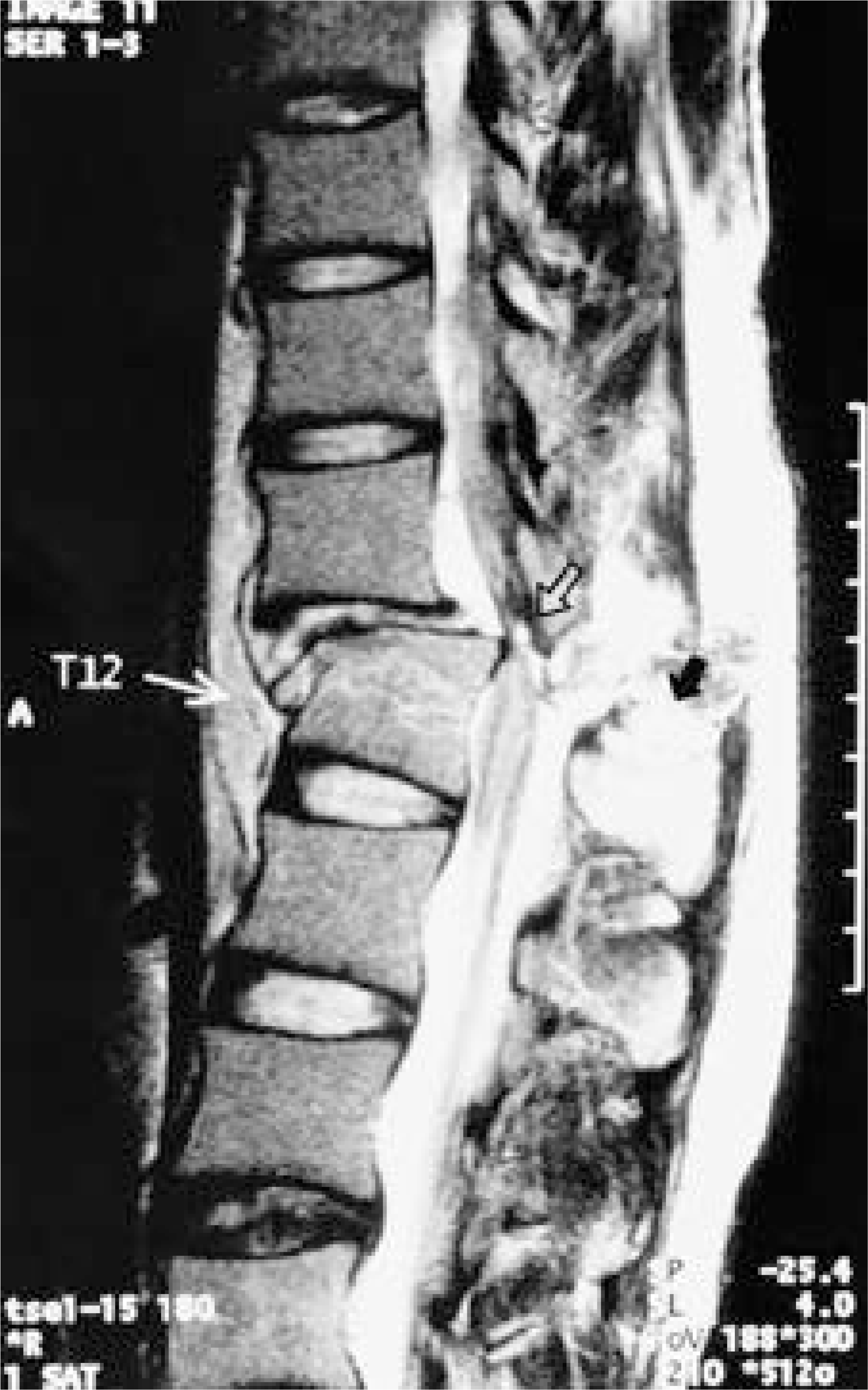
Table 1.
MR signal changes in vertebral trauma
 XML Download