

 ePub
ePub Citation
 Print
Print


INTRODUCTION
Superior mesenteric artery (SMA) embolism is a life-threatening vascular emergency that requires rapid revascularization of mesenteric blood flow as well as early diagnosis and is the most frequent cause of acute mesenteric ischemia, which is associated with a high mortality rate [1]. Although previous reports have described the successful use of thrombolysis with local urokinase infusion to the SMA, prolonged infusion times and increased total drug dosage have a potential for continued intestinal ischemia and bleeding complications [2-6]. We report a patient with acute SMA embolism who was treated with revascularization of the early SMA main branch with aspiration and high-dose urokinase bolus followed by continuous infusion with low-dose urokinase.
CASE REPORT
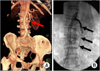
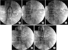
A 74-year-old female was admitted to the hospital with abdominal pain 5 hours in duration. The abdomen was soft and slightly tender to palpation, and atrial fibrillation was evident on the electrocardiogram. Laboratory data on admission were within normal values. She had been diagnosed with hypertrophic cardiomyopathy with obstruction 15 years earlier, also suffered from acute myocardial infarction and right renal infarction due to embolism 6 years prior. Computed tomography (CT) revealed the occlusion of SMA with high suspicion of acute mesenteric ischemia without any evidence of bowel necrosis (Fig. 1A). At that time, we decided on thrombolytic therapy because the patient had not had clinical evidence of bowel necrosis and had not had other contraindications to thrombolytic therapy. All procedures were performed in a surgical operation room with C-arm. At first, femoral access was gained using an 8 Fr introducer. Heparin was administered to achieve an activated clotting time of 250 to 300 seconds. Anatomy of SMA was established by abdominal aortogram in the anterior-posterior and lateral views. Selective catheterization of the SMA with a 5 French (Fr), Omni catheter (Cordis Co., Miami, FL, USA) was performed, and a 0.035-inch superstiff guidewire (Cook Medical Inc., Bloomington, IN, USA) was inserted through selective catheter into the SMA. The 0.035-inch superstiff guidewire could be advanced through the thrombus. Then, the selective catheter was exchanged with an 8 Fr, 55 cm contralateral guiding catheter (Cordis Co.). Aspiration was applied manually via the 5 Fr Multi-purpose catheter (Cordis Co.). Several passes of the aspiration catheter were performed to achieve complete clot removal. Although effective aspiration of the clot was not achieved, a proximal SMA trunk was recanalized. Next, a Benson starting 0.035-inch guidewire (Cordis Co.) was advanced through the thrombus followed by the 8 Fr guiding catheter. Intra-arterial urokinase (300,000 IU), 3 amples of papaverin and heparinized saline were injected via 8 Fr guiding catheter. Partially, the abdominal complaints resolved within 20 minutes after the start of aspiration thrombectomy and thrombolysis. Angiographic visualization of the SMA (Fig. 2A) showed recanalization of the main trunk of the SMA. Repeat angiography after 4 hours showed distal migration after aspiration and thrombolysis (Fig. 2B). A multi-side holes catheter (Cook Medical Inc.) was inserted at the occluded branch by distal migration and continuous intra-arterial infusion of urokinase (50,000 IU/hr) and papaverin (30 mg/hr) was initiated. Then, angiography showed near complete reperfusion of the peripheral mesenteric flow (Fig. 2D). The third angiography after 20 hours demonstrated almost complete reperfusion of the peripheral mesenteric flow (Fig. 2E). At this time, local infusion was stopped, and the patient was heparinized with low-molecular weight heparin (40 IU/BID). But, the patient complained of abdominal pain. Repeat CT revealed a thinned small bowel wall by ischemic injury without bowel infarction. The next day, the patient made an uneventful recovery. The patient improved completely after bowel rest for 5 days. Warfarin therapy was initiated.
DISCUSSION
Acute thromboembolic occlusion of the SMA leads to intestinal infarction and is associated with a mortality rate of around 65% [1]. Therefore, early diagnosis and appropriate treatment are required for a good prognosis [1]. Surgery, such as SMA embolectomy, SMA bypass, and resection of necrotic bowel, has been attempted, but the mortality rates have been reported ranging from 35 to 100% [7,8]. Thrombolytic therapy has resulted in good outcomes during the early stages of the disease, although this therapy is not recommended after intestinal necrosis has developed [4] and hemorrhage risk is increased [2-6]. There are several techniques for infusion of the thrombolytic agent; the more commonly used is the McNamara protocol for peripheral arterial or graft occlusions, which involves a high-dose infusion [9]. Unfortunately, this thrombolytic therapy can require prolonged infusions during which ischemia may continue, leading to intestinal necrosis. Recently, devices for thrombus aspiration in coronary arteries have been developed for patients with acute coronary syndrome. Although these devices can also be applied for treatment of acute thromboembolic SMA occlusions, they are not available in our institute. We tried recanalization of the arteries by aspirating the thrombi using a 5 Fr multi-porpose catheter (Cordis Co.) under an 8 Fr guiding catheter (Cordis Co.) with a wire of 0.035 inches in diameter. Although a large amount of fresh thrombi had not been aspirated effectively, her abdominal pain reduced and angiography demonstrated recanalization of the main trunk on SMA after aspiration and thrombolysis for 20 minutes. Distal migration of the thrombus partly dissolved and broken by aspiration and thrombolysis is one of the disadvantages of this procedure. Even through distal embolization was revealed in angiogram after 4 hours, this patient was relieved of abdominal pain after thrombolysis and aspiration; and the occluded branch by distal migration was clearly delineated through thrombolytic therapy by selection of 5 Fr multi-side Hole catheter for 16 hours. In this patient, we selected thrombus aspiration and thrombolysis as a primary treatment modality instead of open surgery. The reasons are as follows: 1) The patient was very old and diagnosed a hypertrophic cardiomyopathy with obstruction 15 years prior; also suffered from acute myocardial infarction and right renal infarction due to embolism before 6 years earlier, so we considered surgery to be too physically demanding. 2) Because only 5 hours had passed after the onset of his symptoms, the patient was a good candidate for effective thrombolysis. 3) Because the small bowel has a rich collateral network supplied from the marginal artery, we thought that the rate of bowel necrosis would be reduced if the main trunk of th eSMA is recanalized as early as possible.
Katsuhiko et al. [10] suggested that occlusions of origins of major branches of SMA during more than 5 hours should be treated by immediate laparotomy instead of by thrombolysis. In our case, the proximal site of the SMA and major branches were occluded, physical examination, laboratory and computed tomography findings suggested that bowel necrosis had not yet developed. Therefore, we considered that thrombus aspiration and thrombolysis was more effective and safer than surgical treatment for this high-risk patient.
In conclusion, emergency endovascular treatment of SMA embolism is a safe and useful technique for restoring blood flow in selective patients with main trunk occlusion of SMA, especially in those who are ineligible for open surgical embolectomy. Vascular surgeons should be aware that endovascular treatment of SMA occlusion can be performed in selective cases. Further well-designed clinical trials are needed to address the clinical effectiveness of endovascular treatment of SMA embolic occlusion.
 XML Download