

 ePub
ePub Citation
 Print
Print


INTRODUCTION
Although circumferential resection margin (CRM) involvement is a strong predictor of local recurrence in rectal cancer patients, the rate of CRM positivity after rectal surgery is still high despite advancements in surgical techniques [1,2]. Selective postoperative chemoradiation has been required for patients with CRM involvement [3]. Recently, an association was reported between neoadjuvant chemoradiotherapy and CRM status [4,5]. Since CRM has proven to be an effective tool for predicting outcomes following surgery for rectal cancer, previous studies used CRM as a clinical endpoint [2,5,6]. Furthermore, it has been suggested that CRM status is more objective than local recurrence as an endpoint for rectal cancer studies [7], and should be included in future staging systems [8].
The oncologic outcome of lower rectal cancer is inferior to that of upper rectal cancer due to its different anatomical and biologic behaviors [9,10] and the association with higher CRM involvement [11]. In contrast to the upper rectum, the lower rectum is not covered by the peritoneum, and thus, the extraperitoneal rectum is in direct contact with the pelvic sidewall within the narrow bony pelvis. Given that a higher incidence of CRM involvement has been found in patients with lower rectal cancer [2,6,11-13], we hypothesized that achieving a clear CRM for extraperitoneal tumors within the narrow pelvic cavity is more difficult than in intraperitoneal rectal cancer. However, limited data is available with regard to the CRM status of extraperitoneal rectal cancer. Therefore, this retrospective study was designed to identify risk factors of CRM involvement in extraperitoneal rectal cancer and to re-evaluate potential candidates for postoperative adjuvant therapy.
METHODS
Clinicopathologic features
From January 2005 to December 2008, 545 rectal cancer patients underwent surgery at Inje University Busan Paik Hospital, Korea. All patients were entered into our prospective colorectal cancer database. The patients selected for this study had a lesion defined intraoperatively by surgeons as having a distal tumor margin below the peritoneal reflection. Patients with rectal cancer located intraperitoneally, not invading submucosa, those with recurrent rectal cancer or metachronous rectal cancer, and those that underwent transanal local resection or received neoadjuvant treatment were excluded. Those that received neoadjuvant therapy were excluded to enable use of select indications for postoperative adjuvant management. Ultimately, 306 patients with rectal cancer were enrolled in the present study. Clinicopathologic features such as age, sex, body mass index, preoperative carcinoembryonic antigen (CEA), type of surgery, operation method, tumor size, location, differentiation, depth of invasion, lymphatic, vascular and perineural invasion, tumor perforation, tumor height and nodal status were collected from the database.
Clinical staging was performed using a combination of digital rectal examination and imaging study (multidetector computed tomography [MDCT] or endorectal ultrasonography) findings. Tumor height was defined as the distance between the tumor caudal margin and the anal verge, and was measured preoperatively by surgeons using rigid sigmoidoscopy. All operations were performed by two colorectal surgeons. Patients were classified as having extraperitoneal rectal cancer based on whether the tumor caudal margin was found to be present below the anterior peritoneal reflection during the operation and on pathologic examination. Preoperative (chemo)radiotherapy was administered to patients presenting with evidence of neighboring organ(s) invasion on MDCT or endorectal ultrasonography.
Radical proctectomy with total mesorectal excision, defined as sharp dissection under direct vision with excision of either the total mesorectum or the subtotal mesorectum, was performed in all 306 cases. Laparoscopic proctectomy was started in December 2006 at our institution. The exclusion criteria for laparoscopic resection were intestinal obstruction, a T4 tumor as determined by MDCT or endorectal ultrasonography preoperatively. Decisions regarding surgical procedures (anterior versus abdominoperineal resection or Hartmann's procedure) were based on clinical factors, such as, tumor proximity to the anal sphincter complex, preoperative sphincter function, and patient preference. Postoperative (chemo)radiotherapy was administered to patients with a threatened or involved CRM (<1 mm).
Pathological evaluation
The surgical specimens were examined grossly and microscopically. In the operating room, a preliminarily macroscopic examination of excised specimens was performed by the surgeons. Tumor perforation was defined as unintended perforation of the tumor irrespective of bowel contents soiling during surgery. The mesorectal surfaces of the resected specimens were painted with Indian ink, and rectal specimens were assessed for mesorectal surface regularity, as follows: Good, intact mesorectum with only minor irregularities in an otherwise smooth mesorectal surface; Moderate, moderate mesorectal bulk, but with irregularities of the mesorectal surface; Poor, little mesorectal bulk with defects down to the muscularis propria. All specimens were fixed in formalin for 48 hours after opening, but areas containing tumors were left unopened to preserve original anatomies and enable CRM to be reliably assessed. Histologic examinations of resected specimens were performed as previously described by Quirke and Dixon [14]. After fixation, resected specimens were sliced transversely through tumors and the mesorectum. Sufficient tissue blocks of primary tumors and suspected metastatic deposits were prepared. Microscopic CRM was measured using a ruler, and CRMs were considered involved when a microscopic tumor was <1 mm from the inked circumferential or radial resection margins. Cancers were staged according to the tumor node metastasis classification (6th edition) [15].
Statistical analysis
Statistical analysis was conducted using the SPSS ver. 12.0 (SPSS Inc., Chicago, IL, USA). Discrete clinicopathological variables were analyzed using the chi-squared test or Fisher's exact test. Risk factors for CRM involvement were identified by logistic regression analysis. Variables with P-values of <0.05 by univariate analysis were entered into a multivariate stepwise logistic regression model to identify independent predictors of CRM involvement. Two-sided P-values less than 0.05 were considered statistically significant.
RESULTS
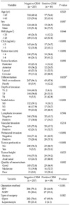
Clinicopathological characteristics of patients are summarized in Table 1. CRM involvement was found in 49 of the 306 extraperitoneal rectal cancer patients (16.0%). Mean patient age was 61 years (range, 28 to 90 years), and there were 169 males and 137 females. CRM involvement was not found to be associated with age, lymphatic and venous invasion. However, CRM involvement was significantly more common in men, overweight patients, in those with a high preoperative serum CEA level, for a tumor measuring ≥4 cm or a tumor located in anal canal, when tumor perforation occurred, for a poorly differentiated tumor grade, and in patients with perineural invasion. In terms of tumor positions, CRM involvement was most frequent in circular, lateral, anterior and posterior tumors, in that order (P = 0.001). T stage was significantly higher in patients with CRM involvement, and patients with lymph node metastasis were found to be significantly more likely to have CRM involvement (P < 0.001). A significant correlation was found between mesorectal quality and the proportion of patients with CRM involvement (P = 0.002). Compared to the rate of 9.7% (22/226) of CRM positivity after sphincter preserving proctectomy, the rate of CRM involvement after non-sphincter preserving proctectomy (NSPP) was 33.8% (27/80) (P < 0.001). A positive CRM was seen in 2 (3.5%) patients who underwent laparoscopic surgery and 47 (18.9%) patients who underwent open surgery, this difference was statistically significant (P = 0.002).
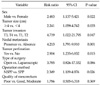
Logistic regression analysis showed that CRM involvement was significantly associated with male sex, larger tumor size, an advanced T stage, nodal metastasis, tumor perforation, and NSPP (Table 2).
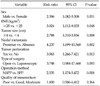
The results of the multivariate analyses of 222 patients with locally advanced tumors (T3-T4 or nodal metastasis) are shown in Table 3. Male sex, overweightedness, larger tumor size, nodal metastasis, and NSPP were found to be significantly associated with CRM involvement. To investigate the relation between rate of CRM involvement and tumor height from the anal verge, we also classified tumors based on preoperative sigmoidoscopy findings into three groups; 0 to 4.0 cm (n = 41), 4.1 to 8.0 cm (n = 180), and 8.0 to 13.0 cm (n = 85). The frequency of CRM involvement was found to increase gradually as tumor level decreased. The highest rate of CRM involvement was for tumors 0 to 4.0 cm from the anal verge (16 of 41, 39.0%), and the lowest rate was for tumors 8.0 to 13.0 cm (8 of 85, 9.4%; P = 0.002) (Table 4).
DISCUSSION
CRM status is an accepted important prognostic factor of local recurrence, a measure of quality of surgery, and an indicator of the need for adjuvant treatment [7,16,17]. The frequency of CRM involvement remains high, and reported rates were in the range 17 to 28% [7,14]. Recent studies [2,5] have reported that the rate of CRM involvement in patients with all types of rectal cancer is somewhat low (range between 5.4% and 12.5%), though this was attributed to the method used for CRM evaluation and the exclusion of patients with distant metastasis.
Different tumor biologies, the technical difficulties of the surgical procedures, and the administration of adjuvant treatment in extraperitoneal rectal cancer [9,10,18,19] suggest the need for studies that focus on the CRM statuses of patients with 'wholly' extraperitoneal rectal cancer. From a technical and anatomical perspective, it is more difficult to achieve a clear CRM for extraperitoneal tumors. However, in previous studies of CRM status in rectal cancer [1,2,6,7,14,16], the data for intraperitoneal and extraperitoneal lesions were combined. Therefore, in the present study, we focused our attention on factors associated with CRM involvement in only extraperitoneal rectal cancer, in the hope that the identification of the risk factors of CRM involvement in extraperitoneal rectal cancer would provide data regarding the indications for adjuvant therapy in patients with these risk factors.
The CRM involvement rate of 16.0% in this study is high compared to the results of multicenter studies focusing on evaluating risk factors and prognostic significance of CRM [2,6,17], which reported CRM positivity for all rectal cancer patients of around 10%. Considering the report that patients with lower rectal cancer have higher rates of non-curative resection compared to those with upper rectal cancer [9], it is assumed to be difficult to achieve negative CRM in 'wholly' extraperitoneal rectal cancer. Therefore, our CRM positivity rate, which is high compared with previous multicenter data [2,6,17], may be explained by technical difficulty associated with curative resection in extraperitoneal rectal cancer.
To our knowledge, the present study is the first to use the anterior peritoneal reflection as a landmark for differentiating extraperitoneal and intraperitoneal lesions based on operative and pathologic findings, rather than on distance from the anal verge. After excluding patients with an intraperitoneal lesion according to our definition, several clinicopathological factors were found to be significantly associated with CRM positivity in extraperitoneal rectal cancer. The predictive factors for CRM involvement were; male sex, larger tumor size (≥4 cm), more than T3, nodal metastasis, tumor perforation, and NSPP. The association between a male sex and greater CRM involvement may be due to difficult surgical access in the narrower male pelvis, which is consistent with a previous study that also reported that male patients were a risk factor for CRM involvement [6]. We also observed that NSPP had an independent effect on CRM involvement, which is supported by previous suggestions that NSPP is a significant risk factor for CRM involvement [1,2,6]. This finding implies that postoperative adjuvant therapy is necessary in patients who underwent NSPP due to the higher rate of CRM positivity. The other factors of tumor size, nodal metastasis, and tumor perforation concur with those reported in previous studies that sought to identify predictive factors of CRM involvement for 'all' types of rectal cancers [2,6,7].
In the present study, the rate of CRM involvement was only 3.5% for patients that underwent laparoscopy as compared with 18.9% for those undergoing open surgery. A previous multicenter trial on conventional versus laparoscopic-assisted surgery in colorectal cancer [20] showed a high positive CRM rate of 16% in the laparoscopic group as compared with a rate of 14% in the open group, thus our positive CRM rate appears to be promising. Concerning the effect of the laparoscopy on CRM positivity, its positive aspect may be the result of a bias. However, a comparison of variables between the laparoscopic and open groups revealed no significant differences with regard to T stage, N stage, or tumor perforation in this study. One plausible explanation for the lower rate of CRM involvement for laparoscopy was that the availability of magnification and a better identification of anatomical structures within deep pelvis during laparoscopy facilitated sharper dissection of the surgical plane [21]. Although laparoscopy was not found to be associated with a lower rate of CRM involvement by multivariate analysis, the suggestion that laparoscopic proctectomy is superior to open proctectomy in terms of reducing the rate of CRM involvement requires further clarification through randomized trials focusing on CRM status.
A few studies [1,9] have demonstrated that tumor height is related to CRM involvement. However, these studies did not assess the rate of CRM involvement with respect to a detailed classification of tumor height. The present study showed that the rate of CRM positivity for tumors located less than 4 cm from anal verge was remarkably high. This finding suggests that a tumor location around the anal canal necessitates postoperative adjuvant therapy regardless of the type of surgical resection, which is in-line with the recommendation that postoperative chemoradiotherapy is necessary for patients with CRM involvement [3]. However, the numbers of the patients with tumors around the anal canal are too small to justify this therapeutic recommendation for postoperative adjuvant treatment.
Some limitations of the present study should be noted. First, oncological outcomes were not analyzed, and therefore, no conclusions could be drawn about the impact of the identified factors on recurrence or survival. Second, although data were prospectively collected, the retrospective nature of this study inherently introduces selection bias.
In conclusion, the rate of CRM involvement after the surgical resection of extraperitoneal rectal cancer was found to be high and to be associated with male sex, larger tumor size, advanced T stage, nodal metastasis, tumor perforation, and NSPP. Given the fact that postoperative chemoradiotherapy is recommended for patients with CRM involvement, further oncologic studies are warranted to ascertain whether extraperitoneal rectal cancer patients with these risk factors should be considered for postoperative adjuvant treatment.
 XML Download