

 Citation
 Print
Print


INTRODUCTION
Gastrointestinal stromal tumors (GISTs) are currently defined as specific mesenchymal neoplasms of the digestive tract containing spindle cells and showing KIT (CD117) positivity [1,2]. Although a GIST may arise anywhere in the gastrointestinal tract, stomach (60 to 70% of cases) and small intestine (30%) are the most common sites [3,4]. Duodenal GISTs comprise a relatively small subset of GISTs with a reported frequency of 6 to 21% of surgically resected GISTs [2,5].
The optimal surgical procedure for duodenal GISTs has not been well defined [5]. The wide margins with extensive lymphadenectomy may not be required in GISTs as GISTs are associated with negligible submucosal spread and lymphatic involvement is rare [6,7]. There are currently very few reports in the English literature addressing the surgical procedures for duodenal GISTs [7]. We report herein two cases of GIST involving the third and fourth portion of the duodenum successfully treated by segmental duodenectomy with end-to-end duodenojejunostomy. This technique should be considered a treatment option for GIST located at the third and fourth portion of the duodenum.
CASE REPORTS
Case 1
A 65-year-old man presenting with abdominal pain was referred to the our hospital. His medical and family history were unremarkable. He had no history of previous abdominal surgery.
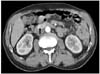
On physical examination, mild tenderness was complained in the right upper quadrant area. Abdominal computed tomography (CT) showed a well-demarcated and enhanced tumor in the third portion of the duodenum, measuring approximately 4.0 cm in diameter. The mass appeared to compress the uncinate portion of the pancreas (Fig. 1). From these radiographic findings, we diagnosed a submucosal tumor of the duodenum.
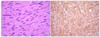
At laparotomy, a 4.0 cm sized solid mass was identifed arising from the pancreatic border of the third portion of the duodenum (Fig. 2A). No evidence of local invasion of the pancreas or of distant metastases was found. After duodenal wall was carefully dissected from the inferior border of the pancreas, segmental resection of the duodenum with adequate grossly free margins and duodenojejunostomy was performed (Fig. 2B). Operative time was 225 minutes and estimated blood loss was 100 mL. Histological examination revealed that the tumor was composed by spindle cells with a mitotic count <5 mitoses/50 high power fields (HPF) (Fig. 3A). Immunohistochemical study revealed positive staining for CD 117 (Fig. 3B). Based on the above findings, the tumor was finally diagnosed as a GIST with low-grade malignancy originating from the duodenum.
The patient had an uneventful postoperative course and no evidence of recurrence has been seen in the 42 months since his operation.
Case 2
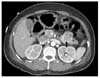
A 49-year-old woman presenting with upper gastrointestinal bleeding and melena was referred to the our hospital. A CT showed a well-demarcated enhancing tumor in the fourth portion of the duodenum, measuring approximately 4.5 cm in diameter (Fig. 4).
At laparotomy, by the Treiz's ligament take-down, a 4.5 cm sized solid mass was identifed arising from the pancreatic border of the fourth portion of the duodenum. The mass was mostly exophytic and did not invade the pancreas. After duodenal wall was carefully dissected from the inferior border of the pancreas and from the superior mesenteric artery, segmental resection of the duodenum with adequate grossly free margins and duodenojejunostomy was performed. Operative time was 90 minutes and estimated blood loss was 50 mL.
Histological examination revealed that the tumor was composed by spindle cells with a mitotic count 0 mitosis/50 HPF. Immunohistochemical study revealed positive staining for CD 117. Based on the above findings, the tumor was finally diagnosed as a GIST with low-grade malignancy originating from the duodenum.
The patient had an uneventful post-operative course and no evidence of recurrence has been seen in the 13 months since her operation.
DISCUSSION
GISTs are uncommon low-grade malignant mesenchymal tumors of gastrointestinal tract believed to originate from the interstitial cells of Cajal [4,8]. GISTs can occur anywhere throughout the gastrointestinal tract but are most commonly found in the stomach or small intestine [3,4]. The small intestine is the second most common site of GISTs, of which approximately 20% are found in the duodenum [8]. Duodenal GISTs most frequently involve the second portion of the duodenum, followed by the third portion, fourth portion, and first portion [2].
For GISTs of the foregut, gastrointestinal endoscopy may be diagnostic whenever the tumor is located in the stomach or in the upper duodenum. On the other hand, GISTs of the distal duodenum may remain undetected at gastrointestinal endoscopy. Alternative diagnostic means include CT, magnetic resonance imaging (MRI), barium study or ultrasonography [9]. However, CT and MRI seem to be the best imaging modalities for assessment of the primary lesion and detection of metastases [10].
Surgical resection with clear margins is the desirable primary treatment of GISTs [6]. As submucosal spread and local lymph node involvement is infrequent in GISTs, wide margins with routine lymph node dissection may not be required [6,7]. Gastric GISTs are adequately treated via wedge resection instead of formal gastrectomy, and there is therefore an evolving role for minimally invasive surgery [4,7,8]. Unlike gastric GISTs, the optimal surgical procedure for duodenal GISTs has not been well characterized [5]. This is because unlike the stomach or small intestine where complete excision with wide margins are relatively straightforward procedures, wide resection of tumors involving the duodenum will almost always entail a pancreaticoduodenectomy (PD) due to the unique and complex anatomy of the pancreaticoduodenal region [7]. Although PD has been reported to be associated with a low mortality rate, it remains a complex surgical procedure associated with significant short and long-term morbidity [5,7]. Limited resection should be considered a viable treatment option for duodenal GISTs when technically feasible, and extensive surgery with significant morbidity and possible mortality such as PD avoided when possible [7,8]. The main concern regarding limited resection is the risk of involved margins and hence the theoretical increased risk of local recurrence. Due to the unique and complex nature of the pancreaticoduodenal region with the proximity of the ampulla, margins of less than 5 mm after resection of duodenal GISTs are usual. Various techniques of limited resection for duodenal GISTs have been advocated depending on the site and the size of the tumors. Wedge resection with primary closure can be performed for small lesions if the resulting lumen is adequate and the ampulla can be preserved [2,7]. Segmental duodenectomy with side-to-end or end-to-end duodenojejunostomy can be performed for larger tumors located at the third and fourth portion of the duodenum [7]. Partial duodenectomy with Roux-en-Y duodenojejunostomy can be performed for larger tumors involving the antimesenteric border of the second and third portion of the duodenum [8]. An aggressive surgical approach may be required for complete removal. Major resection via a PD or a pancreas-sparing duodenectomy is indicated when the tumors are located at the second portion of the duodenum [5]. In this report, we have described two cases of GIST involving the third and fourth portion of the duodenum successfully treated by segmental duodenectomy with end-to-end duodenojejunostomy. This technique should be considered a treatment option for GIST located at the third and fourth portion of the duodenum.

 XML Download