

 PDF
PDF ePub
ePub Citation
 Print
Print


Abstract
Purpose
Although screening MMG leads to increase of early small breast cancer, axillary lymph node metastasis is still an important prognsotic factor in these patients. The aim of this study is to evaluate the incidence and predictors for axillary lymph node metastasis in patients with invasive breast carcinoma of 1 cm or less.
Methods
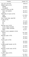
A retrospective analysis was made of 144 patients who underwent resection of primary tumor and axillary procedures between January 1999 and August 2009 for breast cancer of 1 cm or less in size. Patients were divided into two groups according to axillary node metastasis and clinicopathologic factors including age, palpable mass during physical examination, location of tumor, multifocality, tumor size, histologic type, extensive in situ component, histologic grade, nuclear grade, lymphovascular invasion, hormonal receptor status, and C-erbB-2 status were compared.
Results
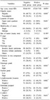
Twenty-eight (19.4%) patients of all 144 patients had metastasis in the axillary lymph node. Three variables such as multifocality (P=0.023), histologic high grade (P=0.033), presence of lymphovascular invasion (P=0.002) were found to be significant in univariate analysis. In a multivariate logistic regression analysis, however, multifocality (P=0.022) and presence of lymphovascular invasion (P=0.007) were independent predictors of axillary lymph node metastasis.
Conclusion
The incidence of axillary lymphnode metastasis of breast cancer 1 cm or less in size was 19.4%. Although the size of invasive breast carcinoma is less than 1 cm, if the tumor presented lymphovascular invasion or multifocality, axillary lymph node dissection might prove better than sentinel node biopsy.

References
1. Ko SS. The Korean Breast Cancer Society. Chronological changing patterns of clinical characteristics of Korean breast patients during 10 years (1996-2006) using nationwide breast cancer registration on -line program: Biannual update. J Surg Oncol. 2008. 98:318–323.
2. Veronesi U, Paganelli G, Viale G, Luini A, Surrida S, Galimverti V, et al. Sentinel - lymph-node biopsy as a staging procedure in breast cancer: update of a randomized controlled study. Lancet Oncol. 2006. 7:983–990.
3. Martelli G, Boracchi P, Michaela de P, Pilotti S, Oriana S, Zucali R, et al. A randomized trial comparing axillary dissection to no axillary dissection in older patients with T1N0 breast cancer: Results after 5 years of follow-up. Ann Surg. 2005. 242:1–6.
4. Veronesi U, De Cicco C, Galimberti VE, Fernandez JR, Rotmensz N, Viale G, et al. A comparative study on the value of FDG-PET and sentinel node biopsy to identify occult axillary metastases. Ann Oncol. 2007. 18:473–478.
5. Lovrics PJ, Chen V, Coates G, Cornacchi SD, Goldsmith CH, Law C. A prospective evaluation of positron emission tomography scanning, sentinel lymph node biopsy, and standard axillary dissection for axillary staging in patients with early stage breast cancer. Ann Surg Oncol. 2004. 11:846–853.
6. Ahn JH, Son EJ, Kim JA, Youk JH, Kim EK, Kwak JY, et al. The role of ultrasonography and FDG-PET in axillary lymph node staging of breast cancer. Acta Radiol. 2010. 51:859–865.
7. Kang HS, Noh DY, Youn YK, Oh SK, Choe KJ. The predictors of axillary node metastasis in 2 cm or less breast cancer univariate and multivaritate analysis. J Korean Breast Cancer Soc. 1999. 2:7–13.
8. Mustafa IA, Bland KI. Indications for axillary dissection in T1 breast cancer. Ann Surg Oncol. 1998. 5:4–8.
9. Silverstein MJ, Skinner KA, Lomis TJ. Predicting axillary nodal positivity in 2282 patients with breast carcinoma. World J Surg. 2001. 25:767–772.
10. Yip CH, Taib NA, Tan GH, Ng KL, Yoong BK, Choo WY. Predictors of axillary lymph node metastases in breast cancer: Is there a role for minimal axillary surgery? World J Surg. 2009. 33:54–57.
11. Kim TH, Bae JW, Kim F, Lee JB, Son GS, Koo BH. Factors related with axillary lymph nodes metastases in T1 invasive ductal carcinomas of the breast. J Breast Cancer. 2006. 9:31–35.
12. Fein DA, Fowble BL, Hanlon AL, Hooks MA, Hoffman JP, Sigurdson ER, et al. Identification of women with T1-T2 breast cancer at low risk of positive axillary nodes. J Surg Oncol. 1997. 65:34–39.
13. Markopoulos C, Kouskos E, Gogas H, Mandas D, Kakisis J, Gogas J. Factors affecting axillary lymph node metastases in patients with T1 breast carcinoma. Am Surg. 2000. 66:1011–1013.
14. Brenin DR, Manasseh DM, El-Tamer M, Troxel A, Schnabel F, Ditkoff BA, et al. Factors correlating with lymph node metastases in patients with T1 breast cancer. Ann Surg Oncol. 2001. 8:432–437.
15. Chen M, Palleschi S, Khoynezhad A, Gecelter G, Marini CP, Simms HH. Role of primary breast cancer characteristics in predicting positive sentinel lymph node biopsy results. A multivariate analysis. Arch Surg. 2002. 137:606–610.
16. Rivadeneira DE, Simmons RM, Christos PJ, Hanna K, Daly JM, Osborne MP. Predictive factors associated with axillary lymph node metastases in T1a and T1b breast carcinomas: Analysis in more than 900 patients. J Am Coll Surg. 2000. 191:1–8.
17. Guarnieri A, Nerri A, Correale PP, Lottini M, Testa M, Mariani F, et al. Prediction of lymph node status by analysis of prognostic factors and possible indications for elective axillary dissection in T1 breast cancers. Eur J Surg. 2001. 167:255–259.
18. Lee JH, Kim SH, Suh YJ, Shim BY, Kim HK. Predictors of axillary lymph node metastases (ALNM) in a Korean population with T1-2 breast carcinoma: Triple negative breast cancer has a high incidence of ALNM irrespective of the tumor size. Cancer Res Treat. 2010. 42:30–36.
19. Bevilacqua J, Cody H 3rd, MacDonald KA, Tan LK, Borgen PI, Van Zee KJ. A model for predicting axillary node metastases based on 2000 sentinel node procedures and tumor position. EJSO. 2002. 28:490–500.
20. Cabioglu N, Ozmen V, Kaya H, Tuzlali S, Igci A, Muslumanoglu M, et al. Increased lymph node positivity in multifocal and multicentric breast cancer. J Am Coll Surg. 2009. 208:67–74.
21. Andea AA, Bouwman D, Wallis T, Visscher DW. Correlation of tumor volume and surface area with lymph node status in patients with multifocal/multicentric breast carcinoma. Cancer. 2004. 100:20–27.
22. Coombs NJ, Boyages J. Multifocal and multicentric breast cancer: Does each focus matter? J Clin Oncol. 2005. 23:7497–7502.
23. Giard S, Chauvet MP, Penel N, Mignotte H, Martel P, Tunon De Lara C, et al. Feasibility of sentinel lymph node biopsy in multiple unilateral synchronous breast cancer: results of a French prospective multi-institutional study (IGASSU0502). Ann Oncol. 2010. 21:1630–1635.
24. Kim HJ, Lee JS, Park EH, Choi SL, Lim WS, Chang MA, et al. Sentinel node biopsy in patients with multiple breast cancer. Breast Cancer Res Treat. 2008. 109:503–506.
25. Ferrari A, Dionigi P, Rovera F, Boni L, Limonta G, Garancini S, et al. Multifocality and multicentricity are not contraindications for sentinel lymph node biopsy in breast cancer surgery. World J Surg Oncol. 2006. 4:79–86.
 XML Download