

 PDF
PDF ePub
ePub Citation
 Print
Print


Abstract
Purpose
The extracelluar domain (ECD) of HER2 may be cleaved from the surface of cancer cells whereby serum HER2 ECD levels can be detected. We explored the correlation between serum HER2 ECD and tissue HER2 status and their relationship with clinicopathological parameters.
Methods
We included 125 patients with stage 0-3 breast cancer. The serum HER2 ECD level was measured by chemiluminescence immunoassay (ADVIA Centaur® system). The tissue HER2 status was analyzed by immunohistochemistry (IHC) and fluorescence in situ hybridization (FISH) in all tumors. We reviewed the medical records retrospectively. The analyzed clinicopathological parameters were age, tumor size, histologic grade, vascular invasion, lymph node involvement, stage, estrogen receptor (ER) and CA 15-3.
Results
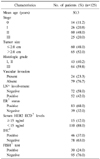
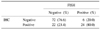
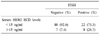
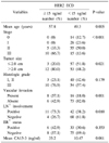
High serum HER2 ECD levels (≥15 ng/ml) were reported in 15 patients (12.0%). For tissue HER 2 status, 30 patients (24.0%) had positive results in FISH and 46 patients (37.0%) had strong positive results in IHC (3+). The specificity of serum HER2 ECD was 92.6% but the sensitivity was only 26.7%. The concordance between serum HER2 ECD and FISH tests was 23.3%. High serum HER2 ECD levels were significantly associated with old age (P=0.005), large tumor size (P=0.021), vascular invasion (P=0.001), lymph node involvement (P=0.010) and advanced stage (P<0.001). ER and CA 15-3 levels were not significantly related with serum HER2 ECD.
Figures and Tables
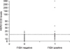
Fig. 1
Distribution of serum HER2 ECD levels in breast cancer patients with FISH positive or FISH negative tissue. The horizontal line indicates the cut-off value of 15 ng/ml.
References
1. Slamon DJ, Clark GM, Wong SG, Levin WJ, Ullrich A, McGuire WL. Human breast cancer: correlation of relapse and survival with amplification of the HER-2/neu oncogene. Science. 1987. 235:177–182.
2. Slamon DJ, Leyland-Jones B, Shak S, Fuchs H, Paton V, Bajamonde A, et al. Use of chemotherapy plus a monoclonal antibody against HER2 for metastatic breast cancer that overexpresses HER2. N Engl J Med. 2001. 344:783–792.
3. Smith I, Procter M, Gelber RD, Guillaume S, Feyereislova A, Dowsett M, et al. 2-year follow-up of trastuzumab after adjuvant chemotherapy in HER2-positive breast cancer: a randomised controlled trial. Lancet. 2007. 369:29–36.
4. Wolff AC, Hammond ME, Schwartz JN, Hagerty KL, Allred DC, Cote RJ, et al. American Society of Clinical Oncology/College of American Pathologists guideline recommendations for human epidermal growth factor receptor 2 testing in breast cancer. J Clin Oncol. 2007. 25:118–145.
5. Zabrecky JR, Lam T, McKenzie SJ, Carney W. The extracellular domain of p185/neu is released from the surface of human breast carcinoma cells, SK-BR-3. J Biol Chem. 1991. 266:1716–1720.
6. Codony-Servat J, Albanell J, Lopez-Talavera JC, Arribas J, Baselga J. Cleavage of the HER2 ectodomain is a pervanadate-activable process that is inhibited by the tissue inhibitor of metalloproteases-1 in breast cancer cells. Cancer Res. 1999. 59:1196–1201.
7. Quaranta M, Daniele A, Coviello M, Savonarola A, Abbate I, Venneri MT, et al. C-erbB-2 protein level in tissue and sera of breast cancer patients: a possibly useful clinical correlation. Tumori. 2006. 92:311–317.
8. Ludovini V, Gori S, Colozza M, Pistola L, Rulli E, Floriani I, et al. Evaluation of serum HER2 extracellular domain in early breast cancer patients: correlation with clinicopathological parameters and survival. Ann Oncol. 2008. 19:883–890.
9. Lennon S, Barton C, Banken L, Gianni L, Marty M, Baselga J, et al. Utility of serum HER2 extracellular domain assessment in clinical decision making: pooled analysis of four trials of trastuzumab in metastatic breast cancer. J Clin Oncol. 2009. 27:1685–1693.
10. Muller V, Witzel I, Luck HJ, Kohler G, von Minckwitz G, Mobus V, et al. Prognostic and predictive impact of the HER-2/neu extracellular domain (ECD) in the serum of patients treated with chemotherapy for metastatic breast cancer. Breast Cancer Res Treat. 2004. 86:9–18.
11. Colomer R, Llombart-Cussac A, Lloveras B, Ramos M, Mayordomo JI, Fernandez R, et al. High circulating HER2 extracellular domain levels correlate with reduced efficacy of an aromatase inhibitor in hormone receptor-positive metastatic breast cancer: a confirmatory prospective study. Cancer. 2007. 110:2178–2185.
12. Lee JS, Min WK, Park EH, Lim WS, Choi SL, Son BH, et al. Correlation between the Her-2/neu status as determined by immunohistochemical analysis and the serum Her-2/neu concentration as determined by the use of ADVIA Cencaur® automated immunoassay in breast cancer patients. J Breast Cancer. 2008. 11:116–124.
13. Kim JW, Kim SY, Lee HS, Woo HD, Son DM, Lim CW, et al. Establishment for reference range of serum HER-2/neu in Korean healthy women. J Breast Cancer. 2006. 9:301–308.
14. Kong SY, Kang JH, Kwon Y, Kang HS, Chung KW, Kang SH, et al. Serum HER-2 concentration in patients with primary breast cancer. J Clin Pathol. 2006. 59:373–376.
15. Kong SY, Nam BH, Lee KS, Kwon Y, Lee ES, Seong MW, et al. Predicting tissue HER2 status using serum HER2 levels in patients with metastatic breast cancer. Clin Chem. 2006. 52:1510–1515.
16. Fornier MN, Seidman AD, Schwartz MK, Ghani F, Thiel R, Norton L, et al. Serum HER2 extracellular domain in metastatic breast cancer patients treated with weekly trastuzumab and paclitaxel: association with HER2 status by immunohistochemistry and fluorescence in situ hybridization and with response rate. Ann Oncol. 2005. 16:234–239.
17. Harris L, Luftner D, Jager W, Robertson JF. C-erbB-2 in serum of patients with breast cancer. Int J Biol Markers. 1999. 14:8–15.
18. Garoufali A, Kyriakou F, Kountourakis P, Yioti I, Malliou S, Nikaki A, et al. Extracellular domain of HER2: a useful marker for the initial workup and follow-up of HER2-positive breast cancer. J BUON. 2008. 13:409–413.
19. Narita T, Funahashi H, Satoh Y, Takagi H. C-erbB-2 protein in the sera of breast cancer patients. Breast Cancer Res Treat. 1992. 24:97–102.
20. Willsher PC, Beaver J, Pinder S, Bell JA, Ellis IO, Blamey RW, et al. Prognostic significance of serum c-erbB-2 protein in breast cancer patients. Breast Cancer Res Treat. 1996. 40:251–255.
 XML Download