

 PDF
PDF ePub
ePub Citation
 Print
Print


INTRODUCTION
Early postoperative assessments of the degree of graft injury are essential to manage and predict the outcome of liver transplantation (LT). Graft injury due to preservation, ischemia, reperfusion, and immunologic activation can be related to clinical graft outcome. However, there are a few definite markers [1] that predict the degree of graft injury.
A multidrug resistance-associated protein (MRP) 2 is a glutathione conjugate in the canalicular membrane of hepatocytes. It is a canalicular multispecific organic anion transporter [2] localized to the hepatocyte canalicular membrane. MRP2 is a conjugate transporting ATPases functioning in detoxification and the defense against oxidative stress.
In a normal liver, MRP2 is demonstrable at canaliculi as a homogenous pattern on immunohistochemical staining using a monoclonal antibody. In animal studies, MRP2 was vulnerable under specific circumstances i.e., ischemic injury due to arterial thrombosis [3], rapid regeneration of the graft [2], obstructive jaundice [4], sepsis [56], or chronic cholestatic liver disease [78910]. MRP2 alterations are related to functional depletion, causing conjugated hyperbilirubinemia after major hepatectomy, chronic cholestatic liver disease, and transplantation, although there would another way of efflux of bile excretion.
MRP2 alteration is a result of liver (or liver graft) damage. MRP2 alteration is associated with functional depletion, causing conjugated hyperbilirubinemia and cholestasis, although there are other pathways of efflux of bile excretion. MRP2 alteration itself can cause cholestasis in several genetic diseases. However, MRP2 alteration in secondary changes does not reflect specific changes of focal anatomic distortion, but instead indicates overall hepatocyte damage leading to cholestatic hepatic damage. Fortunately, MRP2 alteration can be reversed with liver regeneration, if damage is not severe or if there is no ongoing damage. Ongoing graft damage slows, halts, or worsens recovery subclinically before presentation.
Therefore, we hypothesized that the degree of graft injury and any ongoing subclinical graft damage may be related to alterations of the expression pattern of MRP2 after LT, as observed on an event-free protocol biopsy.
In the present study, we investigated the pattern of MRP2 expression after LT compared to the normal liver of live donors. We then evaluated the relationship of this pattern of MRP2 expression with the graft outcome.
METHODS
This study included adult recipients who underwent LT and subsequent event-free protocol graft biopsy within 2 months after LT. The following were excluded: recipients who underwent an event-driven liver biopsy, ABO incompatible LT, retransplantation, or multiorgan transplantation. Finally, 41 recipients were enrolled in this study over 2 years.
Among these 41 recipients, 19 patients underwent living donor LT (LDLT) (46.3%) and the other patients underwent deceased donor LT (DDLT). The intraoperative biopsy samples were available for 15 matched donors among the 19 LDLT pairs. The liver biopsy from living donors were routinely performed just after exploration of the abdomen during donor operation in order to confirm the quality of the liver, and these remnant biopsy samples were used as the control group. Therefore, a total of 56 paraffin-embedded liver tissues, i.e., 41 tissues of the recipients and 15 tissues of the donors were evaluated with immunohistochemical staining using monoclonal antibodies for MRP2 (M2 II-6, Chemicon, Temecula, CA, USA) [11].
This study was performed in accordance with the ethical standards laid down in an appropriate version of the 2000 Declaration of Helsinki as well as the Declaration of Istanbul 2008. The Institutional Review Board at Seoul National University Hospital (H-1208-090-422) approved this study, and we followed the study protocol of the pathology department for paraffin embedded samples. Informed consent was waived by the institutional review board of Seoul National University Hospital.
The expression pattern of MRP2 in the donor liver and graft
The normal MRP2 staining pattern observed in donors was homogeneous canalicular staining (Fig. 1A). In recipients, the canalicular MRP2 staining was subclassified into 3 types; type C0 (homogenous canalicular staining) (Fig. 1B), type C1 (focal canalicular staining) (Fig. 1C), and type C2 (no canalicular staining) (Fig. 1D). Type C0 showed a pattern similar to a normal donor liver, whereas types C1 and C2 showed patterns different from a normal donor liver.
Post-LT management
Immunosuppression consisted of basiliximab induction and maintenance of calcineurin inhibitor and steroids. Mycophenolate mofetil was employed in cases of toxicity from calcineurin inhibitors or renal impairment. The corticosteroid was tapered to discontinuation by 6 months after LT. In our center, we performed the protocol biopsy of the graft within 2 months, regardless of graft function.
The definitions used for complications were adapted from the Clavien-Dindo grading system for negative outcomes [12]. Patients routinely underwent liver ultrasound or computed tomography scans at 1, 4, 12, and 24 months. Patients were followed for a mean of 58.3 ± 31.9 months (range, 5–93 months).
Statistical analysis
All values are expressed as means ± standard deviations. The categorical variables were compared using the Fisher exact test, and the continuous variables were compared via nonparametric Mann-Whitney U-test. We employed the Kaplan-Meier method with the log-rank test for the analysis of patient survival and cumulative risk of posttransplantation complications. A P-value of <0.05 was regarded as statistically significant. Statistical analyses were conducted using SPSS ver. 17.0 (SPSS Inc., Chicago, IL, USA).
RESULTS
Patients' characteristics
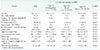
The recipients (Table 1) included 31 men and 10 women, ranging in age from 37 to 64 years (mean, 52.1 ± 7.2 years). Korean Network for Organ Sharing Status was 1 in 1 patient (2.4%), 2A in 2 patients (4.9%), and 2B or less in 38 patients (92.7%). The mean medical Model for End Stage Liver Disease (MELD) score, which was calculated solely based on liver disease severity without an additional point for hepatocellular carcinoma (HCC), was 18.8 ± 8.0. Hepatitis B virus-associated liver disease was the most common original liver disease (n = 33, 80.5%), followed by hepatitis C virus in 5 patients (12.2%) and other diseases in 3 patients. HCC was diagnosed in 26 patients (63.4%). The mean operation time was 515.4 ± 148.9 minutes (range, 290–910 minutes). The mean cold ischemic time was 382.1 ± 299.7 minutes (range, 41–890 minutes). There were no cases of split LT and ABO incompatible LT.
The 15 living donors were healthy prior to the operation. They had no underlying liver disease or medical history. Their median age was 27 years (range, 19–44 years). There were 13 male donors. The mean graft versus recipient weight ratio (GRWR) was 0.96% ± 0.21% (range, 0.58%–1.30%) in LDLTs. The GRWR was not measured in DDLTs.
The expression pattern of MRP2 in the recipients
The mean time to the event-free protocol biopsy of the recipients was 4.1 ± 2.0 weeks after LTs. The expression pattern of MRP2 in the graft showed alterations in 34 recipients (82.9%), and type C0 staining was only noted in 17.1% of the patients (n = 7). Type C1 was noted in 36.6% of the patients (n = 15), and type C2 was noted in 46.3% of the patients (n = 19).
The pretransplant factors associated with the score of alteration of MRP2 in the graft
The preoperative characteristics of the donors, grafts, and recipients showed on Table 1. There were no differences among the patient groups according to the different patterns of posttransplant MRP2 alterations. This included age (P = 0.882), sex (P = 0.257), original liver disease (P = 0.761), accompanying HCC before transplantation (P = 0.624), type of the graft (P = 0.390), Korean Network for Organ Sharing Status (P = 0.797), MELD score (P = 0.104), GRWR in cases of LDLTs (P = 0.238), cold ischemic time (P = 0.975), donor age (P = 0.951), and the time of liver biopsy (P = 0.606).
The posttransplant factors associated with the score of MRP2 alteration in the recipients
The operation time was significantly longer in patients with type C2 staining (562.6 ± 162.9 minutes) than in patients with type C0 staining (393.8 ± 52.7 minutes) (P = 0.038).
Posttransplant hospital stay was longer in patients with type C2 staining (56.7 ± 89.4 days) than in patients with type C0 (29.0 ± 13.0 days) and type C1 staining (44.5 ± 67.8 days). However, it was not statistically significant (P = 0.477).
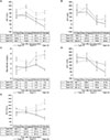
The laboratory findings at the time of biopsy were not different among these 3 groups, including the total bilirubin, AST, ALT, ALP, and gamma-glutamyl transpeptidase (GGT) levels (P > 0.05). The serum levels of AST, total bilirubin, and ALP were more elevated in the C2 group than in the C0 group at 6 months and one year after LT, but this was not statistically significant (P > 0.05) (Fig. 2).
The posttransplant complications and graft survival, and the score of MRP2 alteration in the recipients
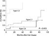
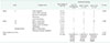
There were significant differences in rates of posttransplant complications according to the type of posttransplant MRP2 alteration (type C0, n = 4, 42.9%; type C1, n = 11, 73.3%; type C2, n = 19, 100%) (P < 0.001) (Table 2). The rates of minor complications were significantly different among the three groups (type C0, n = 1, 14.3%; type C1, n = 4, 26.7%; type C2, n = 10, 52.6%) (P = 0.018). The rates of major complications were significantly different among these 3 groups (type C0, n = 2, 28.5%; type C1, n = 5, 33.3%; type C2, n = 17, 89.4%) (P = 0.007). The rates of complications were not different among these 3 groups at the time of biopsy, but the cumulative risks of the complications were increased according to the type of MRP2 alteration (P = 0.003) (Fig. 3). However, no significant factors were associated with posttransplant complications in multivariate analysis.
The overall graft survival rates (C0, 85.7%; C1, 73.3%; C2, 89.5%) (P = 0.453), and the overall patient survival rates (C0, 85.7%; C1, 80.0%; C2, 84.2%) (P = 0.927) were not different according to the type of the posttransplant MRP2 alteration.
DISCUSSION
This research attempted to clarify the following points: First, whether any changes in MRP2 patterns occur during the early post-LT period, and second, if yes, whether there are factors associated with alteration of MRP2.
MRP2 alteration is observed in several genetic disease leading to hyperbilirubinemia (in humans with Dubin-Johnson syndrome and in GY/TR EHBR rats) [1314], in obstructive jaundice [15], in severe fatty liver grafts [16], and in secondary liver injury (macro- or microangiopathy, biliary complications, toxic bile salts, infection, or rejection in the liver graft) [1234567891017181920212223]. In genetic disease lacking MRP2 transporter as well as BSEP, MRP2 alteration can lead to jaundice. In contrast, in hepatic injury due to obstructive jaundice, ischemia, or sepsis, MRP2 alteration is a kind of stigma of hepatic injury, and can represent the degree of injury. In this study, MRP2 alteration after LT could be caused by preservation, ischemia/reperfusion injury, or other insult such as a longer operation time. Long operation time might indicate a difficult operation, leading to a negative impact on graft quality. Unfortunately, we did not collect some intraoperative data, because a half of the recipients underwent LT at other institutes and were only followed up in our institute. MRP2 alteration is a result of liver graft damage, and would not be associated with increased operation time nor posttransplant complication.
For this reason, MRP2 alterations were noted in more than 80% of the recipients in the routine protocol biopsy after LT, i.e., alteration of the canalicular staining would be related to graft damage due to preservation and ischemic/reperfusion injury that every graft undergoes.
In addition, MRP2 alteration can reflect ongoing subclinical liver injury as well as previous liver graft damage. A liver graft usually recovers from preservation and ischemia/reperfusion injury within 1 month after transplantation; thus, MRP2 alteration should improve by that time if there is no additional problem. Persistence of MRP2 alteration in event-free protocol biopsy indicates that graft damage was too severe to completely resolve within 1 month, or that there is new subclinical damage. Type C2 alteration on biopsy before clinical signs such as cholestasis could be interpreted as a predictor of a worse outcome. In this study, the post-LT complications were not clinically obvious at the time of event-free protocol biopsy at a median of 4 weeks after transplantation. For this reason, MPR2 alteration (type C1 or 2) in a liver graft represents any kind of graft damage regardless of cause, and can predict a worse outcome than in cases without MRP2 alteration (type C0). Thus, valuation of MRP2 in an event-free protocol biopsy can predict the degree of graft injury, and can indicate the prognosis related to graft quality after LT. In this study, according to the time course, the rate of complications was higher in the type C2 group than in the other groups. For example, one patient showed normal intrahepatic biliary structure by cholangiography but his event free biopsy showed type C2 MRP2 alteration, without evidence of acute cellular rejection at 1 month after transplantation. Two months later, he developed from cholangitis and follow-up cholangiography showed a nearly necrotic intrahepatic biliary tree (Supplementary Fig. 1).
Furthermore, several studies confirmed that MRP2 alteration in inherited cholestatic liver disease and in obstructive cholestatic disease could lead to vicious cycles of MRP2 alterations, with severe liver damage due to a build-up of pressure and cell lysis caused by accumulation of bile salts [131415]. For this reason, MRP2 alteration could indirectly lead to posttransplant graft damage via cholestasis or more severe complications.
Recent studies using the hepatobiliary contrast agent, Gd-EOB-DTPA, in dynamic contrast-enhanced MRI of the liver revealed that the MRP2 pathway was related to contrast agent excretion into the biliary tree as well as bile salts [2425]. One patient in our study showed type C2 MRP2 alteration on protocol biopsy at one month after transplantation. One month later, the patient complained of itching and hyperbilirubinemia. Follow-up MRI with Gd-EOB-DTPA showed no contrast excretion into the biliary tree, but there was no stricture in MR cholangiography in T2-weighted images (Supplementary Fig. 2).
The abnormal liver function test results, especially idiopathic hyperbilirubinemia after liver surgery, could be related with the alteration of the bile transporter system [26]. Most patients with idiopathic hyperbilirubinemia after liver surgery recovered spontaneously after the liver restored new hepatocytes, and a relief of bile salt retention by stimulating canalicular transport and bile secretion [25272829]. In this study, AST, total bilirubin, GGT and ALP levels were higher in the type C2 group than in the type C0 group. These laboratory values were related to the bile transporter system.
The pattern of MRP2 alteration was not associated with both patient and graft survival outcomes in this study. This might be related to the bias associated with retrospective selection of patients who underwent protocol biopsy. If we enrolled every recipient with event-driven biopsy, the biopsy results would show more aggressive changes than those in this cohort of patients. The severity of MRP2 alteration would be related to graft survival outcomes. In this study, we investigated the pattern of MRP2 expression after LT in event-free biopsy. Thus, patients who could not survive but who might have severe graft damage were not enrolled in this study. Further research would be helpful to reveal the pattern of MRP2 alteration and survival outcome.
In summary, MRP2 alteration was observed in 82.9% of early posttransplantation liver grafts. The pattern of MRP2 alteration was associated with longer operation time and post-LT complications. These MRP2 alterations would represent the significance of early posttransplantation graft damage without full recovery or with ongoing graft damage, leading to sequential posttransplant complications. Further prospective study of large cohort is required to validate this finding.

 XML Download