

 PDF
PDF ePub
ePub Citation
 Print
Print


INTRODUCTION
Many patients have been suffering from severe liver diseases such as hepatocellular carcinoma, liver cirrhosis from chronic viral hepatitis and alcoholic liver disease. So many treatments have been developed for decades for the patients having serious liver diseases. Liver transplantation is an effective therapeutics options for chronic liver diseases. However, it is limited by donor organ shortage, surgical complications and the risk of rejection [1]. Recently, artificial livers have been considered as an alternative for liver transplantation. The major obstacle to this is that primary hepatocytes cannot be cultured for a long time. Commonly hepatocytes are differentiated into fibroblastlike after isolated 3–5 days and turned on the cell death program [2]. Hence, long-term culture of hepatocytes is the main problem that has to be resolved to be able to make bionic livers.
Currently, 3-dimensional (3D) printing technology plays an important role in tissue engineering. Principle of 3D printing is that a computer recognizes a 3D model and slices it into a series of 2-dimensional (2D) sections, which in then printed from bottom to the top. This method has the advantage that it can print any desired shape. It can be used in biological applications since several cellular matrixes, such as collagen, alginate, etc., are available to which cells can attach.
Many reports suggested that 3D cultures are superior to 2D cultures in previous studies [3]. In this report, we printed primary hepatocytes using a 3D bio printer and compared their functions. By using the 3D bio-printing, drawback of primary hepatocytes that make long-term culture difficulty can be resolved. And it can be one of the possible options for bioartificial liver.
METHODS
Isolation of mouse primary hepatocytes
Primary hepatocytes were isolated from 6–8 week mouse livers by a 2-step collagenase perfusion method [4]. Briefly, A and B solution had almost same compositions except presence of ethylenediaminetetraacetic acid (EDTA) in solution A. EDTA in solution A was calcium chelating agent and solution B was comprises of many compounds including collagenase and trypsin inhibitor for isolate primary hepatocytes from liver tissues. After sequential flows of solution A for 5 minutes and solution B for 8 minutes at 37℃, livers were removed and chopped up in a dish. After percoll treatment, we obtain 4 × 10⁷ hepatocytes with 80%–90% viability. The primary hepatocytes were cultured in Williams' Medium E (Gibco, Grand Island, NY, USA) supplemented with 0.1µM dexamethasone, 0.5% penicillin/streptomycin, 1% ITS, 5mM GlutaMAX (Gibco) and 15mM HEPES (N-2-hydroxyethylpiperazine-N-2-ethanesulfonic acid) at 37℃ in a 5% CO2 incubator. The medium was changed every 2 days.
Fabrication of cells-alginate constructs
The cells-alginate constructs were fabricated using 3D bio-printing system (Korea Institute of Machinery and Materials, Daejeon, Korea). The 3D printing system consisted of a computer-aided system with a 3-axes stage, syringe, nozzle, and pressure controller. To fabricate the hydrogel constructs, 3% alginate was dissolved in cell medium solution. Cells-alginate hydrogels were ejected through the nozzle layer-by-layer. Hydrogel porous patterns of strand thickness and space were controlled by pressure and velocity. 4 × 10⁷ hepatocytes were blended with 3% alginate solution and 1% calcium chloride. The 3D bio-printed structures measured 25 mm × 25 mm and consisted of 7 layers. After printing, the scaffolds were incubated in 5% calcium chloride solution for 10 seconds cross-linking and washed with phosphate buffered saline (PBS) (WELGENE, Gyengsan, Korea). After washing, the alginate scaffolds with primary hepatocytes were incubated in culture medium.
Quantitative real-time polymerase chain reaction
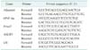
Total RNAs were isolated with TRIzol Reagent (Ambion, Foster City, CA, USA) with chloroform. 1-µg samples of RNA were reverse transcribed using a Transcriptor First Strand cDNA Synthesis Kit (Roche, Mannheim, Germany). Real-time polymerase chain reaction (PCR) was conducted using 1 µg of cDNA, 10 µg of quantitative PCR PreMix (Dyne bio, Seongnam, Korea), and oligonucleotide primers. The primers are shown at Table 1 (mouse Albumin, HNF-4α, Foxa3, ASGR1 and U6). The PCR cycles consisted of 40 cycles of 95℃ for 20 seconds and 60℃ for 40 seconds. Melting curves were constructed to characterize the PCR products.
Hematoxylin & eosin staining
Alginate scaffolds were embedded in Optimal Cutting Temperature Compound (Sakura Finetek, Tokyo, Japan) and sectioned at 4-µm thickness. Sections were fixed with 95% ethanol for 5 minutes. After fixation, sections were washed with DW for 5 minutes. After washing, sections were stained with hematoxylin for 3 minutes and washed with DW for 1 minute. Next, sections were counterstained with eosin for 1 minute and washed with running water for 5 minutes and sections were dehydrated with 50% ethanol for 3 minutes, 70% ethanol for 3 minutes, 95% ethanol for 3 minutes and 100% ethanol for 3 minutes. Finally, sections were immersed with xylene and mounted with mounting solution (Southern Biotech, Birmingham, UK).
Immunofluorescence
Alginate scaffolds were sectioned as above, fixed in acetone for 15 minutes and dried for 5 minutes. After washing with PBS buffer they were incubated with primary antibodies, anti-goat serum albumin antibody (ab19194, Abcam, Cambridge, MA, USA) and anti-mouse monoclonal cytokeratin 18 antibody (ab668, Abcam), at 4℃ overnight each was diluted 1:100. After incubation, sections were washed 3 times with PBS. The sections were then incubated with Hoechst 33342 (H3570, Life Technologies, Eugene, OR, USA) was diluted 1:10,000, followed by secondary antibodies, 594 donkey anti-goat (A11058, Life technologies) and 488 donkey anti-mouse (A21202, Life technologies) at room temperature for 1 hour each diluted 1:250. After incubation, sections were washed 3 times with staining solution and PBS. Finally, sections were mounted with mounting solution.
RESULTS
Isolation of primary hepatocytes & 3D alginate scaffold application
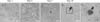
For 3D printing of primary hepatocytes, we isolated mouse hepatocytes by collagenase perfusion method. Perfusion solutions A and B were injected through the mouse portal vein (Fig. 1A, B). The following day, polygonal and binuclear hepatocytes could be seen attached onto collagen coated dishes (Fig. 1C).
In general, primary hepatocytes cannot be cultured for more than 2 days without loss of function. Therefore, to see whether hepatocytes could multiply for longer in the 3D structures, 4 × 10⁷ hepatocytes were mixed with 3% alginate solution and 1% calcium chloride. The 3D bio-printed structures measured 25 mm × 25 mm and had 7 layers (Fig. 1D, E).
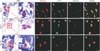
The 3D printed hepatocytes were observed for 14 days in hepatocyte culture medium. On day 1, the primary hepatocytes were present in twos and threes within the alginate construct (Fig. 2A, B), but by day 3 they were had formed aggregates that expanded in time. These phenomena cannot present on 2D culture system. Seven days after culture, primary hepatocytes were assembled more and more as time passes (Fig. 2C–E). And this aggregates were found at many locations in alginate scaffold. These results suggest that primary hepatocytes printed with alginate, can be cultured for more than 2 weeks and create in vivo like hepatocyte masses.
Expression of hepatic genes in the 3D constructs
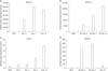
A key question concerning 3D printing of primary hepatocytes is whether they maintain hepatic function. We therefore examined the expression of four major marker genes by quantitative real-time PCR albumin, hepatocyte nuclear factor 4 alpha (HNF-4α), forkhead box protein A3 (Foxa3), and asialoglycoprotein receptor 1 (ASGR1). As shown in Fig. 3, expression of these genes increased gradually up to day 14, though expression of ASGR1 fell on day 14 (Fig. 3D).
We also stained the 3D printed hepatocytes with hematoxylin & eosin staining (H&E), and examined the production of hepatic-specific proteins by immunofluorescence analysis. H&E staining showed that the primary hepatocytes maintained their normal appearance up to day 14 (Fig. 4A, B, G, H, M, N). Albumin and cytokeratin 18 could also be demonstrated up to day 14 (Fig. 4C–F, I–L, O–R). Taken together these findings indicate that functions of isolated primary hepatocytes are maintained in the 3D alginate constructs, and suggest the possibility of manufacturing artificial livers using 3D bio-printing techniques.
DISCUSSION
Researches of primary hepatocytes have been briskly proceeded to detect hepatic uptake and metabolism [5]. The hepatic functions of primary hepatocytes such as secretion of albumin, synthesis of urea, and expression of CYP3A4 have usually been used in drug screening study [6]. But primary hepatocytes quickly decrease hepatic functions when they are isolated from the tissues [78]. Previous studies have demonstrated the effect that culture conditions such as the culture media, supplements of media, extracellular matrix and collagen gel sandwich methods [9].
Recently, many researcher use 3D bio-printing for improve culture systems. 3D printing considers a diverse of processes for manufacturing 3D objects from a 3D models or electronic data sources. First, 3D models or electronic data sources are split to many slices and stacking by the layers. In the near future, we can make bio-printed livers by 3D printing technology [10]. For applications of therapeutic into human, the typical process of bio-printing would cover the isolation and enlargement of human cells beforehand printing the scaffold. Scaffolds printed bio-printing could eventually be used as devices of therapeutic such as a platform of testing for drug screening and discovery, or an in vitro model system for disease [3].
Alginates are naturally obtainable glycans that easily form hydrogels [11]. They consist of α-L-glucuronic acid monomers and (1-4)-linked β-D-mannuronic acid that form polymer chains [12]. Divalent cations such as Ca2+ promote crosslinking by binding between the regions of sequential α-L-guluronic acid. This makes alginate scaffolds maintain the desired shape. Alginate has been use for 3D cell culture and cell transplantation and is bio-degradable. Alginate scaffolds are reported to be suitable for studying cell-ligand interactions owing to the low protein assimilation of the anionic polysaccharide [13].
Primary hepatocytes form characteristic aggregate when cultured in organ systems [14]. Up 7 days, primary hepatocytes cultured in the 3D alginate scaffolds existed as single cell. After 7 days, they show gradually agglomerated, and their morphology suggested such as albumin synthesis [14]. These findings indicated that primary hepatocytes in printed 3D scaffolds were stable and retained hepatic-function.
After isolation, primary hepatocytes quickly dedifferentiate in 2D culture system, losing hepatic function and remaining viable for only a short time. Expression of hepatic genes therefore steadily declines [15]. However, our investigation has shown above that hepatocyte function is retained for up to 14 days in the 3D alginate scaffolds.
Up to now, the collagen sandwich method has been the best technology for culturing primary hepatocyte. The system is better than 3D bio-printing, for observation of morphological changes, while retention of function is similar in the 2 systems.
In conclusion, the advantages of 3D bio-printing are the ability to control the cell distribution, its high scalability and the ability to form desired shapes. This technique has enormous potential in tissue engineering and for make micro unit of complex in bio-medical area.

 XML Download