

 PDF
PDF ePub
ePub Citation
 Print
Print


INTRODUCTION
Different mechanisms are involved in the pathogenesis of chronic spontaneous urticaria (CSU).12 In the early 90s, Grattan et al.3 demonstrated the presence of immunoglobulin (Ig) G with anti-IgE function in chronic urticaria. Two years later, the same research group found IgG against the high affinity IgE receptor and also demonstrated that these IgG autoantibodies could induce basophil activation.4
After demonstrating IgG autoantibodies in CSU, Bar-Sela et al.5 suggested that IgE may have an auto-reactivity role in urticaria by demonstrating IgE against thyroid peroxidase (TPO). However, while reproducing the results with IgG autoantibodies in multiple studies has been possible, the study of auto-reactive IgE has been more difficult. Studies from Concha et al.6 indicate that the detection of IgE specific for TPO was an occasional finding, and the results suggest that these antibodies were unlikely to play an important pathogenic role in urticaria. With the depletion of IgG prior to the measure of IgE, or with new innovations in IgE detection techniques, some recent studies789 have shown that anti-TPO IgE in patients with CSU is more frequent than previously described. However, if these IgE autoantibodies can trigger urticaria or not in patients with CSU has not been proven and the clinical role of this IgE has been little studied.
In this study, we investigated the clinical impact of IgE autoantibodies against TPO in CSU. For this purpose, we followed 3 steps:
1. Estimated the prevalence of anti-TPO IgE in our population.
2. Evaluated the interaction of anti-TPO IgE with basophil cells for the induction of CD203c expression.
3. Evaluated if TPO can induce urticaria on the skin of patients with anti-TPO IgE and if this response could be transferred to healthy subjects.
MATERIALS AND METHODS
Study population
This is a cross-sectional study with 3 groups based on their clinical status: the CSU, healthy control and autoimmune thyroid disease (ATD) groups. The CSU population was recruited from the Urticaria Research of Tropical Impact and Control Assessment (URTICA) cohort (ClinicalTrials.gov number: NCT01940393).1011 The CSU group included patients older than 12 years with CSU, defined as the recurrence of hives, with or without angioedema, for at least 6 weeks.12 Patients could be receiving their treatment with antihistamines but not with anti-IgE antibodies. The exclusion criteria were the following: systemic diseases or pharmacologic treatment that could cause hives. The impact of CSU on the quality of life was assessed with the dermatology life quality index (DLQI) and the urticaria activity score (UAS) was used for the disease activity.11 For skin tests, patients suspend antihistamines 7 days before the autologous serum skin test (ASST), intradermal, skin prick test (SPT) or passive transfer of serum.
The healthy control group consisted of people older than 12 years without historical autoimmune diseases, chronic urticaria or history of acute urticaria in the previous 2 years.
The ATD group included patients with Hashimoto's thyroiditis according to the American thyroid association; 13 the presence of thyroid autoantibody above the normal range (anti-TPO IgG ≥ 5.0 IU/mL and/or anti-thyroglobulin ≤ 100.0 IU/mL) in addition to thyroid dysfunction (free thyroxine ≤ 0.1 mUI/L and/or thyroid-stimulating hormone ≥ 5 mIU/L) was considered as ATD.7
TPO recombinant
The rTPO was obtained using Escherichia coli strains BL21 (DE3) as an expression vector (details are presented in the Supplementary Data S1).
Determination of total IgE, anti-TPO IgE and anti-TPO IgG
Total IgE levels in serum samples were determined using a flouroenzyme immunoassay (ImmunoCap System, Uppsala, Sweden).
The sera used for the quantification of sIgE were previously depleted of IgG by immunoaffinity depletion. The cut-off value for serum specific IgE level to TPO was defined as the mean and 3 standard deviations of absorbance values from 40 healthy controls without urticaria or autoimmune diseases. Subsequently, this cut-off point was confirmed with the complete control population (70 additional patients). Results were expressed in optical density (OD). Absorbance at 405 nm was determined using spectrophotometer (Supplementary Data S1).
The same protocol was followed to determine anti-TPO IgG, except for the IgG depletion and that the human sera adsorbed with E. coli lysate were diluted 1:50 and the secondary antibody was alkaline phosphatase-conjugated anti-IgG (Pharmigen, San Diego, CA, USA).
CD203c expression
Peripheral blood basophils were obtained from subjects from each group. We used the CD203c expression protocol previously reported by Shin et al.9 (Supplementary Data S1).
The percentage of CD203c expression was defined as the percentage of basophils expressing more CD203c than the critical point, which was ≥10.0% of the basophils incubated with buffer only, similar to previous studies.14 To evaluate the response of the basophils of each group to the stimulus with serum of subjects from another group, we performed incubations at room temperature for 4 hours of the mixed samples and subsequently performed the BAT according to what was described above.
Intradermal and SPT
Recombinant TPO was used for serum and skin tests. The protein solutions were treated with LPS-Free (Toxin eraser Kit, Genscript Cat. No. L00338) and were prepared at 25.0 ng/µL in 50.0% glycerol, 0.4% phenol, and filtered using a 0.22-µm filter. Result interpretations were made according to international recommendations for skin tests and intradermal tests:151617 a 3-mm wheal over the negative control was considered positive. As a control, saline was incubated with sepharose-protein G to exclude the release of skin irritants from sepharose-protein G.1819 Atopic sensitization to the principal allergenic sources in this population20 was evaluated with the SPT.
Passive transfer of serum
In order to prevent infections or other complications, before taking the test sample, donors were screened and evaluated for different infections (HIV 1-2, Hepatitis B, Hepatitis C, syphilis, Chagas), the determination of the ABO group, antigen D (Rh) and, screened for anti-erythrocytic antibodies and their identification.
The protocol for serum skin testing was similar to that used for ASST.15 Venous blood was collected into sterile glass tubes (BD Vacutainer, Franklin Lakes, NJ, USA) without anticoagulant or other additives. After collection, blood was allowed to clot at room temperature for 30 minutes before separation of serum by centrifugation at 500 g at 16°C for 15 minutes. The volar forearm skin was used after cleansing with antiseptic, leaving 3–5 cm gaps between each of the injections. A palpable bleb of fluid within the papillary dermis was performed with the injection. Three superficial intradermal injections were given using 0.05 mL of fresh undiluted serum from donor to minimize risk of sample contamination. After 24 hours, the SPT and the intradermal test with TPO (10 µg/50 µL and 1 µg/50 µL, respectively) were carried out in 2 of the 3 points where the passive transference had been done before. In the third point, saline solution was injected as negative control. SPT with histamine (10 mg/mL) was used as positive control. A test was considered positive when a wheal larger than 3 mm over the negative control after 30 appeared.
Ethical considerations
The Ethics review committee of the Universidad de Antioquia and the “IPS Universitaria” (university health care services provider and hospital), approved the study (Code number CCEI-5311-2016) according to the principles of the declaration of Helsinki. Written informed consent was obtained from all the participants or in the case of children, it was obtained from their parents. For the passive transfer tests, the same informed consent was made but with an addendum explaining the need to perform the previous control tests to avoid the infection risk.
Statistical analysis
Statistical analyses were performed using IBM SPSS Statistics for Windows, version 21.0 (IBM, Armonk, NY, USA). The mean was reported for descriptive variables. The Mann-Whitney U test was used to compare anti-TPO IgE levels. Pearson's χ2 test was used to evaluate the differences among groups. Differences between proportions were analyzed using Pearson's χ2 test. Correlations were assessed with the Spearman coefficient (r).
Given the results of previous studies evaluating the presence of anti-TPO IgE,89 we considered that a sample of at least 70 patients with CSU, 30 patients with ATD, and 50 healthy subjects would be adequate to ensure a power of 90.0% and an alpha error of 0.05 for the primary outcome measure (presence of anti-TPO IgE). A P value of < 0.05 was considered statistically significant.
For comparisons among the 3 study groups (CSU, ATD, and control group), we use the Kruskal Wallis test, for quantitative variables (e.g., IgE levels, IgG levels); when this test showed a significant difference (P ≤ 0.05), a multiple comparison test, using the same analysis, can be performed to compare the differences between each possible pair of groups.
Because experiments with the basophil activation test (BAT) were performed before and after exposure to TPO, a generalized linear model of repeated measures was used to compare intra- and inter-group measures and the analysis of covariates.
RESULTS
Population characteristics
One hundred patients with CSU, 30 with ATD and 110 healthy subjects were recruited. Age and sex have a similar distribution between the groups. The CSU group had a higher prevalence of atopy and total IgE than the ATD or healthy control groups (P = 0.03). Positive ASST was higher in CSU than the other 2 groups (P = 0.04). The CSU group had a higher atopy prevalence (42.0%) than the ATD (33.3%) or control groups (23.6%). Levels of total IgE are presented in Table 1.
Table 1
Population characteristics

Data are shown as number (range), number (%), or mean ± standard deviation. A P ≤ 0.05 (bold numbers) was considered statistically significant in sociodemographic and clinical parameters among 3 groups according to Kruskal Wallis test.
CSU, chronic spontaneous urticaria; ATD, autoimmune thyroid disease; Ig, immunoglobulin; TPO, thyroid peroxidase; N/A, no apply; ASST, autologous serum skin test; DLQI, dermatology life quality index; UAS, urticaria activity score.
Prevalence of anti-TPO IgE
Anti-TPO IgE was detected in all 3 groups (Fig. 1). More patients with CSU had positive anti-TPO IgE compared to the other groups and concentration levels were also higher in the CSU group (CSU O.D.: 0.294 ± 0.147; ATD O.D.: 0.244 ± 0.253; control O.D.: 0.163 ± 0.117). Total IgE level was higher among CSU patients with sIgE to mites (273.0 ± 108.0) compared to CSU patients without it (229.0 ± 125.0), but the difference was not statistically significant. Taking into consideration that atopy and total IgE levels were higher in CSU patients, we adjusted this analysis for these 2 variables. Although patients with atopy and total IgE levels over 300 IU/mL had a higher frequency of anti-TPO IgE (P = 0.05), no direct correlation was observed between total IgE and anti-TPO IgE. Patients with anti-TPO IgE had a mean UAS (3.0 ± 2.0 vs. 2.0 ± 1.0) and DLQI (10.0 ± 6.0 vs 7.0 ± 7.0) higher than patients with (−)anti-TPO IgE, but the difference was not statically significant (P = 0.09) (Table 2).
Fig. 1
Anti-TPO IgE levels in each group.
TPO, thyroid peroxidase; Ig, immunoglobulin; O.D., optical density; CSU, chronic spontaneous urticaria; ATD, autoimmune thyroid disease.
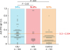
Table 2
Comparisons of CSU with anti-TPO IgE and anti-TPO IgE

Data are shown as number (range), number (%), or mean ± standard deviation. Comparisons of clinical characteristics of CSU patients with positive and negative anti-TPO IgE. A P ≤ 0.05 (bold numbers) was considered statistically significant.
CSU, chronic spontaneous urticaria; Ig, immunoglobulin; TPO, thyroid peroxidase; ASST, autologous serum skin test; DLQI, dermatology life quality index; UAS, urticaria activity score; SPT, skin prick test; IDT, intradermal test.
Among CSU patients and healthy subjects, there were some subjects with anti-TPO IgG and no clinical diagnosis of autoimmune thyroiditis. An additional subanalysis was done after excluding these patients and comparing these groups with the ATD group, but no significant statistical differences were found.
BAT
We observed a similar concentration of basophils in total blood in the 3 groups, being slightly lower in the CSU group (23 cells/mL) compared to the ATD group (32 cells/mL) on the healthy control group (36 cells/mL), without statistical differences.
For the BAT, basophils were obtained from 4 groups of subjects; CSU (+)anti-TPO IgE, ATD (+)anti-TPO IgE, control (−)anti-TPO IgE and control (+)anti-TPO IgE.
With the different dilutions of TPO used (dilution 0.01, P = 0.04; dilution 0.1, P = 0.01; dilution 1, P < 0.01) (Fig. 2A), CD203c expression from patients with CSU and anti-TPO IgE significantly increased compared to the other groups.
Fig. 2
Basophil activation test with TPO.
TPO, thyroid peroxidase; Ig, immunoglobulin; CSU, chronic spontaneous urticaria; ATD, autoimmune thyroid disease.
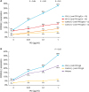
Basophils of control subjects with (+)anti-TPO or (−)anti-TPO IgE and no urticaria or ATD did not have a significant CD203c expression when stimulated with TPO. With 0.1 and 1 µg/mL dilutions, the ATD group had a CD203c expression, which was significantly lower than the CSU group and was not significantly different from control subjects with (+)anti-TPO or (−)anti-TPO IgE (Fig. 2). When the basal levels of CD203c were compared among patients of the CSU group, a higher expression of basal CD203c was observed in the group with anti-TPO IgE (P = 0.05) (Table 2).
When we mixed a pool of serum from CSU patients with anti-TPO IgE and basophils from a control subject with (−)anti-TPO IgE (Fig. 2B), CD203c expression was significantly increased (P = 0.04) after stimulation with TPO (1 µg/mL). A similar result was observed when basophils from ATD patients were used (P = 0.05).
For the following experiments combining serum and basophils from different subjects, only the dilution of 1 µg/mL TPO was used: CSU basophils with sera from healthy control subjects with anti-TPO IgE induced the expression of CD203c after stimulation with TPO (1 µg/mL) (15.5%), but it was lower than with their own serum or with anti-TPO IgE serum from another CSU patient (23%). CSU basophils with sera from healthy control subjects with (−)anti-TPO IgE did not induce the expression of CD203c after TPO stimulation (5.0%).
Prevalence of anti-TPO IgG
The CSU group had a higher frequency of anti-TPO IgG than the control subjects (28.0% vs. 18.1%, P = 0.04), but it was lower than the ATD group (93.3%). CSU patients with anti-TPO IgE exhibited significantly higher anti-TPO IgG levels than those with (−)anti-TPO IgE (28.2 ± 42.3 vs. 20.7 ± 62.0, P = 0.03) (Table 2).
Levels of anti-TPO IgE and IgG had a significant correlation in the CSU and healthy control groups. Taking into consideration that this correlation could occur because most of these subjects had a negative result for IgG and IgE, we evaluated the IgG and IgE correlation using only subjects with anti-TPO IgE, and there was a significant correlation in ATD (r = 0.548) and CSU (r = 0.643) for IgE and IgG (Table 1).
Twelve patients in the CSU group had Hashimoto's thyroiditis; 10 had anti-TPO IgG and 3 of these patients also had anti-TPO IgE. All subjects in the healthy control group had normal thyroid function, even those with positive IgG or IgE against TPO. We did not find that any clinical parameter related with active thyroid disease, like the presence of anti-TPO IgG, could predict the presence of anti-TPO IgE.
Skin response to TPO
The CSU group alone showed positive SPT to TPO (Fig. 3A), and intradermal skin reaction to TPO in this group was higher than the other groups (P = 0.01) (Fig. 3A). All subjects with positive SPT had positive serum anti-TPO IgE and positive intradermal test. Six of the 9 patients with CSU and positive intradermal test also had anti-TPO IgE (Table 2). Three of the 4 control subjects and the ATD patients with positive intradermal test had anti-TPO IgE. Among the subjects with positive skin tests in the 3 groups, there was a moderate correlation (r = 408; P = 0.05) between the wheal size in the skin tests and the levels of sIgE against TPO.
Fig. 3
Skin tests with TPO.
TPO, thyroid peroxidase; CSU, chronic spontaneous urticaria; ATD, autoimmune thyroid disease; SPT, skin prick test.
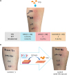
We did passive intradermal transfer of anti-TPO IgE serum from 1 CSU patient to 2 healthy subjects with negative anti-TPO IgE in serum as well as negative SPT, intradermal test and ASST (Fig. 3B). The CSU serum used was negative for anti-TPO IgG. After 24 hours, we repeated intradermal test and SPT with TPO, and subjects presented positive reactions. In these 2 subjectss, after 2 and 4 weeks we repeated skin tests with TPO at the same point previously injected with anti-TPO IgE serum from a CSU patient; the skin test remained positive after 2 weeks, but it was negative after 4 weeks.
We did not observe clinical changes or alterations in thyroid function among patients with ATD who underwent skin tests with TPO, and no changes were observed either among the 2 subjects who participated in the passive transfer after 2 months of follow-up.
DISCUSSION
Different mechanisms of self-reactivity are involved in the development of CSU82122232425 and some studies described a strong association between self-reactivity and autoimmune diseases, especially thyroid diseases.26272829 It is necessary to identify auto-reactive proteins in CSU.30 The presence of anti-TPO IgE in CSU has led us to assume that this auto-reactive IgE may play a role in the development of CSU or can, at least, be an indicator of inflammation. To evaluate the causal role of anti-TPO IgE in CSU, we first calculated the prevalence of anti-TPO IgE in our population. We observed that 34% of patients with CSU had anti-TPO IgE and it was higher than in ATD patients (16.6%) or healthy subjects (8.1%). Nevertheless, anti-TPO IgE seems to be important only in some patients with CSU; the presence of this autoantibody in subjects without urticaria and the presence of urticaria in subjects without this autoantibody indicate that anti-TPO IgE is not the only factor influencing the development of the disease and that different inflammatory cells or other substances of the serum could have a relevant role.31
The direct detection of anti-TPO IgE in a wheal performing a biopsy in the acute phase of urticaria could help clarify the clinical relevance of anti-TPO IgE in urticaria. However, the technique for detecting this low serum antibody in fresh skin can be complex. Although BAT has limitations because it is an “in vitro” technique, it allows us to evaluate the capacity of anti-TPO IgE to activate basophils, which shares many characteristics with mast cells in terms of IgE activation. Mast cells play a prominent role in urticaria,32 but recent studies using basophils have provided useful information to understand this disease. Rauber et al.33 demonstrated that in CSU, different basophil phenotypes can be categorized after stimulating basophils through FcεRI. BATs using CD63 or CD203c showed that different allergens have similar sensitivity, with a slightly higher sensitivity to CD203c.343536 We used the protocol with CD203c, which has previously been tested with TPO; Shin et al.9 suggested that anti-TPO IgE plays a pathogenic role in aspirin-exacerbated cutaneous urticaria in terms of the expression of CD203c. Some studies suggested that basophils from 10.0% to 20.0% of healthy individuals are non-releasers to FcεRI-dependent stimuli37 and that basophils from chronic urticaria patients seem to have some changes in the FcεRI-signaling pathway.38 Taking into account these results and that we observed an expression of CD203c among patients with ATD but without having urticaria, it is necessary to evaluate if basophils from patients with autoimmune thyroiditis have their particular characteristic phenotypes.
These cell phenotypes are associated with different clinical characteristics, pointing to basophils as important players in CSU's physiopathology. We observed that patients with CSU and anti-TPO IgE had a higher CD203c expression than the 2 other groups. The lack of activation through anti-TPO IgE in ATD and healthy control groups suggested that in patients without urticaria, some factors can antagonize the effect of anti-TPO IgE, considering the results of Rauber et al.33 that these groups may be have a high proportion of hypo-reactive basophils. Similar to Shin et al.9 in aspirin urticaria, we also observed that among patients with CSU, despite having anti-TPO IgE, not all samples had CD203c. To clarify whether this lack of basophil response was due to the basophil type or perhaps to another factor present in the serum, we evaluated the basophil auto-reactive capacity by mixing basophils and serum from the 3 groups. We observed that when we use CSU anti-TPO serum IgE, basophils from ATD or control subjects express CD203c, but at a lower intensity than patients with CSU and only at the highest concentration of TPO. In addition, when we incubated basophils of CSU patients with anti-TPO IgE from control subjects, we observed expression of CD203c, but lower than what observed with its own anti-TPO IgE serum or with anti-TPO IgE serum from another CSU patient. Based on these observations, it seems that the different types of basophils may play a role in the type of response to TPO or that other factors from sera can prevent this activation at least partially. Inhibition experiments are needed in order to answer these questions.
As expected, IgG levels were higher in the ATD group, but the anti-TPO IgG levels were higher in the CSU group than in the healthy control group. Although previous studies have established the coincidental occurrence of autoimmune diseases and CSU,39 there are no current data to determine the basis for this link. Due to the design of the study, we cannot state with certainty if thyroid diseases in the CSU group were more or less frequent than in the healthy control group. However, we observed that within the CSU group, patients with anti-TPO IgE had higher levels of anti-TPO IgG than those with (−)anti-TPO IgE, although the CD203 expression baseline was higher in the anti-TPO IgE group. Our results suggest that anti-TPO IgG could interfere with anti-TPO IgE. This hypothesis is supported by 2 results: (1) In patients with ATD and positive anti-TPO IgE, anti-TPO IgG levels were higher compared to ATD patients who did not have IgE and the activation of basophils was lower compared to the CSU group. (2) In CSU patients with positive IgE anti-TPO but negative skin test results, anti-TPO IgG levels were higher than in CSU patients with negative IgE (data not shown). As suggested by Mozena et al.,32 it is plausible that urticaria and ATD are pathogenically linked, possibly reflecting a shared epitope targeted by different autoantibodies. It is possible that in patients with ATD and high levels of anti-TPO IgG and anti-TPO IgE, this IgE could play a role in worsening Hashimoto's thyroiditis. However, in patients with ATD, there must be some differences in the effector mechanism of anti-TPO IgE regarding what happens in patients with CSU because a smaller number of patients presented skin exacerbation with TPO and had less reactivity in BAT. Additional experiments are required with a larger number of patients to confirm this hypothesis.
The SPT and intradermal tests are excellent tools to evaluate the clinical impact of allergens on the skin.1140 The usefulness of skin tests in CSU has been limited in part by the lack of knowledge of mechanisms associated with the disease, which does not allow scientists to choose the right protein to be tested. The ASST has been the most commonly used diagnostic tool in urticaria, although it has excellent sensitivity but quite low specificity.15 With the growing evidence of the possible role of TPO in a group of patients with CSU, we tested whether the stimulation of skin with TPO could induce the formation of wheals in patients with CSU. We observed a strong correlation between in vitro and in vivo results. SPT had better specificity than the intradermal test, but the sensitivity of the intradermal test was better. With the passive transfer of serum from a CSU patient to the skin of 2 control subjects, we demonstrated that anti-TPO IgE plays a central role in the pathogenesis of wheal in CSU, at least in a group of patients. The standardization of skin tests with TPO will provide a easy and simple tool to identify patients in whom urticaria is due to this IgE auto-reactivity. Although at the moment anti-TPO IgE measurement is conducted only in some specialized centers, it will be generalized to all the clinical laboratories. However, according to our results, the advantages of the skin test (rapid implementation, avoids puncture, almost immediate results), can be supported by anti-TPO IgE measurement in serum.
Our study has some limitations. As mentioned earlier, we did not directly evaluate different phenotypes of basophils that could answer some of new questions arising from these results. In addition, we did not evaluate how other serum elements, such as complement factors, could play a role in auto-IgE pathogenesis. Although the case-control design of the study was appropriate for the main objective of the study, the lack of follow-up did not allow for evaluating whether IgE or IgG levels are dependent on the exacerbation of urticaria. Despite these limitations, the study has some strengths. First, the different experiments carried out show that anti-TPO IgE can induce the disease in some patients with CSU.Secondly,, this study could be considered a preliminary effort for future research in the technological development of new diagnostic tools for urticaria.
In conclusion, since anti-TPO IgE is present in patients with CSU and ATD as well as in healthy subjects, it is not a specific biomarker for CSU. However, IgE to TPO plays a pathogenic role in cellular effector activation and skin exacerbation in a subgroup of CSU patients, and this effect can be transferred to healthy subjects.
 XML Download