

 PDF
PDF ePub
ePub Citation
 Print
Print


INTRODUCTION
Chronic rhinosinusitis (CRS) is a common disease that affects 4%–10% of the global population and is generally characterized by chronic inflammation of the mucosa of the nose and paranasal sinuses that persists for a minimum of 3 months. On the basis of whether visible nasal polyps (NPs) are present in the middle meatus, CRS can generally be classified into 2 distinct subtypes: chronic rhinosinusitis with nasal polyps (CRSwNP) and chronic rhinosinusitis without nasal polyps (CRSsNP).12 The tissue inflammatory response in CRSwNP patients is characterized by prominent eosinophilia with a Th2-skewed response, which reflects a complex interplay between environmental factors (bacteria, virus, fungi, etc.) and the host response.2 Based on the extent of tissue eosinophilia, it has been proposed that CRSwNP can be subclassified into eosinophilic (ECRS) and non-eosinophilic (non-ECRS) subtypes, each of which is characterized by distinct degrees, therapeutic strategies and prognoses.34 Pathogenic mechanisms of CRS have recently become the focus of intense investigations, and significant progress has been made. Defects in the innate immune function of nasal epithelial cells have been reported to play a role in the initial inflammatory response by producing and releasing cytokines like interleukin (IL)-25, IL-33 and thymic stromal lymphopoietin (TSLP). These cytokines subsequently recruit and activate eosinophils, neutrophils, mast cells, basophils and innate lymphoid cells (ILCs), which could further contribute to a chronic inflammatory response and directly activate adaptive immune cells including T and B-cells.56
In recent years, B-cells are emerging as a critical component of the adaptive immune response and are known to play several important roles in many inflammatory disorders and at mucosal sites.78 In addition to producing immunoglobulins that contribute to disease pathogenesis, B-cells can function as antigen-presenting or regulatory cells and produce a variety of cytokines and chemokines that can influence inflammation.79 Increased accumulation and activation of B-cells, driven by up-regulated expression of B-cell chemokines and activating factors, have been demonstrated in NPs.710 Accumulating data have demonstrated that B-cell class-switch recombination (CSR) to immunoglobulin (Ig) E and IgA can occur in NPs and subsequently mediate activation of local mast cells and eosinophils, respectively, in response to antigen exposure.7911 Importantly, increased local IgE and IgG levels have been demonstrated to be associated with poorly controlled disease in patients with CRSwNP, even after surgical intervention.1213 Collectively, these studies suggest that polyp tissue might provide a supportive environment for B-cell survival and immunoglobulin production, which can play important roles in the pathogenesis of CRSwNP.
Hrd1, also known as synoviolin, is a membrane-spanning protein on the endoplasmic reticulum (ER). As a ubiquitin ligase, Hrd1 is involved in recruiting misfolded cytoplasmic proteins and disease-related mutant proteins for ubiquitination and degradation by the endoplasmic reticulum-associated degradation (ERAD) machinery.14 It has been reported that Hrd1 is often highly expressed in synovial fibroblasts in patients with rheumatoid arthritis (RA), and proinflammatory cytokines including tumor necrosis factor (TNF)α and IL-1β are responsible for inducing its overexpression in synovial fibroblasts.1516 Intriguingly, sustained high-level expression of Hrd1 in whole peripheral blood was related to a decreased clinical response in RA patients treated with TNF blockade.17 In addition, overexpression of Hrd1 in transgenic mice leads to advanced arthropathy caused by reduced synoviocyte apoptosis.15 We postulated that Hrd1 exerts anti-apoptotic effects via the ERAD pathway in autoimmune disease. Consistent with our hypothesis, we recently identified that Hrd1 is a positive regulator of T-cell activation and differentiation and that knockdown of Hrd1 in human CD4+ T-cells inhibits activation and differentiation to Th1 and Th17 cells.18 Additionally, we found that Hrd1 plays a critical role in protecting mature B-cell populations from activation-induced apoptosis via degradation of the death receptor Fas/CD95.19 These studies highlight the important role of Hrd1 in T-cell activation and B-cell immunity in peripheral immune responses. However, little is known about the role of Hrd1 during CRSwNP pathogenesis and whether its expression in the periphery might contribute to the generation of pathogenic antibody responses. This study aims to more fully assess whether Hrd1 might play a role in the nasal mucosa of patients with CRSwNP.
MATERIALS AND METHODS
This study was approved by the Ethics Committee of Xin Hua Hospital Affiliated to Shanghai Jiao Tong University School of Medicine (approval No. XHEC-D-2018-018), and written informed consent was obtained from each subject. Ninety-four patients with CRSwNP, 23 patients with CRSsNP and 22 normal control subjects were recruited from the Department of Otolaryngology, Head and Neck Surgery of Xinhua Hospital, Shanghai Jiaotong University School of Medicine. Patients with CRSwNP and CRSsNP were diagnosed on the basis of clinical history, nasal endoscopy and computed tomography (CT) results in accordance with the European Position Paper on Rhinosinusitis and Nasal Polyps (EPOS) guidelines.1 To evaluate the correlation between Hrd1 expression and the severity of CRSwNP, Lund-Mackay CT scores were assessed in patients with CRSwNP. The Lund-Mackay scoring system was used to grade the severity of CRSwNP. Each sinus group was assigned a numeric grade: 0 = no abnormality, 1 = partial opacification and 2 = total opacification. During endoscopic surgery, the following nasal tissues were collected: polyp tissue from patients with CRSwNP and uncinate process tissue from patients with CRSwNP and those with CRSsNP. The control group comprised excessive hypertrophic uncinate process from patients undergoing septoplasty for anatomic variations and middle turbinate tissue from patients with traumatic neuropathy undergoing endoscopic optical decompression. The atopic status of the patients and normal control subjects was evaluated by allergen skin prick tests. Asthma was diagnosed by a pneumologist based on clinical history and evaluation of airway responsiveness. None of the subjects used oral/nasal steroids or other medications (e.g., antibiotics or antileukotrienes) within 1 month before sample collection. Patients with an established immunodeficiency, acute infection or allergic fungal sinusitis were excluded from this study. According to eosinophil levels, the CRSwNP patient population was divided into ECRS and non-ECRS subgroups.20 Briefly, the cutoff value separating ECRS from non-ECRS was set at 8 eosinophils/high-power field (HPF); a polyp with ≥ 8 eosinophils/HPF was defined as ECRS. The demographic data of all subjects enrolled in this study are included in Table.
Table
Subjects' characteristics
ECRS, eosinophilic chronic rhinosinusitis with nasal polyp; non-ECRS, non-eosinophilic chronic rhinosinusitis with nasal polyp; CRSsNP, chronic rhinosinusitis without nasal polyps; M, male; F, female; SPT, skin prick test; qRT-PCR, quantitative real-time polymerase chain reaction; IHC, immunohistochemical; WB, western blot; ELISA, enzyme-linked immunosorbent assay; PBMC, peripheral blood mononuclear cell; NP, nasal polyp.
The tissues were divided into 3 portions. The first was stored immediately in RNA-stabilizing solution (RNAlater; Tiangen, Beijing, China) for subsequent RNA extraction; the second was fixed with 4% paraformaldehyde overnight and then embedded in paraffin for immunohistochemical (IHC) staining. The third was stored immediately at −80°C for Western blot analysis and protein isolation. In addition, dispersed NP cells and matched peripheral blood mononuclear cells (PBMCs) were collected for flow cytometry and/or in vitro assays.
Quantitative real-time polymerase chain reaction (qRT-PCR)
Hrd1, CD19, CD20, CD138 and B-cell activating factor (BAFF) mRNA expression levels were evaluated by using qRT-PCR analysis as previously described.2021 Briefly, total RNA was extracted with TRIzol reagent (Invitrogen, Carlsbad, CA, USA), according to the manufacturer's instructions. Reverse transcription was performed, in which cDNA for quantitative PCR was synthesized from 2 μg of total RNA using an oligo (dT) 18 primer and M-MLV reverse transcriptase (Takara, Dalian, China). RNA integrity and the success of the reverse transcription reaction were monitored by PCR amplification of β-actin transcripts. Messenger RNA expression was determined by using an ABI PRISM 7500 Detection System (Applied Biosystems, Foster City, CA, USA) with SYBR Premix Taq (Takara). The primer sequences for each gene are listed in Supplementary Table S2. The qRT-PCR amplification protocol consisted of 40 cycles of a denaturation step at 95°C for 15 seconds and an annealing/extension cycle at 60°C for 45 seconds. Melting curve analysis was used to control for amplification specificity. The mean cycle threshold (Ct) values were normalized to those of β-actin, and the relative mRNA levels of the target genes were analyzed using the 2−△△Ct method. Experiments were performed in triplicate for each data point.
IHC staining
IHC staining for Hrd1 (Santa Cruz Biotechnology, Santa Cruz, CA, USA), CD19, CD20 and CD138 (all from Abcam, Cambridge, MA, USA) was performed as described elsewhere.20 Briefly, the IHC staining was performed using the Envision method. Paraffin-embedded human nasal tissues were cut into 4-μm sections and placed onto glass slides. The sections were rehydrated, and antigen retrieval was performed using protease digestion for 5 minutes. After blocking endogenous peroxidase with 3% hydrogen peroxide and 1% BSA, the sections were incubated overnight at 4°C in the presence of an anti-Hrd1 (1:200; Santa Cruz Biotechnology), anti-CD19 (1:100; Abcam), anti-CD20 (1:200; Abcam) or anti-CD138 (1:8,000; Abcam) antibody according to the manufacturer's instructions. This was followed by incubation with a secondary antibody and then with a horseradish peroxidase-labeled streptavidin complex (Zhongshanjinqiao, Beijing, China). Immunostaining was considered positive when brown cells were observed after treatment with 3% 3,3′-diaminobenzidine reagent. As a control, the sections were incubated with the isotype-matched IgG antibody at appropriate concentrations. The sections were examined by using an Olympus CX40 Microscope (Olympus Optical, Hamburg, Germany). The number of positive cells was counted in 5 HPFs (×400 magnification) by 2 independent observers in a blinded manner, and the relative positive cell rate was calculated as positive cell per total inflammatory cell.
Western blotting
The Hrd1 protein level was determined by using Western blot analysis as previously described.21 Briefly, the tissues or cells were dissociated on ice and homogenized in a radioimmunoprecipitation assay (RIPA) lysis buffer (Beyotime, Shanghai, China) containing 50 μL/mL protease inhibitor cocktail (Sigma-Aldrich, St. Louis, MO, USA) and 1 mM phenylmethylsulfonyl fluoride (PMSF) (Beyotime). The protein concentration in the supernatants was determined by the BCA method. Samples containing 20 µg of protein were boiled and subjected to sodium dodecyl sulfate-polyacrylamide gel electrophoresis (SDS-PAGE) on 10% Tris-glycine gels and electrophoretically transferred onto a polyvinylidene fluoride membrane. The membrane was blocked with 5% fat-free milk in Tris-buffered solution containing 0.05% Tween-20 for 1 hour at room temperature and then incubated with rabbit anti-human Hrd1 (1:5,000; Santa Cruz Biotechnology) overnight at 4°C. The membrane was washed and incubated with a horseradish peroxidase-linked secondary antibody (1:2,000, Beyotime) and finally processed by using an ECL chemiluminescence reaction kit (Millipore, Billerica, MA, USA), followed by exposure to medical film. The band density of the target protein relative to that of β-actin was quantified with Bio-Rad Quantity One 1-D Analysis Software (Bio-Rad Laboratories, Hercules, CA, USA).
Enzyme-linked immunosorbent assay (ELISA)
BAFF protein levels in nasal extracts were measured by ELISA, as previously described.22 In brief, freshly obtained samples were weighed, placed in phosphate buffered saline (PBS; 1 mL of PBS/100 mg of tissue) containing 50 μL/mL protease inhibitor cocktail (Sigma-Aldrich), and homogenized on ice for 1 minute. The suspension was then centrifuged at 4,000 rpm for 20 minutes at 4°C. The supernatants were stored at −80°C for ELISA. The cytokine concentrations were measured with commercially available ELISA kits (R&D Systems, Minneapolis, MN, USA), according to the manufacturer's protocols. The detection limit for BAFF was 6.44 pg/mL. For convenient analysis, all values lower than the detectable limit were considered zero. To reduce errors, the measured levels were normalized to the total protein levels.
Measurement of immunoglobulin
Protein levels of immunoglobulins in tissue homogenates were detected by using Bio-Plex suspension chip technology (Bio-Rad Laboratories) according to the manufacturer's instructions. Briefly, tissues samples were weighed, placed in PBS (1 mL of PBS/100 mg of tissue) containing 50 μL/mL protease inhibitor cocktail (Sigma-Aldrich), and homogenized on ice for 1 minute. The suspension was then centrifuged at 4,000 rpm for 20 minutes at 4°C, and the supernatants were harvested for later analysis. The samples were analyzed for total immunoglobulin isotype levels on a Bio-Plex MAGPIX System (Bio-Rad Laboratories). Total IgG was calculated as the sum of the 4 subclasses, as previously described.23 Values were normalized to total protein levels. The detection limit for the Bio-Plex assay is shown in Supplementary Table S3.
Flow cytometric analysis
The Hrd1 expression level in B-cells was determined by using flow cytometry. Briefly, fresh polyp tissues and healthy middle turbinate tissues were cut into pieces of approximately 1 mm and digested in incomplete RPMI-1640 medium (HyClone, Logan, UT, USA) with 2 mg/mL collagenase I (Sigma-Aldrich) and 5 U/mL DNase I (Takara) for 1 hour at 37°C. The digested fragments were filtered through a cell strainer with a pore size of 70 µm to remove any undigested tissue. Mononuclear cells were then obtained from the dispersed cells by using Ficoll-Paque (GE Healthcare, Arlington Heights, IL, USA) density gradient centrifugation at 2,200 rpm for 20 minutes, and the cells were then washed twice with Hank's solution. To obtain a reliable number of cells for analysis, 1–5 × 10⁵ dispersed nasal tissue cells or PBMCs were suspended in complete RPMI-1640 medium containing 10% fetal bovine serum (FBS), 100 IU/mL penicillin, 100 IU/mL streptomycin and 1% L-glutamine. PBMCs of matched patients with CRSwNP and control subjects were isolated by using Ficoll-Paque density gradient centrifugation.
For surface staining, cells were incubated with anti-CD19-AF488 (1:20; BioLegend, San Diego, CA, USA) and anti-CD45-PE-Cy5 (1:5; BD Biosciences, San Jose, CA, USA) at 4°C in the dark for half an hour. After fixation and permeabilization with permeabilization/fixation buffer (BD Biosciences, San Jose, CA, USA), these cells were stained with a monoclonal antibody against Hrd1 (1:500; Abcam). The stained cells were washed twice before analysis with a FACSAria II cytometer (BD Biosciences). The gating strategy for lymphocytes in nasal tissues and PBMCs is indicated in each figure.
Dispersed polyp cell (DPC) culture and stimulation
Primary DPCs were randomly collected from patients with CRSwNP by using enzymatic digestion to establish in vitro cultures. Briefly, fresh polyp tissues were washed with PBS 3 times, cut into pieces of approximately 1 mm, digested in incomplete RPMI-1640 medium (HyClone) with 2 mg/mL collagenase I (Sigma-Aldrich) and 5 U/mL DNase I (Takara) and placed in a 37°C/5% CO2 incubator for 1 hour. After repeated vortexing, the cell suspensions were filtered through 70-μm cell strainers to remove undigested tissue. The cells were then transferred into RPMI 1640 medium with L-glutamine (HyClone) supplemented with 10% FBS and 1% penicillin/streptomycin and cultured on a 12-well plate in a humidified incubator infused with 5% CO2 at 37°C overnight. The cells were cultured in the presence of IL-1β (5 ng/mL; R&D Systems), lipopolysaccharide (LPS) (100 ng/mL; Invitrogen) and Poly (I:C) (10 μg/mL; Sigma-Aldrich), with or without dexamethasone (Dex) (1 μg/mL; Sigma-Aldrich). After 20 hours, the cells were harvested for qRT-PCR and Western blot analyses.
Statistical analysis
Data are expressed as medians and interquartile ranges, and statistical comparisons between the groups were made by the nonparametric Mann-Whitney U test; Spearman's rank correlation test was used when appropriate. Comparisons between multiple groups were made using one-way analysis of variance (ANOVA), with Tukey's correction for multiple comparisons. For in vitro assays, the data are expressed as the mean ± standard error of the mean. Calculations were done with GraphPad Prism version 5.0b software (GraphPad Software, San Diego, CA, USA). Significance was declared at P < 0.05.
RESULTS
Hrd1 is differentially expressed in polyp tissues from patients with ECRS and those with non-ECRS
To evaluate whether Hrd1 would be altered in polyp tissues from patients with CRSwNP, we first determined its mRNA levels in the nasal mucosa. As shown in Fig. 1A, Hrd1 mRNA levels were significantly higher in NPs from patients with ECRS than in those from patients with non-ECRS (P = 0.005) or in uncinate tissues from patients with CRSsNP (P = 0.011) and normal controls (P = 0.01). To confirm this observation at the protein level, we measured Hrd1 protein expression in nasal tissues by using IHC staining and Western blot analysis. As indicated in Fig. 2A, Hrd1 exhibited strong cytoplasmic staining that was mainly located in inflammatory cells in the subepithelial areas of NPs. By contrast, we found that Hrd1 immunoreactivity was quite weak in normal tissues. The relative cell count (positive cell per total inflammatory cell) of Hrd1-positive cells was significantly increased in NPs from patients with ECRS compared with NPs from patients with non-ECRS (P < 0.045) and nasal tissues from patients with CRSsNP (P < 0.043) or from control subjects (P < 0.001) (Fig. 2B). Moreover, the relative cell count of Hrd1+ cells was significantly higher in polyp tissues from patients with non-ECRS than in polyp tissues from control subjects (P = 0.005; Fig. 2B). Western blot analysis of Hrd1 protein expression in nasal tissues produced similar results (Fig. 2I and J). Interestingly, the Hrd1 protein levels of NPs were positively correlated with the CT scores of matched ECRS or non-ECRS patients (P = 0.002, r = 0.607; P = 0.001 and r = 0.666, respectively; Fig. 2K).
Fig. 1
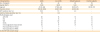
Hrd1 mRNA expression in nasal tissues. Hrd1 (A), CD19 (B), CD20 (C), CD138 (D) and BAFF (E) mRNA levels were significantly enhanced in polyp tissues from patients with ECRS compared with values seen in the other subgroups. Results are expressed as medians (interquartile ranges).
BAFF, B-cell activating factor; ECRS, eosinophilic chronic rhinosinusitis with nasal polyp; non-ECRS, non-eosinophilic chronic rhinosinusitis with nasal polyp; CRSsNP, chronic rhinosinusitis without nasal polyps.
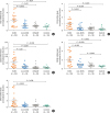
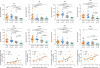
Fig. 2
Increased Hrd1 protein levels and accumulation of B cells and plasma cells in NPs. Representative IHC staining of Hrd1 (A), CD19 (C), CD20 (E), and CD138 (G) (magnification, × 400) in nasal tissues. Relative positive cells of Hrd1 (B), CD19 (D), CD20 (F), and CD138 (H) were significantly increased in polyp tissues from patients with ECRS relative to those in other subgroups. Representative Western blotting results (I), densitometric analysis (J). Positive correlation between relative Hrd1 protein levels in the polyp tissues and Lund-Mackay CT scores of the matched patients (K). ELISA of BAFF in nasal tissues (L). Positive correlation between BAFF and Hrd1 protein levels in the matched NPs (M). Bars = 20 μm.
NP, nasal polyp; ECRS, eosinophilic chronic rhinosinusitis with nasal polyp; non-ECRS, non-eosinophilic chronic rhinosinusitis with nasal polyp; CRSsNP, chronic rhinosinusitis without nasal polyps; HPF, high-power field; CT, computed tomography; ELISA, enzyme-linked immunosorbent assay; BAFF, B-cell activating factor.
B-cells and plasma cells are differentially accumulated in NPs from patients with ECRS and those with non-ECRS
To first assess the accumulation of B-cells and plasma cells in the NPs of patients with CRSwNP, we examined the expression of the B-cell markers CD19 and CD20, the plasma-cell marker CD138, and BAFF in nasal tissues. We observed strong cytomembrane staining of CD19, CD20 and CD138, which were mainly located in inflammatory cells in the subepithelial areas of polyp tissues (Fig. 2C, E, and G). Both the mRNA and protein levels of these molecules were significantly elevated in NPs from patients with ECRS compared with NPs from patients with non-ECRS or uncinate processes from CRSsNP patients or control subjects (P < 0.05) (Fig. 1B-D, 2D, F, and H). We next assessed BAFF mRNA and protein levels by using qRT-PCR and ELISA. As indicated in Fig. 1E, BAFF mRNA levels were significantly increased in polyp tissues from patients with ECRS compared with polyps from patients with non-ECRS (P = 0.032) or uncinate processes from control subjects (P = 0.019). BAFF protein levels were significantly higher in polyp tissues (P < 0.05) from patients in both CRSwNP subsets than in uncinate processes from CRSsNP patients or control subjects (Fig. 2L). Interestingly, its protein levels correlated positively with Hrd1 protein levels in the matched polyp tissues (P = 0.003, r = 0.627; Fig. 2M). However, we did not observe an obvious difference in BAFF protein levels between the 2 CRSwNP subsets.
Enhanced Hrd1 levels in B-cells in NPs and matched PBMCs
To examine and characterize Hrd1 expression in B-cells within polyp tissues and PBMCs, we assessed its levels using flow cytometry (Fig. 3A and C). We found a significant increase in the average Hrd1 levels in B-cells of polyp tissues from patients with ECRS compared with B-cells of tissues from patients with non-ECRS (P = 0.021) or control subjects (P = 0.002). Additionally, the average Hrd1 levels were significantly higher in B-cells of polyp tissues from patients with non-ECRS than in B-cells of middle turbinate tissues from normal controls (P = 0.012; Fig. 3B). However, there was no difference in the B-cell Hrd1 level in PBMCs from the matched patients between the 2 CRSwNP subsets (data not shown), though PBMCs from patients with CRSwNP did show enhanced B-cell Hrd1 levels compared with control subject (P = 0.003; Fig. 3D).
Fig. 3
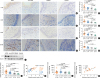
Enhanced Hrd1 expression in B-cells from polyp tissues and matched PBMCs. Gating strategy and representative flow plots for nasal tissues (A). MFI levels of Hrd1 in CD19+ B-cells were significantly increased in polyp tissues from patients with ECRS compared with those in in polyps from patients with non-ECRS, middle turbinates from control subjects (B). Gating strategy and representative flow plots for PBMCs (C). MFI levels of Hrd1 in CD19+ B-cells were significantly increased in PBMCs from patients with CRSwNP compared with those in PBMCs from healthy controls (D).
PBMC, peripheral blood mononuclear cell; MFI, mean fluorescence intensity; ECRS, eosinophilic chronic rhinosinusitis with nasal polyp; non-ECRS, non-eosinophilic chronic rhinosinusitis with nasal polyp; CRSwNP, chronic rhinosinusitis with nasal polyps.
Increased local immunoglobulin levels in NPs
As the effector functions mediated by secreted immunoglobulins are dependent on their isotype, we set out to evaluate the levels of different antibodies present in the nasal tissues (Fig. 4). We found up-regulated local IgE levels in polyps from patients with ECRS compared with polyps from patients with non-ECRS (P < 0.001) and uncinate tissues from patients with CRSsNP (P = 0.001) or control individuals (P = 0.004). Moreover, the levels of total IgG, IgG1, IgG2, IgG3, IgG4, IgM, and IgA were increased similarly in both patients with ECRS and those with non-ECRS compared with control subjects (P < 0.05). Interestingly, we found the Hrd1 protein levels correlated positively with IgM, IgG, IgG1, and IgG2 levels in the matched polyp tissues (P < 0.05; r = 0.542, r = 0.714, r = 0.508 and r = 0.537, respectively).
Fig. 4
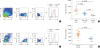
Increased local immunoglobulin levels in polyp tissues. Protein levels of IgA, IgE, IgM, IgG and IgG subclasses in tissue homogenates from ECRS, non-ECRS, CRSsNP, and control tissue are detected by using the Bio-Plex assay. Positive correlations are found between relative Hrd1 protein levels and IgM, IgG, IgG1, and IgG2.
Ig, immunoglobulin; ECRS, eosinophilic chronic rhinosinusitis with nasal polyp; non-ECRS, non-eosinophilic chronic rhinosinusitis with nasal polyp; CRSsNP, chronic rhinosinusitis without nasal polyps.
IL-1β, LPS, and Poly(I:C) differentially regulate Hrd1 expression in DPCs from patients with ECRS and those with non-ECRS in vitro
To determine the critical roles of Hrd1 in innate immune response, we attempted to establish a direct relationship between Toll-like receptors (TLRs) and innate immunity as well as between proinflammatory cytokines and innate immunity in the nasal mucosa. Thus, we investigated the effects of TLR activation and IL-1β stimulation on Hrd1 expression in DPCs in vitro. As indicated in Fig. 5A-C, Hrd1 mRNA and protein levels were significantly increased in DPCs from patients with ECRS after stimulation with IL-1β, LPS and Poly(I:C) (P < 0.05). However, when DPCs from patients with non-ECRS were stimulated with IL-1β, LPS, or Poly(I:C), we did not observe obvious changes in Hrd1 mRNA and protein expression (Fig. 5D-F). We also examined the effect of Dex on Hrd1 mRNA and protein expression in DPCs in vitro. After incubating cells with Dex, Hrd1 mRNA and protein levels were significantly down-regulated in the absence or presence of TLR agonists and proinflammatory cytokines (IL-1β, LPS and Poly(I:C); P < 0.05; Fig. 5).
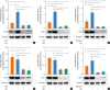
Fig. 5
Hrd1 mRNA and protein levels in cultured DPCs in response to IL-1β, LPS, Poly(I:C) and Dex. mRNA expression and representative western blot results of DPCs lysates from ECRS patients after 20-hour stimulation (A-C). mRNA expression and representative western blot results of DPCs lysates from non-ECRS patients after 20-hour stimulation (D-F). Results represent mean values from 3 independent experiments. Data are expressed as means (standard error of the means).
DPC, dispersed polyp cell; IL, interleukin; LPS, lipopolysaccharide; Poly(I:C), polyinosinic-polycytidylic acid; Dex, dexamethasone; ECRS, eosinophilic chronic rhinosinusitis with nasal polyp;non-ECRS, non-eosinophilic chronic rhinosinusitis with nasal polyp.
*P < 0.05; †P < 0.01; ‡P < 0.001.
DISCUSSION
It is well known that CRSwNP is characterized by type 2 inflammation and eosinophilia.234 However, recent evidence has suggested that the local proliferation and activation of B-cells and plasma cells are of central pathogenic importance in airway diseases including CRSwNP. Studies have demonstrated an increase in B-cell chemoattractants, B and plasma cell numbers, and antigen-specific IgE in the nasal mucosa of CRSwNP patients,791024 and more recently increases in BAFF have also been demonstrated by other study groups and ours.1025 This chemokine is thought to not only promote B-cell survival, proliferation, and maturation but also facilitate immunoglobulin CSR, a process central to IgA production, eosinophil activation, and subsequent polyp formation.91026 Although numerous studies have demonstrated the critical roles of B-cells in the pathophysiology of CRSwNP, precise molecular mechanisms underlying the inflammatory pattern in patients with CRSwNP have not yet been completely elucidated.
In the present study, we have expanded on our earlier work and found that B-cells exhibited eosinophilic accumulation in NPs from patients with ECRS.25 Polyp tissues from patients with ECRS not only showed increased expression of BAFF but also contained significantly more B-cells and plasma cells compared with NPs from patients with non-ECRS or uncinate tissues from control subjects (Figs. 1 and 2). Whether these B-cells enter the tissue as naive cells and become activated or if they enter as memory cells primed to respond within the tissue is not yet clear. Previous studies have demonstrated that eosinophils not only produce a variety of proinflammatory molecules, including BAFF, that can contribute type 2 inflammation but also play an important role in the long-term maintenance of plasma cells and have been shown to activate T-cells during inflammatory responses.102728 Further studies are needed to determine whether B-cells can be activated within NPs themselves and what mechanisms might be involved.
Hrd1 is known to exert antiapoptotic effects in a variety of immune responses.171819 However, little is known about the expression of Hrd1 and its possible roles in the polyp tissues of CRSwNP patients. In the current study, we found a significant increase in both Hrd1 gene and protein expression in NPs from patients with ECRS compared with NPs from patients with non-ECRS and uncinate tissues from CRSsNP patients or normal controls (Fig. 1A, Fig. 2A, B, I and J). We also showed that Hrd1 relative protein levels correlated with disease severity indicated by Lund-Mackay CT scores (Fig. 2K). In addition, we observed increased Hrd1 expression in B-cells in polyp tissues and matched PBMCs from CRSwNP patients (Fig. 3). As we previously showed that Hrd1 has an anti-apoptotic function in B-cell development,19 it is reasonable to speculate that Hrd1 expression could play an anti-apoptotic role in NPs. Interestingly, we found that Hrd1 mRNA and protein levels were strongly increased in polyp tissue homogenates (Fig. 1A, Fig. 2A, B, I and J), as well as in B-cells in NPs, but not PBMCs (data not shown) (Fig. 3B and D), from ECRS patients compared with those from non-ECRS patients. These data might suggest that Hrd1 expression is associated with eosinophil recruitment and localization in polyp tissues in patients with CRSwNP. Several groups have shown that immunoglobulins potentially play an important role in the pathogenesis of NPs.101124 In this study, we found that IgA, IgG and IgM, but not IgE, were similarly enhanced in eosinophilic and non-eosinophilic polyps compared with uncinate tissues from control subjects (Fig. 4). These findings were in accordance with those reported by Zhang et al.29 Moreover, we found that local Hrd1 protein levels in NPs were positively associated with IgM, IgG, IgG1, and IgG2 levels in the matched patients with CRSwNP (Fig. 4). Therefore, increased Hrd1 expression might contribute to immunoglobulin production in polyp tissues. Collectively, the studies described here further suggest that Hrd1 plays a potential role in disease pathogenesis, although future studies are needed to confirm whether Hrd1 is involved in activating local plasma-cell responses in patients with CRSwNP.
The innate immune response is the first line of host defense and is responsible for immediate recognition and control of microbial invasion. The cytokines IL-25, IL-33, and TSLP released by damaged epithelial cells subsequently recruit and activate eosinophils, basophils, mast cells and ILCs, which promote type 2 inflammation. These innate immune cells and released cytokines further contribute to the chronic inflammatory response and directly activate adaptive immune cells including T and B-cells.56 One of the primary families of molecules responsible for innate immunity in humans are the TLRs, expressed by epithelial cells and immune cells in the nose and sinuses, could activate expression of inflammatory mediators and host defense molecules.30 Among them, abnormalities in the immune response or expression of TLR3 and TLR4 in CRSwNP have been reported by other study groups and ours.2131 Activation of TLR3 or TLR4 can lead to the activation of nuclear factor-κB (NF-κB), which results in the expression of proinflammatory cytokines that are critical for both innate and adaptive immune responses.32 In this study, Hrd1 mRNA and protein levels were significantly up-regulated in eosinophilic DPCs after stimulation with IL-1β, Poly(I:C) (TLR3 ligand) and LPS (TLR4 ligand) in vitro. However, we did not observe obvious changes in the Hrd1 mRNA and protein levels in non-eosinophilic DPCs after treatment with the same stimuli. Our findings suggest that Hrd1 participates in the proinflammatory process and innate immunity and that the eosinophilic response might play a role in inducing Hrd1 expression in patients with CRSwNP. B-cells express MHC class II molecules, along with co-stimulatory molecules, and thus play a key role in the activation of T-cell responses.3233 Although the immune regulatory function of Hrd1 has been reported in some studies,181934 the observed positive effects of Hrd1 on B-cells in cultured eosinophilic DPCs after TLR agonist and proinflammatory cytokine stimulation in this study cannot be ignored. Notably, we found that Dex decreased Hrd1 mRNA and protein expression, independent of the presence or absence of IL-1β, Poly(I:C) and LPS in both eosinophilic and non-eosinophilic DPCs. Although the underlying molecular mechanisms require further characterization, this finding demonstrates that Dex treatment decreases proinflammatory cytokine-induced Hrd1 expression. Therefore, Hrd1 might represent a potential therapeutic target for patients with CRSwNP.
In summary, to the best of our knowledge, this is the first report of differential expression of Hrd1 in NPs from patients with ECRS and those with non-ECRS. We showed that NPs from patients with ECRS had abundant accumulation of B-cells and plasma cells. Not surprisingly, we also observed significantly increased levels of several immunoglobulins in NPs. Finally, we showed that Hrd1 was differentially expressed in B-cells in eosinophilic and non-eosinophilic polyp tissues and participated in the innate immune response. These findings indicate that the differential expression of Hrd1 in NPs might facilitate a better understanding of the pathogenesis involved in these 2 CRSwNP subsets and provide novel insights into the development of improved therapeutic interventions.
 XML Download