

 PDF
PDF ePub
ePub Citation
 Print
Print


INTRODUCTION
Urticaria that is present for greater than 6 weeks is arbitrarily considered to be “chronic” based on the observations that acute, self-limited episodes of urticaria tend to subside in 1–3 weeks, and that by assuming a cutoff at 6 weeks, the likelihood that some exogenous cause of the pathogenic process is very low and thus differs from causes of acute urticaria.1 Acute urticaria is typically caused by an identifiable agent such as an allergic reaction to a food or drug, or associated with viral illnesses as is often the case in children. Conversely, an exogenous cause for the chronic circumstance is virtually never found.
Urticaria lasting greater than 6 weeks is divided into 2 general groups; namely, inducible or spontaneous.2 Inducible urticarias are, perhaps, more accurately described as intermittent urticarias because the frequency is dependent on the particular stimulus. In this category are physical urticarias e.g., cold urticaria and dermatographism.3 Others include local heat urticaria, generalized heat urticaria (more commonly called cholinergic urticaria), solar urticaria, and aquagenic urticaria. One inducible physical urticaria that differs from all of these is delayed pressure urticaria.4 Here there is an interval of hours between the time of application of the stimulus and the beginning of the rash. The lesions are long-lasting i.e., 12–36 hours, there is an inflammatory reaction on skin biopsy, and it responds to corticosteroids, but not antihistamines. This is the reverse of all the other inducible urticarias listed which appear a few minutes after the stimulus, disappear within 30 minutes to 2 hours, have no cellular infiltrate on biopsy, and generally respond to antihistamines, but not corticosteroids.5
The second major category is chronic spontaneous urticaria (CSU). Lesions appear unpredictably, are present most days of the week, can occur on virtually any part of the body, are associated with angioedema (but not laryngeal edema) in 40% of patients, and respond to corticosteroids; however, the dose and duration required is too great to recommend. Their use for anything other than a brief course to ameliorate severe episodes is unnecessary. About half the patients respond (i.e., are improved significantly) to antihistamines and the remainder are resistant, regardless of dose.
PATHOGENESIS OF CSU
For an extensive discussion of our current knowledge and historical perspectives, review articles can be consulted.567 I will present a summary of current concepts including some areas that remain controversial. Since there is no exogenous stimulus or “cause,” neither foods, drugs, food additives, or other chemicals are relevant which is why routine food skin testing or radioallergosorbent test (RAST) is not recommended for evaluation. A pseudoallergen elimination diet8 remains in the “controversial” group and is not employed by this author. Since infectious processes are not the cause, routine dental X-rays, sinus films, stool cultures, or liver function tests are not recommended. An area of remaining controversy is testing for Helicobacter pylori, or antibiotic therapy to eliminate it, if found. This author considers any association to be spurious because a positive antibody test or even a positive examination of gastric contents is very common in the general population, and the proper double-blind studies needed to prove the point have never been done.
Given the aforementioned discussion, the evaluation of a typical patient with CSU with no other health issues would be a complete blood count (CBC), differential, erythrocyte sedimentation rate (ESR) and/or C-reactive protein (CRP), and little else. A prominent eosinophilia would suggest checking a stool sample for ova and parasites. A prominently elevated ESR/CRP is seen in autoimmune diseases, infections, malignancy, and any of these might be pursued if other symptoms or signs suggest further evaluation.2
There is an increased incidence of antithyroid antibodies in CSU-both immunoglobulin G (IgG) antiperoxidase and IgG antithyroglobulin9 with an incidence of about 25%.10 A subpopulation of such patients may have clinically significant Hashimoto's thyroiditis.11 This is the main autoimmune association of CSU, but most guidelines do not recommend obtaining a T3, T4, thyroid-stimulating hormone (TSH), or antithyroperoxidase as a routine. I prefer to include them and if hypothyroidism is present, it should be treated. On the other hand, patients with positive anti-bodies in the face of normal thyroid function should not receive thyroid supplementation. A very high incidence of immunoglobulin E (IgE) antithyroperioxidase has been reported in patients with CSU12 but controls utilizing patients with Hashimoto's thyroiditis, where IgG antithyroid antibodies approach 100%, also have IgE antithyroid antibodies in the absence of urticaria. Thus IgE antibodies do not segregate with having urticaria. There is also a high incidence of positive antinuclear antibody (ANA)'s in patients with CSU,13 typically with a low titer and a speckled pattern. This has little significance which is why a routine ANA determination is not recommended recommended unless hives are accompanied by symptoms of systemic lupus erythematosus (SLE) or other connective tissue disease. Further the incidence of finding a true vasculitis in patients undergoing skin biopsy is less than 1%, thus skin biopsies are not recommended as a routine. The presence of petechiae, purpura, arthritis or severe arthralgia, elevated ESR/CRP, and lesions that last an unusually long time (36 hours or more) are circumstances in which a skin biopsy is reasonable. It is occasionally done when patients are refractory to treatment but this is rarely productive in the absence of the abnormalities listed. An exception would be when, on observing the rash, one is not sure whether it is or is not urticarial.
Among the possibilities for the etiology of CSU is that it is an autoimmune skin disease, or at least a subpopulation of patients could be considered as such. Depending on the authors, 35%–40% of patients have an IgG antibody to the α subunit of the high affinity IgE receptor (IgG anti-FcεRIα)1415 while an additional 5%–10% have IgG anti-IgE.1617 These are functional and can be shown to induce histamine release from blood basophils or cutaneous mast cells.18 Basophil activation is augmented by complement,10 which appears due to the formation of C5a.19 While a small percentage of normal subjects have such antibodies,2021 and SLE patients may have a positive antibody assay in the absence of urticaria,22 the association with urticaria far exceeds these.20 While such a determination (for example the Chronic Urticaria [CU] Index) is available commercially, it is considered to be a research tool and guidelines do not support its determination for routine evaluation. Even though positive tests do not dictate therapy, I typically include IgG anti-FcεRIα and antithyroid antibodies for interest regarding the possibility of an autoimmune etiology, or at minimum, a striking association with autoimmunity. Nevertheless, treatment is not dependent on the result.
Saini and associates232425 have made novel observations regarding abnormal basophil numbers and function in CSU. In general, patients have basopenia which might represent migration of basophils to the skin during urticarial episodes. Basopenia reverses when urticaria remits or is successfully treated.25 In addition, the basophils of a sizeable subpopulation of patients (roughly half) are hyporesponsive when stimulated through the IgE receptor.23 Thus stimulation of basophils of such patients with an anti-IgE made in rabbits causes significantly less histamine release than is obtained employing basophils from normal controls. This functional abnormality appears to reverse (i.e., the cells paradoxically release more histamine) when patients improve/remit.25 The abnormality has been attributed to increased levels of phosphatases (e.g., SHP-1 is Src homology tyrosine phosphatase-1) in patients' basophils thereby dephosphorylating factors whose phosphorylated forms are important for cell secretion.23 Thus far one cannot distinguish whether this abnormality is a key to the cause of the urticaria or is secondary to other pathogenic mechanisms that are operative.
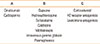
Additional abnormalities noted are high levels of metalloproteinases26 in the plasma of patients with CSU as well as increased fibrin split products and prothrombin fragment 1–22728 suggesting activation of the coagulation cascade and fibrinolysis even though there is no clinical abnormality in hemostasis or thrombosis in this disorder. A summary of major immunologic associations observed in patients with CSU is given in Table 1.
TREATMENT OF CSU
A new guideline has been approved as of December 2016 and endorsed thus far by the European Academy of Allergy and Clinical Immunology (EAACI) and the World Allergy Organization (WAO), but has not yet been published. The prior iteration is now out-of-date2 and many changes were made. I will refer to an earlier publication entitled: Therapy of chronic urticaria: a simple, modern approach29 which, like the new guideline, emphasizes the utility of omalizumab and cyclosporine for antihistamine-resistant patients (Table 2).
Antihistamines remain the initial approach to therapy and approximately 50% of patients respond sufficiently to require no further treatment. Yet one must bear in mind that at last half such patients are unresponsive so that the statement that “most patients with CSU respond well to antihistamines” might be false. Nevertheless, second-generation antihistamines are effective and carry fewer side-effects, such as sedation and mucosal dryness, compared to their first-generation counterparts. These antihistamines were first approved for allergic rhinitis where 1 tablet/day usually suffices, but that is often not the case for urticaria. Thus a rapid increase to 4 tablets/day is recommended.30 Also, this is true for most inducible urticarias31 as well as CSU. My preference is to increase to 4 tablets/day quickly and if it works, decrease the dose after a few weeks rather than increasing one pill at a time. If 4 tablets/day is not satisfactory, the patient is likely to be in the antihistamine-resistant group.
It is clear that the most effective agent, with best side-effect profile for CSU, is Xolair® (omalizumab; Novartis Pharmaceuticals Corp., East Hanover, NJ, USA). A proof-of-concept study demonstrated striking efficacy in severe, autoimmune-associated CSU32; it was placebo controlled and single-blind. This was followed by a dose-escalating phase 2 trial33 and a study of patients with IgE antiperoxidase antibodies34 each of which demonstrated efficacy. These were placebo-controlled and double-blind. Finally, 3 phase 3 trials totaling over 900 patients revealed a response rate of 60%–70%, a complete response rate of about 40%, and effective doses of 150 or 300 mg per injection without regard to body weight or IgE level.353637 It was clear that the 300 mg dose/month was more effective than the 150 mg dose, but that after 6 months, most patients reverted to the placebo level.36 Thus 6 months of treatment did not affect the natural course of the disease but suppressed the symptoms. Pruritus was proportionately reduced as the number of hives diminished. While omalizumab carries a 2%–3% incidence of anaphylaxis when employed for asthma, anaphylaxis was not observed in the urticaria trials. This should be the drug of choice for antihistamine-resistant cases since failure of antihistamines was a criterion for most of the aforementioned studies and one36 consisted of patients who failed H1-antagonists, H2-antagonists, and leukotriene antagonists.
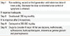
The next issue is what to do for antihistamine-resistant patients who fail to respond to omalizumab. This subpopulation of patients represents about 15%–20% of the total. The most effective of the remaining possibilities is cyclosporine; a conclusion which emerged from double-blind, placebo-controlled studies3839 plus subsequent reports40 and personal observations. Like omalizumab, the response rate is 60%–70%, but one has to monitor patients carefully because it does have potential toxicity. A typical dose for an adult is 200 mg/day with monitoring of blood pressure and renal function every 6 weeks assuming they are normal when the medication is instituted. It is preferable to a host of alternatives, to be addressed below, because its efficacy is far greater. Employment of agents whose efficacy is questionable or minimal in severe patients because they have little or no toxicity makes no sense. Prior guidelines have stated “complete control” as a goal and this may not be attainable when one looks beyond antihistamines, omalizumab, and cyclosporine. The percent success rate i.e., significant (but not complete) improvement with these 3 agents has been estimated to be 93%29 (Table 3). It is predicated on the fact that a lack of response to any one agent has no implication for responsiveness to another.
I will briefly address the reliability of other therapeutic possibilities. H2-antagonists have never been shown to improve urticaria beyond that achievable with H1-antagonists. Their use was based on studies showing that a histamine-induced wheal and flare reaction in the skin can be blocked further by addition of an H2-antagonist once H1-receptor blockade has been achieved.41 This does not translate into clinical efficacy in CSU and H2-antagonists were eliminated in the last EAACI guideline2 but retained in American guidelines.1
Leukotriene antagonists are also employed as adjunctive agents when H1-receptor blockade has been achieved. There is literature pro42 and con,43 but no large-scale double-blind placebo-controlled study has been done. My personal experience is that patient's refractory to antihistamines will not respond to leucotriene antagonists, so I am opposed to their general use as treatment of CSU. They might have some role in patients whose urticaria is exacerbated by aspirin44 or other non-steroidal anti-inflammatory agents although eliminating them, if possible, is a better approach. Acetaminophen can be safely substituted.
A more promising possibility is perhaps dapsone. While there are many case reports claiming success, proper double-blind placebo-controlled trials are limited. Perhaps the best and most recent trial demonstrated a response rate of 30%–40% for dapsone45 with a placebo response of 10%. While the success rate is far lower that of cyclosporine or omalizumab, a conundrum is the 10% placebo response when we know the placebo response rate in CSU is about 25%–30%.353637 Thus the result is questionable and the increment from the usual response rate is small. In fact, the most recent publication suggests that the success rate of the pseudoallergen-free diet46 approximates that of the placebo rate.
Other agents considered in earlier guidelines include sulfasalazine, methotrexate, and plaquanil. The study examining sulfasalazine was not properly controlled47 and it seems unlikely that a sulfa drug combined with an aspirin derivative (salicylate) is likely to help urticaria. Methotrexate has never really been studied; however, the few times I have tried it, there was no effect. Plaquenil® (hydroxychloroquine; Concordia Pharmaceuticals Inc., Oakville, Canada) is particularly effective for the hypocomplementemic urticarial vasculitis syndrome,48 a rare type of cutaneous vasculitis, but its efficacy for CSU is largely anecdotal.49
Corticosteroids are effective for CSU, but its use should be limited to short courses for acute amelioration of severe urticaria/angioedema episodes and should not be employed for chronic use because the side effects outweigh the efficacy. When employed in the past, the doses were too high and sustained for too long. Isolated angioedema can be treated with 40 mg prednisone on 2 successive days and then discontinue it without any taper. A severe urticarial episode with or without angioedema can be treated with 40 mg/day for 3 days; then decrease by 5 mg each day for a total of 10 days. That is sufficient time for the approach outlined above to be instituted.

 XML Download