

 PDF
PDF ePub
ePub Citation
 Print
Print


INTRODUCTION
Mast cells are the key effector cell type in IgE-mediated immediate hypersensitivity and allergic disorders.1,2 Mast cells bound by antigen-specific IgE via the high-affinity receptor for IgE (FcεRI) must encounter multivalent antigen for their activation.3 Activated mast cells secrete preformed proinflammatory mediators, such as histamine, serotonin, nucleotides, proteases, and TNF-α, and synthesize and secrete lipid mediators (such as leukotrienes and prostaglandins), various cytokines and chemokines. In contrast to this traditional view, we and others showed that IgE binding to FcεRI in the absence of specific antigen engenders several biological outcomes in mast cells: upregulation of cell surface expression of FcεRI,4-6 survival,7,8 increase in histamine content,9 histamine release, leukotriene release, receptor internalization, DNA synthesis,10 increased responses to compound 48/80 and substance P,11 increase in filamentous actin content,12 membrane ruffling,13 adhesion to fibronectin,14 and migration.15 These effects are due to binding to FcεRI by monomeric, but not aggregate, IgE molecules.7,8,12 FcεRI cross-linking appears essential for mast cell activation induced by monomeric IgE,16 similar to that by IgE+antigen or IgE+anti-IgE.17,18
Not all IgE molecules can induce all the activation events in the absence of antigen: IgEs display a wide spectrum of heterogeneity in their ability to induce the production and secretion of IL-6 and TNF-α, two tested cytokines produced by mouse mast cells, with HC IgEs at one extreme end and PC IgEs at the other.10 More extensive receptor aggregation can be induced by HC IgEs than by PC IgEs. Consistent with such a difference, strong HC IgEs can induce each of the tested activation events, whereas PC IgEs induce only a limited set of activation events, and to a lesser extent.19 Polyclonal IgE from mice and humans suffering from atopic dermatitis (AD) can enhance survival and cytokine production in mast cell cultures, indicating that monomeric IgE effects are operative in polyclonal situations as well.20
Compared to numerous reports on effects of monomeric IgE on mouse mast cells, effects of human IgE on human mast cells have been less studied. However, using human cord blood derived mast cells (CBMCs), Gilchrest et al.21 reported that the chemokine I-309 RNA and protein levels were upregulated not only in response to IgE+anti-IgE stimulation but also by IgE alone and these responses were further augmented in the presence of IL-4. Cruse et al.22 also reported that human monomeric myeloma IgE stimulated cultured human lung mast cells to release histamine, leukotriene C4, and IL-8 in the absence of a specific antigen. Matsuda et al.16 showed that human monomeric IgE in the absence of specific antigen enhanced IL-8 and monocyte chemoattractant protein 1 (MCP-1) production in cultured human mast cells, and this response was augmented by preincubation of the cells in IL-4. However, it is still unclear whether the effects of monomeric HC versus PC IgEs exist in the human system. Since heterogeneity of human IgEs in the ability to prime basophils to stimulation with histamine-releasing factor (HRF) has been reported,23 it is possible that some highly allergic patients might produce HC IgEs, which may activate mast cells or basophils in the absence of antigen.
Our recent study demonstrated that ~30% of the tested mouse IgE mAbs bind to HRF and that HRF+HRF-reactive IgE can activate mast cells.24 Since HRF has two IgE-binding sites and can be present as a dimer, it is assumed that IgE-bound receptors can be cross-linked by HRF. Similar situations could occur if receptor-bound IgE react with multivalent (auto)antigen. Therefore, we investigated whether HC IgEs react with various antigens.
MATERIALS AND METHODS
IgE preparations
Mouse monoclonal IgEs were purified by ammonium sulfate precipitation from serum-free culture supernatants of hybridomas or peritoneal exudates of hybridoma-bearing mice, followed by DEAE column chromatography.25 Some ELISA experiments were performed with hybridoma culture supernatants. Sources of IgEs: H1 DNP-ε-26 (26) and H1 DNP-ε-206 (206) from Fu-Tong Liu, University of California, Davis; AR40EA, BE1BD (BE1BD2D5), BE2, BE4 (BE4C2C3), DNP48BC, DNP48BD, HB30, and R25 from R.P.S.; C38-2, C48-2, and 27-74 from BD Biosciences; SPE-7 from Sigma-Aldrich; IGELa2 from American Type Culture Collection (ATCC) (TIB142).
Human IgEs: SKO-007 monoclonal IgE was purified by ammonium sulfate precipitation from serum-free culture supernatants of hybridomas (ATCC: CRL-8033-1). HE1 monoclonal IgE from a hybridoma was purchased from Diatec Monoclonals AS (Oslo, Norway). IgEs derived from multiple myeloma patients (BS-κ and ART) were purchased from The Binding Site Inc. (San Diego, CA, USA) or Athens Research & Technology (Athens, GA, USA). IgE from the multiple myeloma patient PS was a gift from Dr. Kimishige Ishizaka, LIAI. All IgE samples were dialyzed extensively against PBS, and ultracentrifuged at 100,000×g for 30 minutes before use to remove possible aggregates.
Sera from AD patients and normal subjects
After the informed consent was obtained, the sera of AD patients diagnosed according to the criteria of Hanifin and Rajka26 were collected. Total numbers of AD patients and normal controls are 41 and 25, respectively. All studies involving human subjects were conducted in accordance with the guidelines of the World Medical Association's Declaration of Helsinki and approved by the Institutional Review Boards of La Jolla Institute for Allergy and Immunology, Kyushu University, University of Occupational and Environmental Health and University of Yamanashi.
Enzyme-linked immunosorbent assay (ELISA)
Calf thymus double-stranded DNA (dsDNA) and single-stranded DNA (ssDNA), β-galactosidase from bovine liver, thyroglobulin from bovine thyroid, insulin from bovine pancreas, LPS (all from Sigma-Aldrich) and recombinant HRF tagged with 6x histidine (all 10 µg/mL in PBS) were coated in 96-well plates for overnight at 4℃. After the wells were blocked with 10% FCS-PBS, mouse IgE molecules (10 µg/mL) were added. Bound mouse IgEs were detected by biotinylated anti-mouse IgE mAb (BD Bioscience PharMingen, San Diego, CA, USA), followed by streptavidin-HRP conjugates. Color was developed and absorbance at 450 nm was measured. In some experiments, human IgEs (10 µg/mL) or sera from healthy subjects and AD patients (1/10 dilution) were incubated in the well. Bound human IgE molecules were detected by biotinylated anti-human IgE mAb (BD Bioscience PharMingen), followed by streptavidin-HRP.
Human mast cell culture
Human CBMCs were generated as described.27 Briefly, CD34+ cells were purified from umbilical cord blood mononuclear cells and cultured in serum-free Iscove's methylcellulose medium (IMDM: Stem Cell Technologies Inc., Vancouver, BC, Canada) containing 200 ng/mL SCF (PeproTech Inc., Rocky Hill, NJ, USA), 50 ng/mL IL-6 (PeproTech Inc.), 1 ng/mL IL-3 (PeproTech Inc.), 1% insulin-transferrin-selenium (Invitrogen, Carlsbad, CA, USA), 50 µM 2-mercaptoethanol, 1% penicillin-streptomycin (Invitroten), and 0.1% bovine serum albumin (Sigma-Aldrich, St Louis, MO, USA). On day 42 of culture, methylcellulose was dissolved in PBS and the cells were then suspended and cultured in IMDM (Invitrogen) supplemented with 100 ng/mL SCF, 50 ng/mL IL-6, and 5% fetal calf serum (Cansera, Rexdale, ON, Canada). After 15 weeks, the purity of the resulting CBMCs was ~98% (c-Kit+FcεRI+ by flow cytometry). The use of human cord blood was approved by the Institutional Review Board of Nihon University Graduate School of Medical Science.
Cytokine measurement
CBMCs were incubated with 10 ng/mL of IL-4 in the presence or absence of 1 µg/mL of IgE for sensitization. After 24 hours, the cells were washed and resuspended in culture medium. The cells (1×10⁵/100 µL) were then stimulated with IgE molecules for the indicated times. After stimulation, supernatants and cell pellets were collected for analysis of IL-8 and IL-6 expression and production. IL-8 production was measured using a human IL-8 ELISA kit (BD PharMingen, San Diego, CA, USA).
Cell survival
CBMCs were incubated with IL-4 in the presence or absence of IgE for sensitization. After 24 hours, the cells were washed and resuspended in SCF-free culture medium. The cells (1×10⁵/100 µL) were then stimulated with anti-IgE, SCF, or IgE molecules for 72 hours. After 72 hours, cell survival was analyzed after staining with Trypan blue.
Real-time RT-PCR
Total cellular RNA was isolated from CBMCs with an RNeasy mini kit (Qiagen, Valencia, CA, USA) according to the manufacturer's instructions. An equal amount of total RNA (100 ng) was used for reverse transcription. Real-time RT-PCR was performed as follows: cDNA (10 ng) was amplified in 20 µL in the presence of 1 µL of "Assay-on-Demand" primer sets for IL-8, IL-6, and GAPDH purchased from Applied Biosystems (Foster City, CA, USA) in 7700 ABI thermal cyclers (Applied Biosystems). Relative expression levels were determined using Ct method.
Measurement of serum HRF
Human sera (10 µL) were precipitated first with acetone. Precipitated materials were dissolved in SDS sample buffer and, together with dilution series of recombinant HRF-His6, analyzed by SDS-PAGE and blotted onto PVDF membranes. The blots were probed with anti-HRF antibody followed by HRP-conjugated secondary antibody. HRF bands were detected by ECL reagent (Perkin-Elmer). The concentrations of HRF in the samples were determined by densitometry.
RESULTS
Mouse HC, but not PC, IgE molecules react with various autoantigens
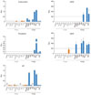
HC IgEs are defined operationally by the ability to induce secretion of IL-6 and/or TNF-α from BMMCs, whereas PC IgEs lack this ability.10 We have recently shown that some of the tested IgEs interact with HRF.24 We extended this line of study by testing whether mouse IgE mAbs interact with other antigens, such as dsDNA, ssDNA, β-galactosidase, thyroglobulin, insulin and LPS. ELISA data (Fig. 1) showed that most HC IgEs react with two or more of these antigens. For example, the most potent HC IgE SPE-7 reacted with dsDNA, ssDNA and thyroglobulin, in addition to DNP (hapten used to generate this mAb) and thioredoxin, which was previously shown to bind to SPE-7 IgE.28 H1 DNP-ε-26 IgE reacted with dsDNA and ssDNA; C38-2 IgE reacted with HRF, dsDNA, ssDNA, β-galactosidase and thyroglobulin; IGELa2 IgE reacted with HRF, β-galactosidase and thyroglobulin. The possibility of non-specific binding was ruled out. For example, interactions of TNP-specific C38-2 and IGELa2 IgEs with HRF were inhibited by TNP-glycine,24 suggesting that the HRF-binding site in IgE overlaps at least in part with the antigen-binding site. By contrast, none of the 13 tested PC IgE mAbs reacted with the antigens under the same conditions. Neither HC nor PC IgEs reacted with insulin or LPS. These experiments clearly demonstrate that most of mouse HC, but not PC, IgEs react with multiple autoantigens.
Human IgE molecules also show the HC vs. PC heterogeneity
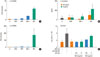
Human CBMCs express low levels of FcεRI on their cell surface.29 Previous studies showed that incubation of human CBMCs with IL-4 enhances FcεRI expression29 and renders the cells sensitive to stimulation with human IgE, which induces the production of chemokines, IL-8 and monocyte chemoattractant protein 1.16 Therefore, we used IL-4-preincubated human CBMCs in this study. To determine whether human IgE molecules show heterogeneity in inducing cytokine production in human mast cells, human CBMCs were incubated with 5 µg/mL of human monoclonal and myeloma-derived IgE molecules for 2 hours, to measure cytokine mRNAs and proteins. As shown previously,16 quantitative RT-PCR showed that IL-8 mRNA expression is induced strongly by a monoclonal (HE1) IgE and weakly by another myeloma IgE, BS-κ (Fig. 2A). We could also show secretion of IL-8 protein induced by the HE1 IgE (Fig. 2B). However, the other tested monoclonal IgE preparations failed to induce IL-8 mRNA. Similarly, only HE1 IgE induced IL-6 mRNA (Fig. 2C). Secreted IL-6 levels could not be measured because CBMCs were cultured in IL-6-containing medium. HE1 IgE, but not SKO IgE, enhanced survival of CBMCs (Fig. 2D). Thus, these results indicate that HE1 is an HC IgE and others are PC IgEs.
Sera from AD patients contain higher levels of HRF and ssDNA- or β-galactosidase-binding IgEs
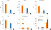
The ability of some IgEs to bind to multivalent autoantigens such as HRF or DNA raised the possibility that mast cells and basophils can be activated by interacting with such an autoantigen via FcεRI-bound IgE. Given the importance of IgE in the pathogenesis of AD, asthma and other allergic diseases30-33 and preferential IgE reactivity with autoantigens in AD,34 we measured levels of IgE that reacted with dsDNA, ssDNA, β-galactosidase, and LPS in sera of AD patients. AD patients had significantly higher serum levels of ssDNA-reactive and β-galactosidase-reactive IgE than healthy controls (Fig. 4A and 4C). Levels of dsDNA-reactive IgE also tended to be higher than healthy controls, although the difference did not reach statistical significance (Fig. 4B). AD patients also had higher serum levels of HRF than healthy controls (Fig. 4D). Four of 34 AD patients had measurable levels of IgE that reacted with HRF (Fig. 4E). Two of these patients had extremely high IgE levels of 48.7 and 384 µg/mL, whereas another patient had 0.17 µg/mL and IgE levels were not known for the other patient. By contrast, none of 25 healthy individuals had detectable levels of anti-HRF IgE. However, we found no increase in anti-LPS IgE titers (OD450 <0.01) in AD or healthy individuals, ruling out the possibility that the measured high O.D. values reflected antigen-non-specific bindings due to high concentrations of total IgE.
We previously showed that polyclonal IgE in AD sera can induce IL-8 production, a signature of HC IgEs, in human CBMCs.20 Therefore, we tested this ability of AD sera (n=8) with high titers (≥0.01 OD450) of dsDNA- and ssDNA-reacitve IgEs, compared to sera from healthy subjects. As shown in Fig. 4F, the AD sera exhibited 4.7- and 7.2-fold higher IL-8-producing activity at 5% and 10% serum concentrations. Therefore, these results strongly suggest that AD patients have autoantigen-reactive IgEs and these IgEs may be HC IgEs.
DISCUSSION
This study demonstrates that most of mouse HC IgEs react with multiple autoantigens. Since some of the autoantigens, e.g., HRF and dsDNA and ssDNA, have multivalency in terms of the IgE-binding ability, it seems reasonable to assume that such an autoantigen can trigger cross-linking of IgE-bound receptors, thus activation of mast cells and basophils, eventually leading to allergic reactions. Indeed, HRF was shown to amplify allergic inflammation by this mechanism in passive cutaneous anaphylaxis and airway inflammation in mice.24 Similar to HRF, dsDNA and ssDNA released from damaged tissues during allergic reactions may contribute to exacerbate allergic inflammation by interacting with HC IgE bound to FcεRI on mast cells and basophils. One of the eight mouse IgE mAbs tested failed to react with autoantigens. This negative result might be due to the fact that the number of autoantigens tested was not exhaustive enough or appropriate autoantigens were not tested.
As encountered by western blotting or other immunoassays on a daily basis, immunologists have long known that an antibody recognizes more than one antigen.35 Particularly, monoclonal antibodies were shown to have multispecificity.36 Conformational diversity of antigen recognition, whereby one antibody (e.g., IgE) sequence adopts multiple structures, can increase the effective size of the antibody repertoire and may lead to autoimmunity.28 Autoimmunity underlies allergic diseases.34 Epidemiological data indicate that the prevalence of allergies and autoimmune diseases have increased in parallel.37,38 Recent studies support the concept that IgE autoreactivity may play a pathogenic role in severe and chronic forms of atopy.34,39 While acute allergic reactions following exposure to exogenous allergens are understood as immediate type inflammation triggered by degranulation of mast cells via allergen-IgE-FcεRI interactions, chronic allergic inflammation with Th1 characteristics can occur and persist in the absence of exogenous allergens. IgE-mediated presentation of autoallergens may activate autoreactive Th1 cells to release proinflammatory cytokines.40,41 Several environmental allergens have striking structural and immunologic similarities with human proteins.42-46 A broad spectrum (≥140 proteins) of IgE-reactive autoantigens were found in AD patients and some of them were shown to have the ability to activate basophils.47 As shown in this study, some AD patients who had high levels of ssDNA- (or dsDNA-) and/or β-galactosidase-reactive IgEs also had high levels of HRF-reactive IgEs, the autoimmune mechanism could be an important part of the disease pathogenesis in this subset of patients. This observation also suggests the heterogeneity of AD pathogenesis, as not all AD patients have increased IgE reactivity to autoantigens. Interestingly, NC/Nga mice spontaneously develop AD-like skin lesions48 and IgE autoantibodies including anti-histone H3 IgE;49 this age-dependent increase in the serum anti-histone H3 IgE was in parallel with the onset of dermatitis, and correlated well with dermatitis severity at 12-16 weeks of age. As global gene expression patterns are similar in lesional skin between NC/Nga mice and human AD patients (Ando et al., in preparation), these data and our present study support the notion that IgE autoreactivity may play a pathogenic role in AD. Thus, further characterization of the role of IgE autoantibodies in the NC/Nga model will be interesting.
This study also demonstrates that human IgE molecules exhibit heterogeneity in the ability to induce cytokine production and survival promotion in human mast cells in a similar way that mouse HC versus PC IgE molecules exhibit differences in inducing various activation events in mouse mast cells.19,50 Thus, a human HC IgE induces cytokine/chemokine production more strongly than do human PC IgEs. However, there are some differences between human and mouse systems. For example, significant survival promotion was seen only with a human HC IgE, in contrast with mouse IgE effects showing that even PC IgEs can promote mast cell survival albeit with weak potency.7,10 Degranulation could not be induced by either type of human IgE. These results could be due to the type of human mast cells used in this study. Indeed, even IgE+anti-IgE-stimulated human CBMCs degranulate only 10%-20% of the granule content (data not shown). This low responsiveness may be related to low expression levels of FcεRI in CBMCs, which can be enhanced by IL-4 stimulation.29 Unlike CBMCs pretreated or not with IL-4, human monomeric myeloma IgE induced dose-dependent histamine release, leukotriene C4 production, and IL-8 synthesis in cultured human lung mast cells.22 That study suggests that cultured human lung mast cells are more responsive to IgE than human CBMCs, although this and our studies were performed using different IgE molecules. Despite the above-mentioned minor differences in monomeric IgE effects between mouse and human, a human HC IgE bound autoantigens such as dsDNA, ssDNA and HRF, like mouse HC IgEs.
In conclusion, HC, but not PC, IgEs exhibit reactivity to various autoantigens. Some of the autoantigens are multivalent, thus potentially capable of activating HC IgE-bound mast cells and basophils. Therefore, these results indicate an autoimmune component in the pathogenic mechanisms of allergic diseases.
 XML Download