

 PDF
PDF ePub
ePub Citation
 Print
Print


INTRODUCTION
Cutaneous leukocytoclastic vasculitis (CLV) is confined to capillaries and post-capillary venules of the superficial plexus.1 The main clinical lesions of CLV are purpuric papules, although other clinical findings secondary to ischemia, including ulceration, may occur.2 Causes of CLV include drugs, infection, connective tissue disease, and malignancy. An estimated 20-30% of all vasculitis cases are attributed to drug administration.3 CLV has rarely been reported in association with tuberculosis or anti-tuberculosis medications.4-6 A case of rifampin-induced Henoch-Schönlein purpura was reported in Korea;7 however, the case presented here is, to our knowledge, the first case of CLV following treatment with rifampin and pyrazinamide.
CASE REPORT
A 38-year-old male presented to our hospital emergency room with palpable purpura of the trunk and extremities. He had been diagnosed with pulmonary tuberculosis 2 months earlier. The patient had a history of idiopathic cardiomyopathy and diabetes mellitus; treatment included daily digoxin 0.125 mg, furosemide 40 mg, candesartan cilexetil 8 mg, glimepiride 2 mg, metformin 500 mg, and voglibose 0.6 mg. When the diagnosis of tuberculosis was confirmed by sputum acid fast bacilli (AFB) staining, standard anti-tuberculosis therapy was initiated with isoniazid (Myambutol®, Yuhan Corp., Suwon, Korea), rifampin (Rifodex®, Chongkundang, Suwon, Korea), ethambutol (Myambutol®, Yuhan Corp.), and pyrazinamide (Pyrazinamid®, Yuhan Corp.). After 1.5 months of anti-tuberculosis therapy, a purpuric, non-blanching rash appeared on the lower and upper extremities. The skin lesions spread progressively over the entire body, and several lesions coalesced, resulting in blisters and ulcers (Fig. 1). The patient had no history of atopy or drug allergy.
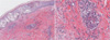
Chest radiographs revealed a speculated nodule and multiple satellite nodules in the left upper lung field; this was suggestive of pulmonary tuberculosis, with no significant changes from the previous radiographic study. However, the sputum AFB smear had become negative. Blood tests showed the following: leukocytes, 7,500×103/µL (neutrophils, 76.4%; lymphocytes, 14.0%; monocytes, 7.6%; eosinophils, 0.8%); hematocrit, 37.8%; hemoglobin, 12.4 g/dL; platelet count, 227,000×103/µL; serum creatinine, 1.1 mg/dL; aspartate aminotransferase, 25 U/L; alanine aminotransferase, 18 U/L; HBsAg negative, anti-HBs antibody negative, anti-HCV antibody negative, syphilis serology negative, and anti-HIV negative. Rheumatoid factor, ANA, and ANCA tests were also negative. Prothrombin time and activated partial thromboplastin time were normal. The fibrinogen degradation product and D-dimer were increased to 5.0 µg/mL (reference range, 0.0-4.0 µg/mL) and 1,225 ng/mL (0-200 ng/mL), respectively. The serum levels of IgG, IgA, IgM, and complement, and the erythrocyte sedimentation rate were within normal limits. The level of C-reactive protein was slightly elevated at 1.12 mg/dL (0.02-0.80 mg/dL). Urinalysis provided no evidence of proteinuria or hematuria. Skin biopsy of the cutaneous purpuric lesions revealed leukocytoclastic vasculitis (Fig. 2).
The anti-tuberculosis medications were stopped after admission due to a clinical suspicion of a drug-induced adverse cutaneous reaction. Levofloxacin (500 mg) was administered as a second-line anti-tuberculosis medication during hospitalization. The patient was treated with prednisolone (20 mg) for 3 days; the dose was reduced on progressive improvement of the vasculitis lesions. We performed rechallenge with each anti-tuberculosis drug after 10 days, as described in Table 1. No adverse reactions were observed after ethambutol (400 mg) and isoniazid (300 mg) were reintroduced. After rifampin (150 mg) therapy, no specific cutaneous findings were noted, but purpura appeared on the right forearm with increasing the dosage to 300 mg. The skin lesion resolved after 3 days of prednisolone treatment (20 mg). Pyrazinamide treatment (1,500 mg) was initiated, and new purpuric lesions were seen on both forearms 3 days post-administration. Consequently, he was treated with isoniazid, ethambutol, levofloxacin, and kanamycin without the recurrence of purpura.
DISCUSSION
Cutaneous adverse reactions to anti-tuberculosis drugs have been reported in up to 5% of treated patients.8 Common cutaneous reactions include pruritus, skin eruptions, maculopapular exanthems, urticaria, pustules, fixed reactions, and erythema nodosum; these reactions can be controlled with the administration of antihistamines.8,9
Cutaneous leukocytoclastic vasculitis is a rare complication of anti-tuberculosis medications. The combination of vasculitis and tuberculosis was first described in 1967.10 There are two general types of pulmonary tuberculosis-related vasculitis: leukocytoclastic vasculitis (a manifestation of pulmonary tuberculosis) and anti-tuberculosis medication-associated vasculitis (particularly with rifampin therapy).4
Direct vasculitic lesions result from the deposition of bacilli. By contrast, microorganisms are not identified in hypersensitivity vasculitis, which may result from the deposition of immune complexes formed by antibodies against Mycobacterium tuberculosis proteins. The existence of such circulating immune complexes has been demonstrated in 56% of patients with active tuberculosis.11 Tuberculids may be a hypersensitivity reaction to M. tuberculosis after it spreads hematogenously from a focus of infection elsewhere. Histologically, a tuberculid is composed of granulomatous inflammation, variable necrosis, and variable vasculitis. The lesions respond dramatically to anti-tuberculosis therapy.12 The possibility of a tuberculosis-associated direct lesion or hypersensitivity vasculitis as the cause of CLV in this patient was unlikely, as the vasculitic lesions developed after 1.5 months of anti-tuberculosis medication. Furthermore, the sputum AFB staining and M. tuberculosis cultures were negative at the time the vasculitis developed.
Several studies have described a relationship between rifampin use and vasculitis onset.5,6 In contrast to tuberculosis-related vasculitis, the skin lesions of rifampin-induced vasculitis typically improve upon withdrawal of the medication. Previous studies have suggested that anti-rifampin antibodies contribute to the vasculitis pathogenesis.13 Although there has been no report of an association between pyrazinamide and vasculitis, the incidence of pyrazinamide-induced hepatotoxicity and other cutaneous adverse reactions is significantly higher than the incidence related to other anti-tuberculosis drugs in Korea and other Western countries.14-16 The development of urticaria or exanthem within 1 to 2 hours of an initial pyrazinamide dose has been reported,17 and pyrazinamide-induced erythema multiforme was confirmed by rechallenge in another study.18 Those studies revealed the causal relationship between the drug and the skin lesions, but no mechanism was documented. Similarly, our patient had a severe cutaneous adverse reaction that improved when the causative drug was withdrawn and recurred after rechallenge. Moreover, there was no cause of the vasculitis except pyrazinamide, and the pathological findings were compatible with leukocytoclastic vasculitis, although the pathogenic mechanism remains unclear.
Risk factors for severe adverse anti-tuberculosis drug reactions include older age, female gender, diabetes mellitus, previous anti-tuberculosis therapy, and a history of hepatitis. Age older than 60 years and a birthplace in Asia are significantly associated with rifampin and pyrazinamide intolerance.14 Our patient had two risk factors: he was Asian and had diabetes mellitus.
We described a case of rifampin- and pyrazinamide-induced CLV confirmed by histology and drug rechallenge. Although rarely seen, rifampin and pyrazinamide should be considered potential causes of drug-induced CLV.


 XML Download