

 PDF
PDF ePub
ePub Citation
 Print
Print


INTRODUCTION
Evaluation of biliary stricture is often challenging in clinical practice, and endoscopic ultrasound (EUS) and endoscopic retrograde cholangiopancreatography (ERCP) are complementary procedures used to evaluate suspected biliary stricture. Pathologic confirmation of malignancy before surgical resection or neoadjuvant therapy is important in such patients. The commonest methods of tissue sampling are ERCP with forceps biopsy and/or brush cytology and EUS-guided fine-needle aspiration.
Several authors have reported that the diagnostic yield of ERCP-guided tissue sampling (ERCP-TS) ranges from 35 to 70% with little improvement in diagnostic yield when brush cytology and forceps biopsy are performed simultaneously.123456 Meanwhile, previous reports for EUS-guided tissue sampling (EUS-TS) using fine-needle aspiration or biopsy provide higher diagnostic yields for solid pancreatic masses, and EUS-TS has been increasingly used to overcome the moderate diagnostic yield of ERCP-TS.789 Furthermore, in patients with suspected biliary stricture, the diagnostic yield of EUS-TS is reportedly superior to that of ERCP-TS when tissue samples are obtained from lesions in the pancreas, lymph node (LN), and others that cause biliary obstruction.10111213
Although EUS-TS has a better diagnostic yield for suspected biliary strictures than ERCP-TS, there is some controversy as to which technique is better for sampling different primary tumor sites. The aim of this study was to compare the diagnostic yields of EUS- and ERCP-TS in patients with suspected biliary stricture at different primary tumor sites.
SUBJECTS AND METHODS
1. Patients
Retrospective analysis was performed on consecutive patients referred for the evaluation of suspected biliary stricture from January 2011 to September 2016. Patients that underwent same-session EUS and ERCP examinations were considered for enrollment. We excluded the followings; 1) those that underwent EUS or ERCP, 2) those that underwent both EUS and ERCP on the same day but without tissue sampling, and 3) those that underwent EUS and ERCP on different days.
2. EUS- and ERCP-TS procedures
EUS was first performed using a curvilinear echoendoscope (GF-UCT240 or GF-UCT260; Olympus Medical Systems, Tokyo, Japan). EUS-TS targeted any solid lesions in the pancreas, bile duct, gallbladder, or LN and periampullary lesions. All EUS-TS procedures were performed without on-site cytopathologic assessment. Specimen were expressed on 1 to 2 slides for alcohol-fixation with Papanicolaou smear. In each case, an additional specimen was placed in a 10% formalin container for subsequent histologic analysis.
ERCP was followed by EUS, if clinically indicated, and performed by the same endoscopist. During ERCP, selective biliary cannulation and cholangiography were performed to identify the level of the bile duct stricture. ERCP-TS was then performed using brush cytology (BC-15C; Olympus, Tokyo, Japan) without stricture dilation before tissue sampling. Cytology brushings were obtained using 10 to-and-fro movements across strictures, and 2 glass slides were then smeared with brushes, which were then fixed in 95% alcohol. Brush tips were cut and placed in 10% formalin for analysis. However, brush cytology was not performed in all patients. An intraductal biliary forceps was then introduced to the distal end of the stricture under fluoroscopy. Biopsies were performed in triplicate and additional biopsies were conducted when no adequate specimen was obtained in any of the three initial trials. EUS- and ERCP-TS procedures were performed by two experienced endoscopists (C. C. and J. M.) who conducted more than 300 EUS- and ERCP-related procedures per year.
A pathologist without prior knowledge of the sampling technique evaluated cytopathologic specimens. Based on histopathologic results, we compared diagnostic accuracies by tissue sampling method and lesion location. Tissue samples were classified as: 1) malignant, 2) suspicious for malignancy, 3) atypical, 4) benign, and 5) inadequate for diagnosis or non-diagnostic.
3. Definitions and outcomes
We considered tissue samples as malignant when the pathologist concluded “suspicious for malignancy” or “malignant”. Failure to collect a specimen by EUS- or ERCP-TS was considered “non-diagnostic”. The final diagnostic standard of malignancy was defined as the followings; 1) a malignant histopathology based a surgical specimen, 2) a malignant cytopathology from EUS- and/or ERCP-TS, or 3) clinical follow-up for more than 12 months with findings consistent with malignancy, such as clinical progression. When no sign of malignancy was noted during follow-up, the lesion was considered benign. Hyperbilirubinemia was defined as a total bilirubin level of ≥2.5 mg/dL.
The primary endpoints of this study were the overall diagnostic accuracies of EUS- and ERCP-TS and differences between these at different primary tumor sites. The secondary endpoints were specimen adequacies of EUS- and ERCP-TS for different target lesions. This study was approved by the Institutional Review Board of Kyungpook National University Chilgok Hospital, Daegu, Korea (IRB No. 2017-01-028).
4. Statistical analysis
The Chi-squared (χ2) or Fisher's exact test were used to determine differences between categorical variables, and the Student's t-test was used to determine differences between continuous variables. McNemar's test was used to compare diagnostic yields and to determine the significances of differences between EUS- and ERCP-TS. Results are presented as means±SDs, and p-value of <0.05 was considered statistically significant. The analysis was conducted using SPSS version 23.0 (IBM Corp., Armonk, NY, USA) and Medcalc (version 18.11; http://www.medcalc.org).
RESULTS
One hundred and twenty-five patients with suspected biliary stricture treated at our institution between January 2011 and September 2016 were considered as the study enrollment. Of these, 32 patients were excluded due to; 1) follow-up loss (n=8), 2) ERCP-TS being performed on the pancreatic duct (n=20) or a periampullary lesion (n=4). Finally, 93 patients were included in the study. No complications, such as pancreatitis, perforation, or hemorrhage, requiring treatment occurred.
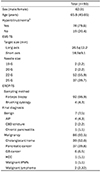
Patient baseline characteristics and final diagnoses are summarized in Table 1. The 93 study subjects (62 males and 31 females; overall median age, 67 years) underwent same-session EUS- and ERCP-TS. Seventy-four patients had hyperbilirubinemia at time of the procedure. Mean solid lesion size targeted by EUS-TS was 26.5±12.2 mm (longest diameter) by 18.9±9.1 mm (shortest diameter). Needle sizes were 19 gauge (G) for 2 lesions, 20G for 2, 22G for 57, and 25G for 37. For ERCP-TS, forceps biopsy was used for 92 lesions and brush cytology for 4.
Malignant tumors were noted in 86 patients; final diagnoses were confirmed in 33 patients through surgery. Forty-nine patients were diagnosed with a malignancy by EUSor ERCP-TS. Malignancy was not detected in two patients using either technique, who were diagnosed by long-term clinical follow-up. Malignancy was also identified in one patient by liver biopsy and in another patient by ultrasound-guided lymph node biopsy.
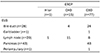
Sites of specimen acquisitions by procedure are summarized in Table 2. For ERCP, 1 sample was collected from the hilar area, 15 samples from common hepatic duct (CHD), and 77 from common bile duct (CBD). For EUS, 28 samples were extracted from bile duct, 1 from gallbladder, 20 from lymph node, 43 from pancreas, and one from periampullary lesion.
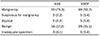
EUS-TS resulted in the detections of malignancy and suspected malignancy in 70 and two patients, respectively, and ERCP-TS resulted in the detections of malignancy and suspected malignancy in 49 and five patients, respectively. Inadequate specimens were obtained from three patients during EUS-TS and from five patients during ERCP-TS (Table 3). No significant difference was noted between the two modalities in terms of specimen inadequacy (EUS-TS: 3.1% vs. ERCP-TS: 5.4%, p=0.687).
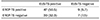
The sensitivities, specificities, accuracies, positive predictive values, and negative predictive values of EUS- and ERCP-TS are present in Table 4. The overall diagnostic accuracy of EUS-TS was superior to that of ERCP-TS (EUS-TS: 90.3% vs. ERCP-TS: 67.7%, p=0.001), and this was especially true for patients with a pancreatic lesion (EUS-TS: 90.7% vs. ERCP-TS: 58.1%, p=0.002). However, no significant differences were observed for the diagnostic accuracies between EUS- and ERCP-TS for bile duct lesions (EUS-TS: 87.8% vs. ERCP-TS: 80.5%, p=0.581) or at other primary sites (EUS-TS: 100% vs. ERCP-TS: 77.8%, p=0.125). Overall diagnostic accuracies of EUS- and/or ERCP-TS were around 92.5%.
Table 5 details cases diagnosed by EUS- and ERCP-TS. As shown in the table, 47 cases were diagnosed when both procedures were used, 30 cases were diagnosed by EUS-TS, 9 cases were diagnosed by ERCP-TS, and seven cases were not diagnosed by either procedure. Of the 30 cases diagnosed by EUS-TS alone, eight were CBD lesions, 18 were pancreatic lesions, and four were other lesions. Of the nine cases diagnosed by ERCP-TS, five were CBD lesions and four were pancreatic lesions.
DISCUSSION
It is difficult to determine the exact cause of a suspected biliary stricture. Preoperative histopathological confirmation and the institution of neoadjuvant therapy are important for determining optimal treatment modalities for this condition. ERCP-TS is conventionally used in these cases, but its reported diagnostic accuracy ranges from 35 to 70%; though this can be slightly increased by performing forceps biopsy and brush cytology.123456 Recently, EUS-TS was shown to have better diagnostic accuracy, that is, between 85% and 93%. Furthermore, EUS-TS is known to be superior to ERCP-TS in patients with a biliary stricture caused by lesions involving the pancreas and LN.714151617 However, few studies have compared the diagnostic yields of these two methods, particularly in patients that undergo initial EUS-TS followed by ERCP-TS at the same time. Previous studies on this topic are limited by incomplete protocols and small cohort sizes, and little information is available on the diagnostic yields of EUS-TS and ERCP-TS for different primary lesion types and locations.
The merits of our study are as follows: 1) all 93 patients included underwent ERCP-TS after EUS-TS, and thus, the experimental protocol did not affect outcomes. Two previous studies reported diagnostic accuracies for EUS-TS and ERCP-TS performed during same sessions as ~20% and ~60%, respectively.1819 Thus, it appears the experimental protocols influence results. And 2) we found results were unaffected by bias secondary to primary lesions. Stenosis caused by biliary or pancreatic lesions occurred in 41 (44.1%) and 43 patients (46.2%), respectively, and thus, the number of patients with a pancreatic or biliary lesion were similar. Previous studies have suggested that pancreatic lesion-induced strictures are more likely to be diagnosed by EUS-TS than by evidence of narrowing caused by bile ducts or other etiologies.71419 Therefore, if a primary lesion has an asymmetric influence on the pancreas or biliary tract, diagnostic outcome are likely to be affected.
In the present study, the diagnostic accuracy of EUS-TS (84.4%) for biliary strictures caused by pancreatic lesions was significantly greater than that of ERCP-TS (51.1%) because EUS-TS directly targets these lesions, whereas ERCP-TS targets sites of bile duct compression.20 The diagnostic accuracies of EUS-TS and ERCP-TS for identifying biliary stricture secondary to a biliary lesion were similar (80.5% vs. 73.2%, respectively). However, EUS-TS detected a bile duct lesion in 11 patients, in which ERCP-TS failed to detect a lesion.
We enrolled patients that underwent initial EUS-TS followed by ERCP-TS during same sessions, because biliary stents inserted during ERCP to decompress biliary obstruction can negatively affect the diagnostic accuracy of future EUS-TS procedures.2122 Therefore, when both procedures are performed during same sessions, initial EUS-TS improves diagnostic yield and relieves obstruction. A small number of studies have reported that same-session EUS-TS and ERCP-TS did not increase complication rates, although procedure times were obviously extended.182023 We concur with these findings as no patient developed a cardiopulmonary complication in the present study. Nevertheless, careful monitoring and caution are required to safely perform EUS-TS and ERCP-TS during same sessions.
Several limitations of our study warrant mention: 1) the study is inherently limited by its retrospective single-center study design. 2) Not all patients underwent brush cytology and forceps biopsy during ERCP-TS. 3) Cholangioscopy is known to improve the diagnostic accuracy of ERCP-TS, but was not performed, and this might have reduced diagnostic accuracy. 4) EUS-TS was performed using different kinds of needles, which may have affected diagnostic accuracies and outcomes. 5) EUS-TS was performed on an LN for CHD and hilar lesions, and thus, it cannot be assumed our results well-reflect diagnostic results for primary lesions. Moreover, a benign finding may indicate that the procedure had been correctly performed for reactive lymphadenopathy secondary to cholangitis. Although EUS-TS returned a benign finding, the final diagnosis was malignancy for all. However, when EUS-TS of an LN returned a finding of benignity, this did suggest a higher probability of a benign final diagnosis. And 6) all procedures were performed by two expert endoscopists, and thus, techniques or practices employed might not accurately reflect those used at other centers. A large-scale prospective multicenter study that adopts the aforementioned diagnostic methods with or without cholangioscopy is required to address these concerns.
In conclusion, we compared the diagnostic accuracies of EUS-TS and ERCP-TS in patients with a suspected biliary stricture. Overall, EUS-TS was observed to be superior to ERCP-TS, especially in cases of pancreatic lesion-induced biliary obstruction. However, the use of EUS-TS alone as a diagnostic aid was limited in such cases. As shown by our results, in patients with pancreatic lesions, nine cases (21%) were diagnosed only by ERCP-TS, and thus, a combination of EUS-TS and ERCP-TS is likely to be more effective than EUS-TS alone for the diagnosis of pancreatic lesions. In cases of suspected malignant primary biliary obstruction, the diagnostic accuracy EUS-TS was found to be non-significantly better than that of ERCP-TS. Furthermore, the present study suggests EUS-TS may be useful in cases of suspected cholangiocarcinoma, though it is not routinely performed in patients with obstruction caused by bile duct cancer. Therefore, we recommend that both, EUS-TS and ERCP-TS be performed in patients with suspected biliary stricture to improve the diagnostic accuracy.

 XML Download