Abstract
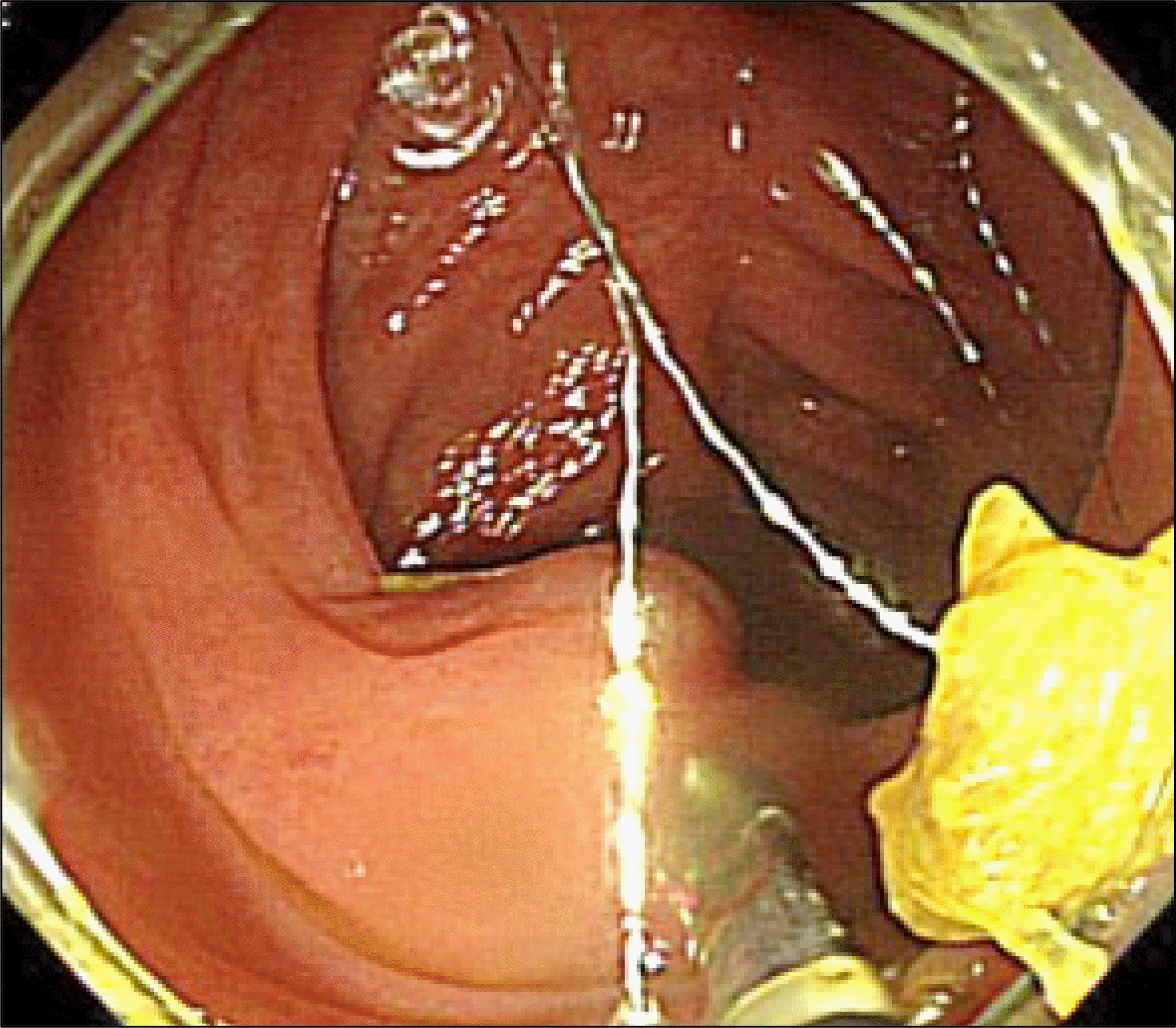
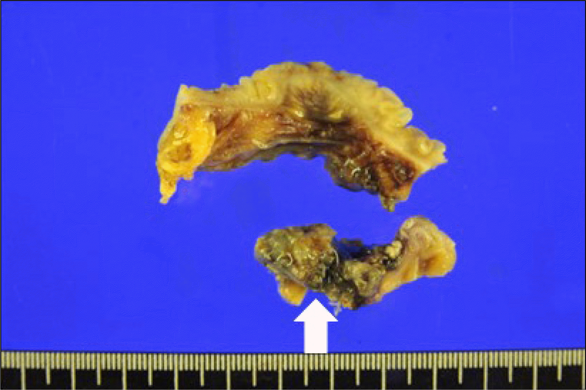
Jejunal variceal bleeding is less common compared with esophagogastric varices in patients with portal hypertension. However, jejunal variceal bleeding can be fatal without treatment. Treatments include surgery, transjugular intrahepatic portosystemic shunt (TIPS), endoscopic sclerotherapy, percutaneous coil embolization, and balloon-occluded retrograde transvenous obliteration (BRTO). Percutaneous coil embolization can be considered as an alternative treatment option for those where endoscopic sclerotherapy, surgery, TIPS or BRTO are not possible. Complications of percutaneous coil embolization have been reported, including coil migration. Herein, we report a case of migration of the coil into the jejunal lumen after percutaneous coil embolization for jejunal variceal bleeding. The migrated coil was successfully removed using surgery.
References
1. Gunjan D, Sharma V, Rana SS, Bhasin DK. Small bowel bleeding: a comprehensive review. Gastroenterol Rep (Oxf). 2014; 2:262–275.

2. Sato T, Yasui O, Kurokawa T, Hashimoto M, Asanuma Y, Koyama K. Jejunal varix with extrahepatic portal obstruction treated by embolization using interventional radiology: report of a case. Surg Today. 2003; 33:131–134.

3. Koo SM, Jeong SW, Jang JY, et al. Jejunal variceal bleeding successfully treated with percutaneous coil embolization. J Korean Med Sci. 2012; 27:321–324.

4. Skipworth JR, Morkane C, Raptis DA, et al. Coil migration–a rare complication of endovascular exclusion of visceral artery pseudoaneurysms and aneurysms. Ann R Coll Surg Engl. 2011; 93:e19–e23.
5. Tekola BD, Arner DM, Behm BW. Coil migration after transarterial coil embolization of a splenic artery pseudoaneurysm. Case Rep Gastroenterol. 2013; 7:487–491.

6. Vangeli M, Patch D, Terreni N, et al. Bleeding ectopic varices–treatment with transjugular intrahepatic portosystemic shunt (TIPS) and embolisation. J Hepatol. 2004; 41:560–566.

7. Sharma B, Raina S, Sharma R. Bleeding ectopic varices as the first manifestation of portal hypertension. Case Reports Hepatol. 2014; 2014:140959.

8. Joo YE, Kim HS, Choi SK, Rew JS, Kim HR, Kim SJ. Massive gastrointestinal bleeding from jejunal varices. J Gastroenterol. 2000; 35:775–778.

9. Yuki N, Kubo M, Noro Y, et al. Jejunal varices as a cause of massive gastrointestinal bleeding. Am J Gastroenterol. 1992; 87:514–517.
10. Akhter NM, Haskal ZJ. Diagnosis and management of ectopic varices. Gastrointest Intervent. 2012; 1:3–10.

11. Carrafiello G, Lagana D, Giorgianni A, et al. Bleeding from peri-stomal varices in a cirrhotic patient with ileal conduit: treatment with transjugular intrahepatic portocaval shunt (TIPS). Emerg Radiol. 2007; 13:341–343.

12. Bühler L, Tamigneaux I, Giostra E, et al. Ectopic varices, a rare cause of digestive hemorrhage. Schweiz Med Wochenschr Suppl. 1996; 79:70S–72S.
Fig. 1.
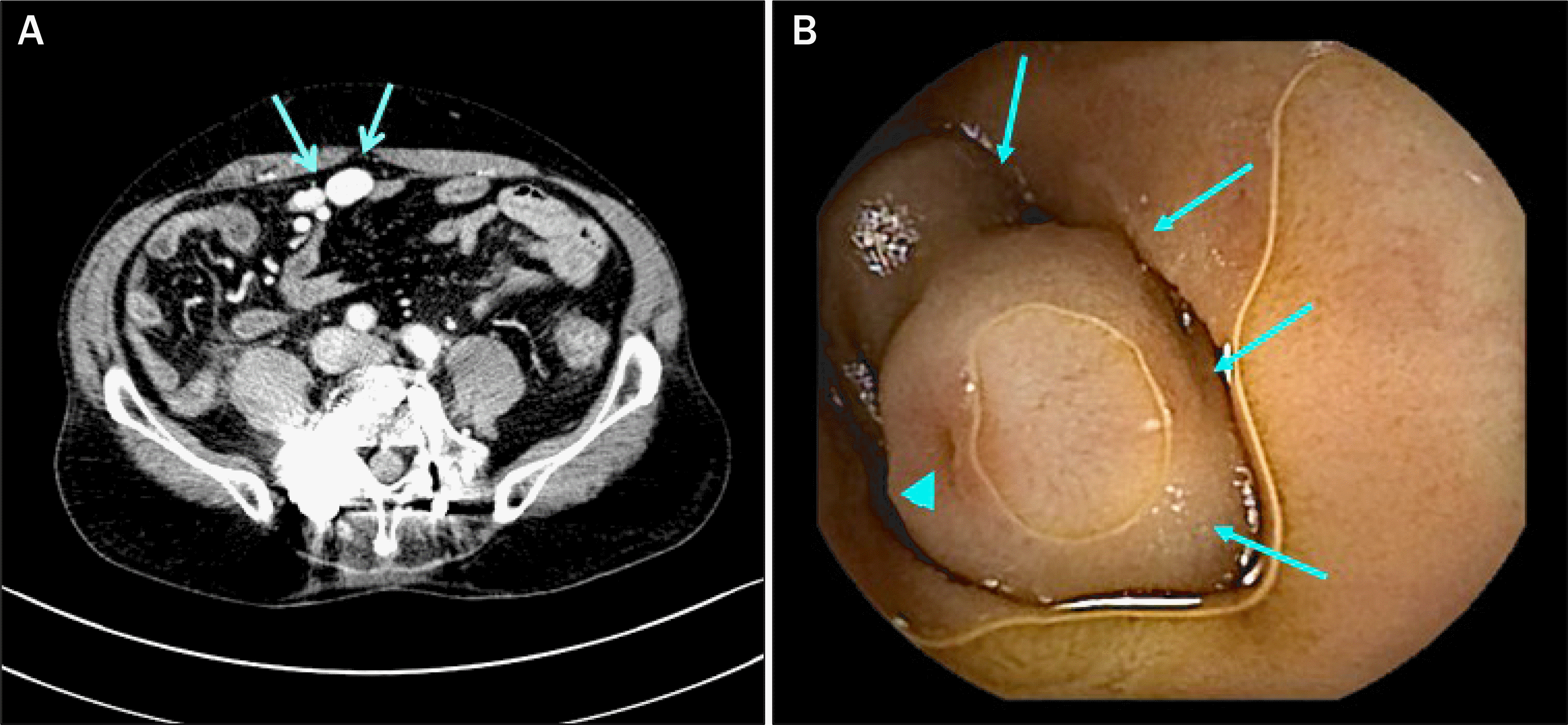
(A) Jejunal varices (arrows) were observed on the portal phase of CT scan. (B) Small bowel video capsule endoscopy revealed a dumbbell-shaped varix (arrows). A dimple on the surface (arrowhead) is suggestive of a recent bleeding stigma.

Fig. 2.
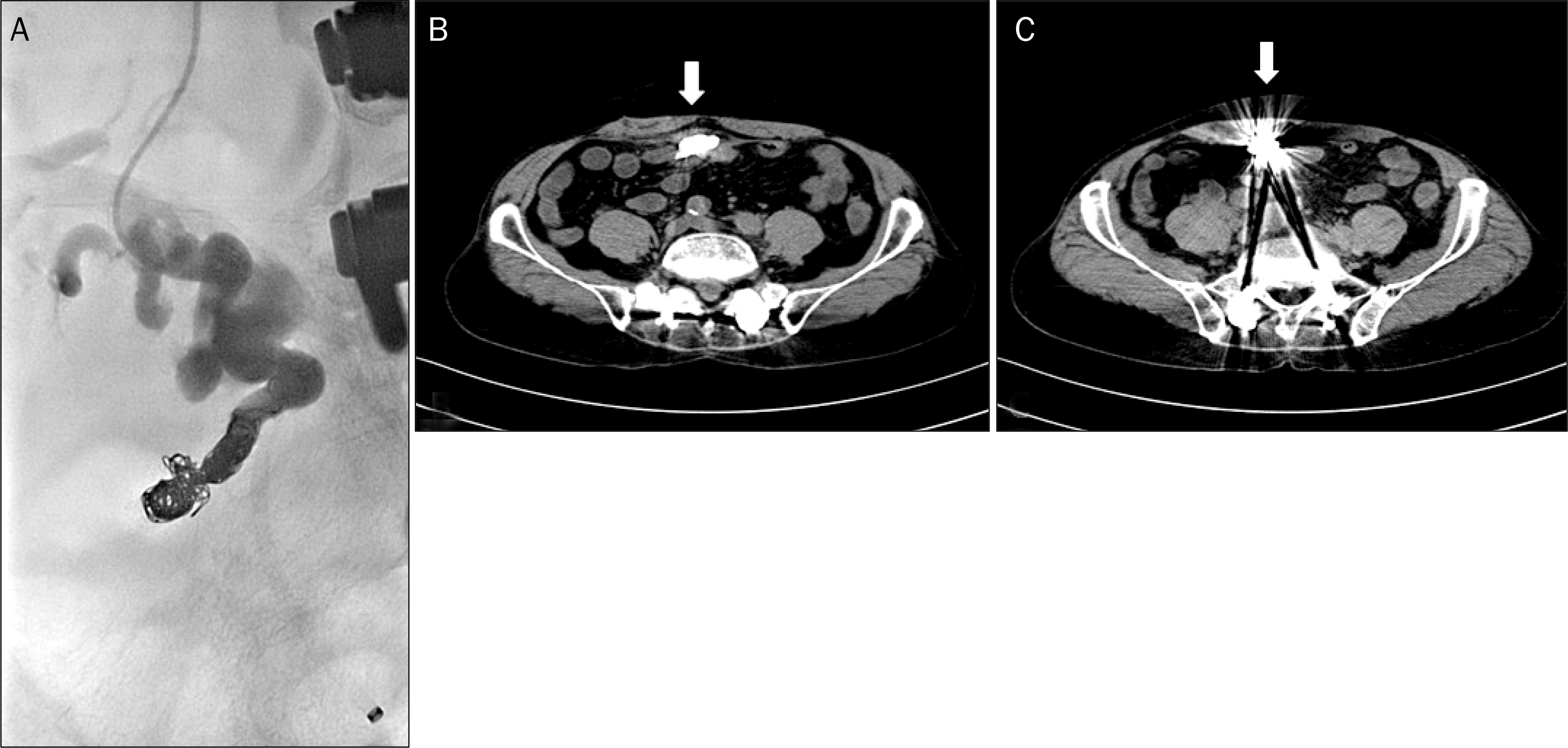
(A). Angiography showing tortuous jejunal varices. Embolization was performed using coil and N-butyl-2-cyanoacrylate (Histoacryl). (B, C) CT scan after embolization showing coil and Histoacryl, and varices were invisible (white arrows).


 PDF
PDF ePub
ePub Citation
 Print
Print


 XML Download