Abstract
Duodenal diverticula are common, but perforated duodenal diverticulum is rare. Because of the disease rarity, there is no standard management protocol for perforated duodenal diverticulum. To properly manage this rare complication, a clear pre-operative diagnosis and clinical disease severity assessment are important. An abdominopelvic CT is an unquestionably crucial diagnostic tool. Perforation is considered a surgical emergency, although conservative treatment based on fasting and broad-spec-trum antibiotics may be offered in some selected cases. Herein, we report two cases of perforated duodenal diverticulum, one case managed with surgical treatment and one with conservative treatment.
References
1. Yin WY, Chen HT, Huang SM, Lin HH, Chang TM. Clinical analysis and literature review of massive duodenal diverticular bleeding. World J Surg. 2001; 25:848–855.

2. Oukachbi N, Brouzes S. Management of complicated duodenal diverticula. J Visc Surg. 2013; 150:173–179.

3. Schnueriger B, Vorburger SA, Banz VM, Schoepfer AM, Candinas D. Diagnosis and management of the symptomatic duodenal diverticulum: a case series and a short review of the literature. J Gastrointest Surg. 2008; 12:1571–1576.

4. de Perrot T, Poletti PA, Becker CD, Platon A. The complicated duodenal diverticulum: retrospective analysis of 11 cases. Clin Imaging. 2012; 36:287–294.

5. Rossetti A, Christian BN, Pascal B, Stephane D, Philippe M. Perforated duodenal diverticulum, a rare complication of a common pathology: a seven-patient case series. World J Gastrointest Surg. 2013; 5:47–50.
6. Miller G, Mueller C, Yim D, et al. Perforated duodenal diverticulitis: a report of three cases. Dig Surg. 2005; 22:198–202.

7. Martinez-Cecilia D, Arjona-Sanchez A, Gomez-Alvarez M, et al. Conservative management of perforated duodenal diverticulum: a case report and review of the literature. World J Gastroenterol. 2008; 14:1949–1951.
8. Coulier B, Maldague P, Bourgeois A, Broze B. Diverticulitis of the small bowel: CT diagnosis. Abdom Imaging. 2007; 32:228–233.

9. Thorson CM, Paz Ruiz PS, Roeder RA, Sleeman D, Casillas VJ. The perforated duodenal diverticulum. Arch Surg. 2012; 147:81–88.

10. Volchok J, Massimi T, Wilkins S, Curletti E. Duodenal diverticulum: case report of a perforated extraluminal diverticulum containing ectopic pancreatic tissue. Arch Surg. 2009; 144:188–190.
11. Favre-Rizzo J, López-Tomassetti-Fernández E, Ceballos-Esparragón J, Hernández-Hernández JR. Duodenal diverticulum perforated by foreign body. Rev Esp Enferm Dig. 2013; 105:368–369.
12. Lee HH, Hong JY, Oh SN, Jeon HM, Park CH, Song KY. Laparoscopic diverticulectomy for a perforated duodenal diverticulum: a case report. J Laparoendosc Adv Surg Tech A. 2010; 20:757–760.

13. Ames JT, Federle MP, Pealer KM. Perforated duodenal diverticulum: clinical and imaging findings in eight patients. Abdom Imaging. 2009; 34:135–139.

14. Barillaro I, Grassi V, De Sol A, et al. Endoscopic rendezvous after damage control surgery in treatment of retroperitoneal abscess from perforated duodenal diverticulum: a techinal note and literature review. World J Emerg Surg. 2013; 8:26.

15. Costa Simões V, Santos B, Magalhães S, Faria G, Sousa Silva D, Davide J. Perforated duodenal diverticulum: surgical treatment and literature review. Int J Surg Case Rep. 2014; 5:547–550.

16. Haboubi D, Thapar A, Bhan C, Oshowo A. Perforated duodenal di-verticulae: importance for the surgeon and gastroenterologist. BMJ Case Rep. 2014. DOI: doi:10.1136/bcr-2014–205859.

Fig. 1.
Abdomino-pelvic CT image in a 53-year-old male in the present case. Axial CT image of perforated duodenal diverticulum with a foreign material demonstrated by a complex collection with fluid and gas surrounding the second portion of the duodenum (arrowheads).
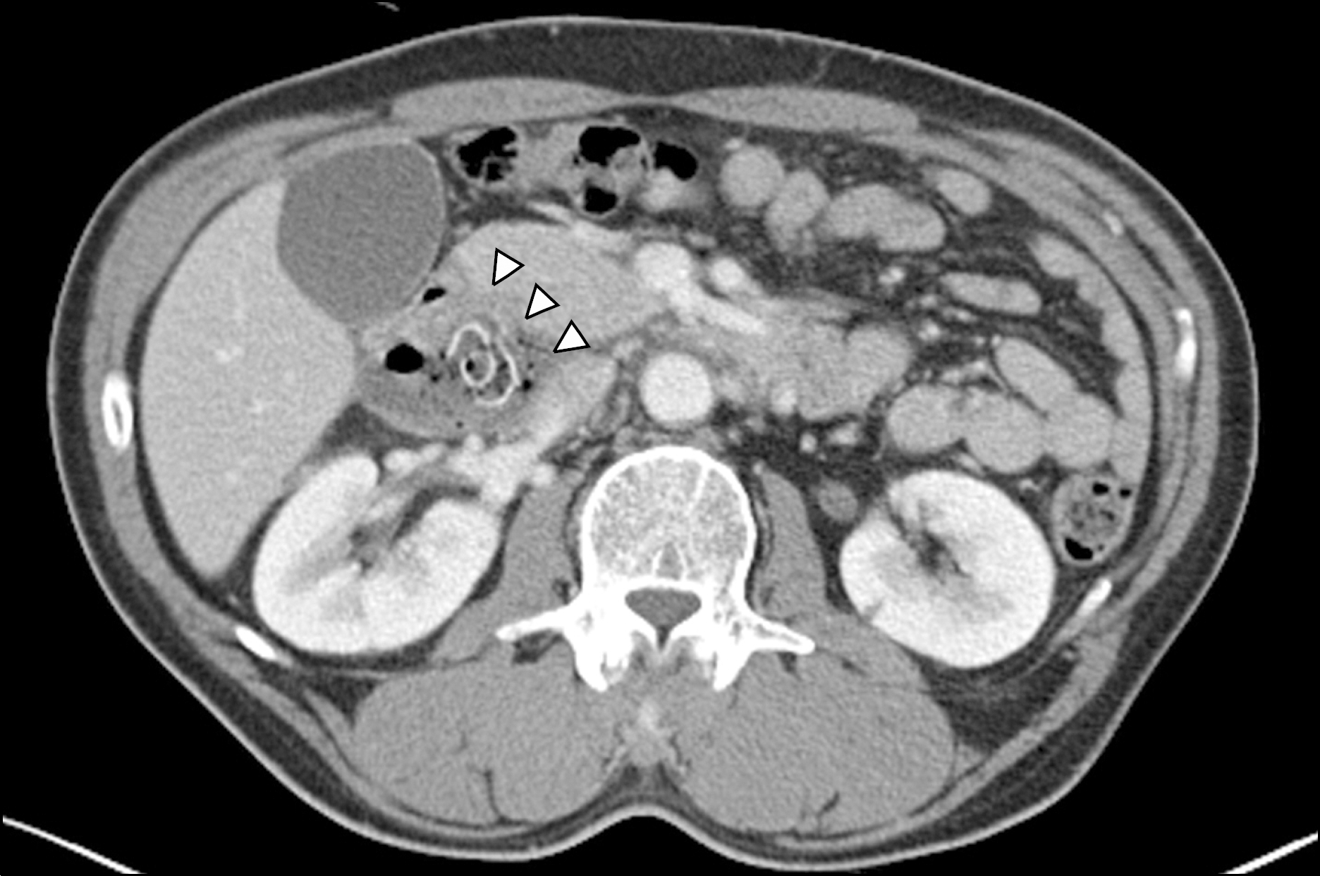
Fig. 2.
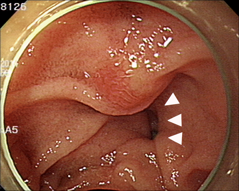
An isolated perforated duodenal diverticulum on an antero-medial side of duodenal 2nd portion. Black arrowheads show the perforation site, and white arrowheads show the stalk of diverticulum.
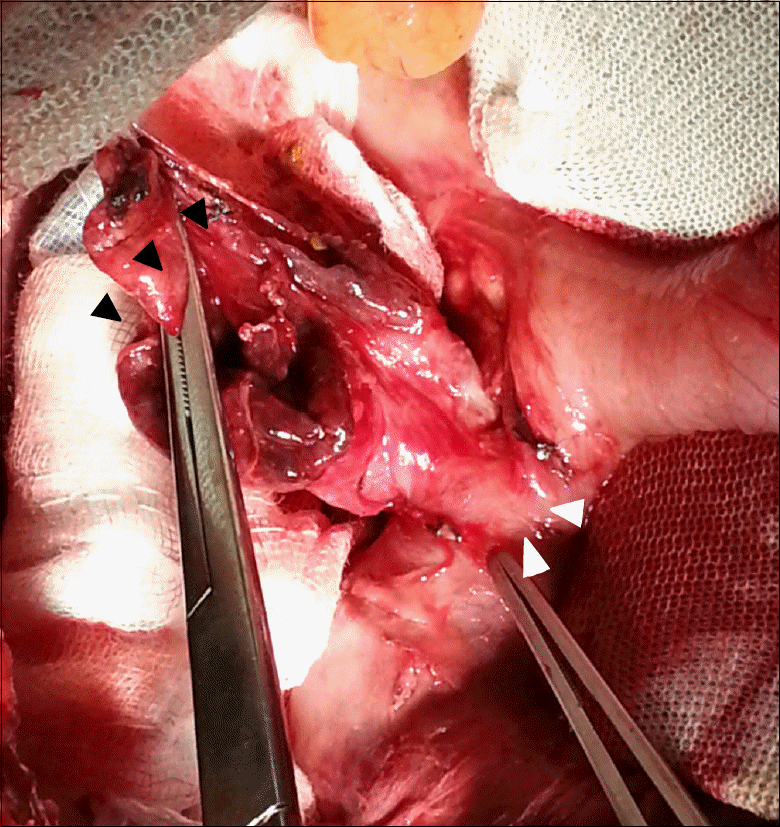
Fig. 3.
Abdomino-pelvic CT image in a 73-year-old male in the present case. Suspected duodenal perforation denoted by retroperitoneal air (arrowheads).
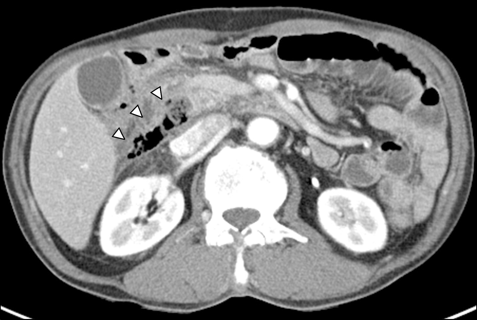
Fig. 4.
Following CT after 18 days in a 73-year-old male in the present case. The axial image shows a near complete disappearance of fluid collection (arrowheads).
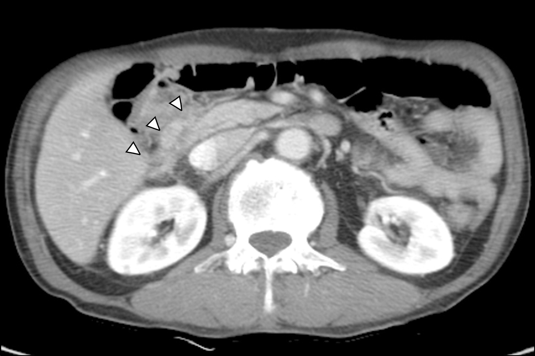
Table 1.
Reported Cases of Perforated Duodenal Diverticulum, 2012–2014


 PDF
PDF ePub
ePub Citation
 Print
Print


 XML Download