Abstract
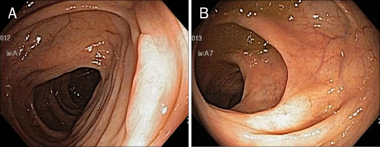
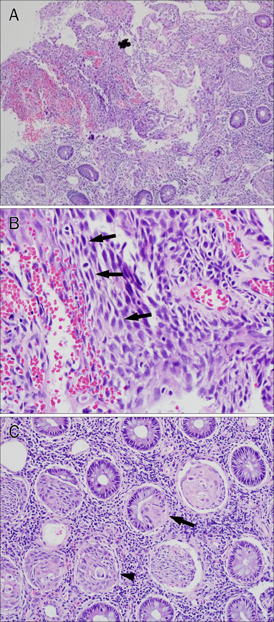
Primary squamous cell carcinoma of the colon is an extremely rare malignancy. A 48-year-old male visited our hospital for screening colonoscopy. Colonoscopic examination showed a 1 cm sized sessile polyp in the ascending colon. The patient underwent endoscopic mucosal resection (EMR) without any complication. The pathologic findings were compatible with squamous differentiation of tumor cells in inflammatory colonic mucosa. The tumor was confined to the mucosa and the margins of the excised tissue were found to be free of the tumor. There were no other primary sites and no distant metastases in the extensive evaluation using a whole body CT scan and PET-CT. Additional surgical resection was not done. Follow-up colonoscopy performed eight month later showed a whitish scar without evidence of local recurrence and follow-up PET-CT demonstrated no evidence of recurrence. Herein, we report a case of primary squamous cell carcinoma of the ascending colon presenting as a sessile polyp which was removed by EMR.
References
1. Dyson T, Draganov PV. Squamous cell cancer of the rectum. World J Gastroenterol. 2009; 15:4380–4386.

2. Woods WG. Squamous cell carcinoma of the rectum arising in an area of squamous metaplasia. Eur J Surg Oncol. 1987; 13:455–458.
3. Ouban A, Nawab RA, Coppola D. Diagnostic and pathogenetic implications of colorectal carcinomas with multidirectional differentiation: a report of 4 cases. Clin Colorectal Cancer. 2002; 1:243–248.

4. Schneider TA 2nd, Birkett DH, Vernava AM 3rd. Primary adenosquamous and squamous cell carcinoma of the colon and rectum. Int J Colorectal Dis. 1992; 7:144–147.

5. Williams GT, Blackshaw AJ, Morson BC. Squamous carcinoma of the colorectum and its genesis. J Pathol. 1979; 129:139–147.

6. Sameer AS, Syeed N, Chowdri NA, Parray FQ, Siddiqi MA. Squamous cell carcinoma of rectum presenting in a man: a case report. J Med Case Rep. 2010; 4:392.

7. Lafreniere R, Ketcham AS. Primary squamous carcinoma of the rectum. Report of a case and review of the literature. Dis Colon Rectum. 1985; 28:967–972.
8. Gould L, Shah JM, Khedekar RR, Burns WA. Squamous cell carcinoma of the splenic flexure of the colon. Dig Dis Sci. 1983; 28:918–922.

9. Nahas CS, Shia J, Joseph R, et al. Squamous-cell carcinoma of the rectum: a rare but curable tumor. Dis Colon Rectum. 2007; 50:1393–1400.

10. Kim TS, Park SO, Ahn JK, et al. A case of squamous cell carcinoma in situ of rectum. Korean J Gastroenterol. 1997; 30:114–118.
11. Park SM, Hur SE, Kwon BJ, et al. Basaloid squamous cell carcinoma of the rectum manifesting as multiple submucosal lesions. Korean J Gastrointest Endosc. 2006; 33:168–172.
12. Shin US, Yu CS, Kim CW, et al. Malignancy associated with inflammatory bowel disease. J Korean Soc Coloproctol. 2009; 25:150–156.

13. Kim NR, Chung DH, Baek JH, et al. Squamous cell carcinoma of the rectum: report of two cases. Intest Res. 2010; 8:172–176.

14. Ocque R, Tochigi N, Ohori NP, Dacic S. Usefulness of immunohistochemical and histochemical studies in the classification of lung adenocarcinoma and squamous cell carcinoma in cytologic specimens. Am J Clin Pathol. 2011; 136:81–87.

15. Pittella JE, Torres AV. Primary squamous-cell carcinoma of the cecum and ascending colon: report of a case and review of the literature. Dis Colon Rectum. 1982; 25:483–487.
16. Dzeletovic I, Pasha S, Leighton JA. Human papillomavirus-related rectal squamous cell carcinoma in a patient with ulcerative colitis diagnosed on narrow-band imaging. Clin Gastroenterol Hepatol. 2010; 8:e47–e48.

17. Al Hallak MN, Hage-Nassar G, Mouchli A. Primary submucosal squamous cell carcinoma of the rectum diagnosed by endoscopic ultrasound: case report and literature review. Case Rep Gastroenterol. 2010; 4:243–249.

18. Frizelle FA, Hobday KS, Batts KP, Nelson H. Adenosquamous and squamous carcinoma of the colon and upper rectum: a clinical and histopathologic study. Dis Colon Rectum. 2001; 44:341–346.
Fig. 1.
Colonoscopic findings. (A) About 10 mm sized sessile polyp is seen in the ascending colon. (B) Follow-up colonoscopy after endoscopic mucosal resection 8 month later shows whitish scar without gross evidence of intraluminal local recurrence.

Fig. 2.
Microscopic findings of the resected specimen (H&E stain). (A) Squamous differentiation of tumor cells is invading lamina propria in the inflammatory colonic mucosa. There is no malignant adenomatous component (×100). (B) Squamous differentiation of tumor cells with interceullular bridge (black arrows) (×400). (C) Atypical squamous metaplasia (black arrow) is seen around tumor cells (arrowhead) and normal colonic epithelial cells (×200).

Fig. 3.
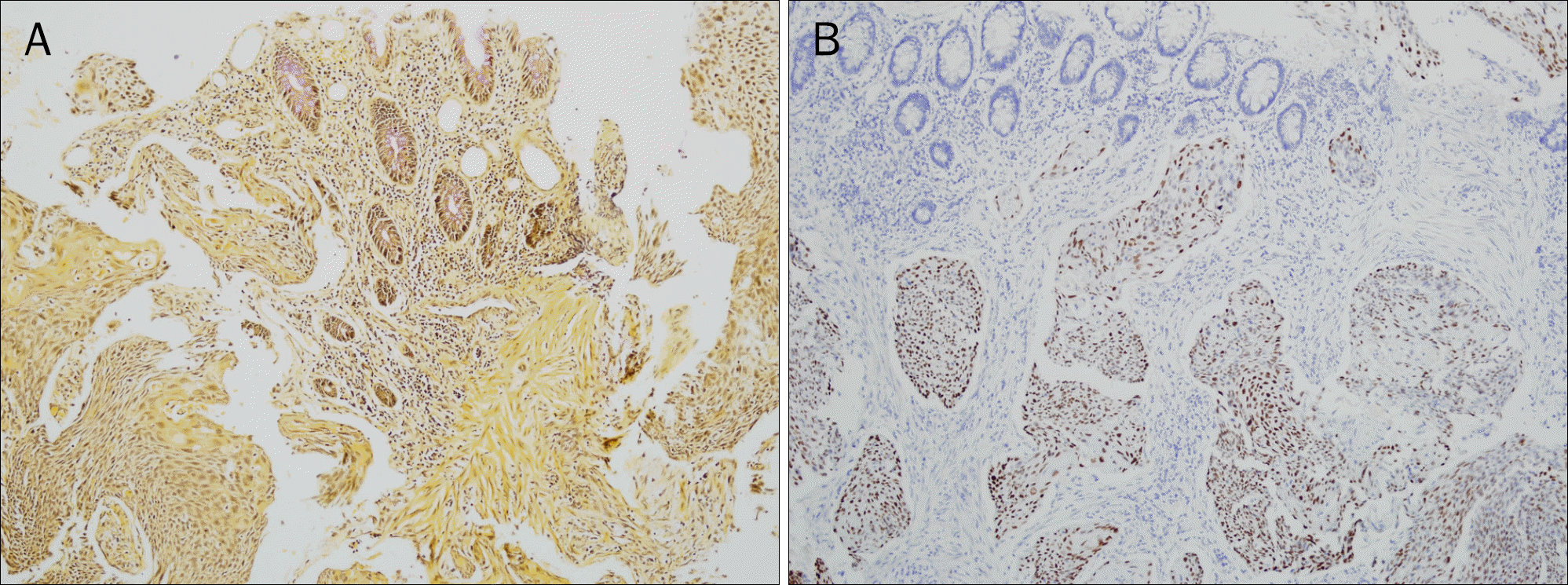
Immunohistochemical stain findings. (A) Mucicarmine stain for mucin shows pink colored stain in normal colon mucosa but negative stain in squamous cell carcinoma, thereby establishing the absence of any glandular or adenomatous component (×100). (B) p63 stain shows diffuse and strong positivity in squamous cell carcinoma (×100).


 PDF
PDF ePub
ePub Citation
 Print
Print


 XML Download