

 PDF
PDF ePub
ePub Citation
 Print
Print


Abstract
After the start of antituberculous treatment, paradoxical worsening of tuberculous lesions has been described. However, abdominal tuberculosis as paradoxical response is relatively rare. This report describes the 26-year-old female who suffered from peritoneal tuberculosis while treating tuberculous pleurisy with antituberculous medications. It was considered as paradoxical response, rather than treatment failure or else. She was successfully managed with continuing initial antituberculous medications. When a patient on antituberculous medications is presented with abdominal symptoms, the possibility of paradoxical response should be considered to avoid unnecessary tests and treatments, which may result in more suffering of the patient. Herein, we report a case of peritoneal tuberculosis as paradoxical response while treating tuberculous pleurisy.
References
1. Demir K, Okten A, Kaymakoglu S, et al. Tuberculous peritonitis–reports of 26 cases, detailing diagnostic and therapeutic problems. Eur J Gastroenterol Hepatol. 2001; 13:581–585.
2. Cheng VC, Ho PL, Lee RA, et al. Clinical spectrum of paradoxical deterioration during antituberculosis therapy in non-HIV-in-fected patients. Eur J Clin Microbiol Infect Dis. 2002; 21:803–809.

3. Blumberg HM, Burman WJ, Chaisson RE, et al. American thoracic society/centers for disease control and prevention/infectious diseases society of America: treatment of tuberculosis. Am J Respir Crit Care Med. 2003; 167:603–662.
4. Al-Majed SA. Study of paradoxical response to chemotherapy in tuberculous pleural effusion. Respir Med. 1996; 90:211–214.
5. Ahn TH, Park MB, Lee KJ, et al. A case report of tuberculous brain abscess and tuberculous peritonitis developing due to paradoxical reactions. Tuberc Respir Dis. 2009; 66:457–462.
6. Cheng VC, Yam WC, Woo PC, et al. Risk factors for development of paradoxical response during antituberculosis therapy in HIV-negative patients. Eur J Clin Microbiol Infect Dis. 2003; 22:597–602.
7. Meintjes G, Rangaka MX, Maartens G, et al. Novel relationship between tuberculosis immune reconstitution inflammatory syndrome and antitubercular drug resistance. Clin Infect Dis. 2009; 48:667–676.
8. Song EJ, Baek DH, Jung JY, et al. Paradoxical response developed during the antituberculous treatment in tuberculous pleurisy. Tuberc Respir Dis. 2008; 64:427–432.
9. Kim JY, Kwon JH, Kim MJ, et al. Paradoxical response during antituberculous treatment for abdominal tuberculosis. J Korean Radiol Soc. 2006; 55:599–605.
10. Kasahara K, Fukuoka A, Murakawa K, et al. Tuberculous peritonitis developing during chemotherapy for pulmonary and intestinal tuberculosis: a case report. Respirology. 2005; 10:257–260.
11. Ha CY, Kim JY, Kim GC, et al. An intraperitoneal tuberculous abscess: Computed tomography (CT) findings and clinical course. Korean J Med. 2008; 74:243–249.
12. Lee JS, Kim KA, Lee WJ, et al. Diagnostic value of ascitic fluid adenosine deaminase activity for diagnosis of tuberculous peritonitis. Korean J Gastroenterol. 2003; 41:126–132.
13. Kim KS, Pyon YJ, Seong KC, et al. A case with chylous ascites fol-lowed by tuberculous peritonitis. Korean J Gastroenterol. 1997; 29:847–852.
14. Jung MK, Lee HJ, Kim MJ, et al. Paradoxical response to chemotherapy in tuberculous pleural effusion. Pediatr Allergy Respir Dis. 2009; 19:71–77.
Fig. 1.
Chest X-ray. (A) The initial chest X-ray showed left sided pleural effusion. (B) On admission, 10 weeks later, left sided pleural effusion was decreased. (C) Left sided pleural effusion was almost resolved 9 months after the start of antituberculous treatment.
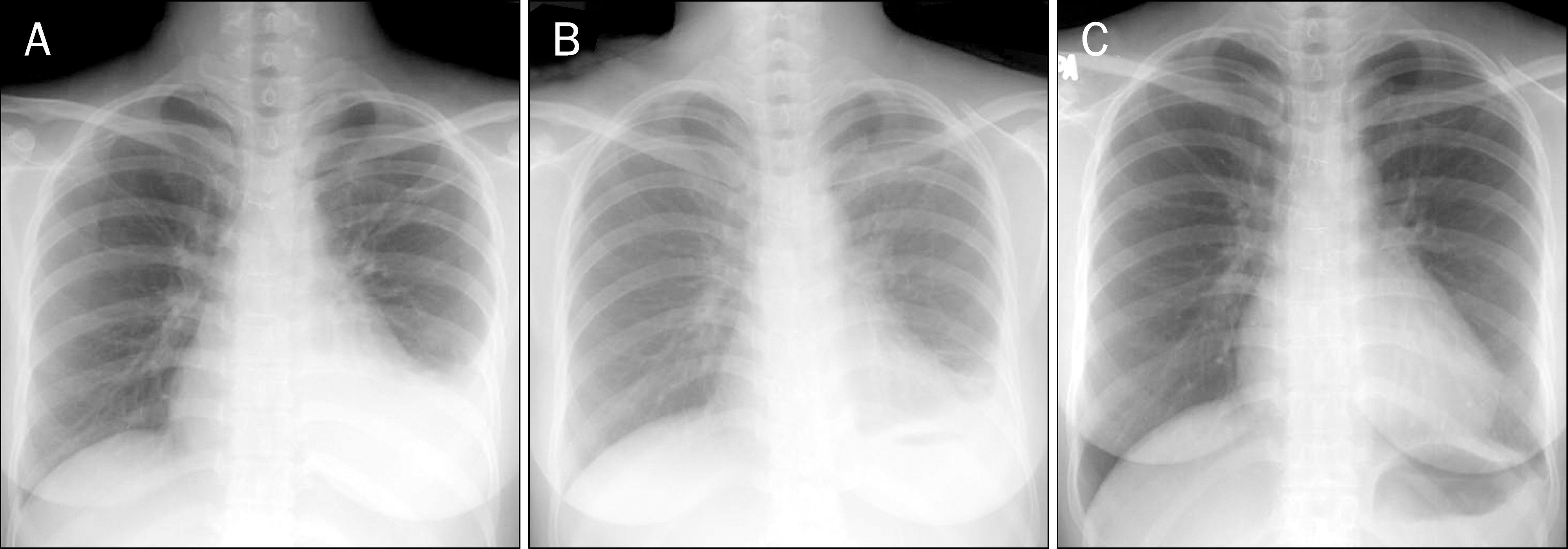
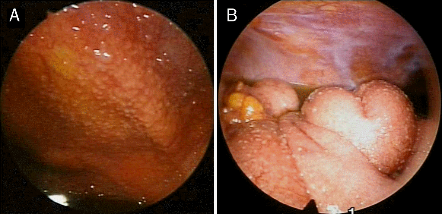
Fig. 2.
Abdominal CT. Ascites and smooth uniform thickening with pronounced enhancement (arrows) and smudged pattern of the peritoneum (arrow heads), known as radiologically characteristic findings of tuberculous peritonitis, were shown (A, B). Ascites and the extent of smudged pattern of peritoneum disappeared 6 months after the start of antituberculous treatment (C, D).

 XML Download