Abstract
Radiation dermatitis can develop after fluoroscopy-guided interventional procedures. Cases of fluoroscopy-induced radiation dermatitis have been reported since 1996, mostly documented in the fields of radiology, cardiology and dermatology. Since diagnosis and treatment of fluoroscopy-induced radiation dermatitis can be difficult, high grade of suspicion is required. The extent of this reaction is determined by radiation dose, duration of exposure, type of procedure, and host factors and can be aggravated by concomitant use of photosensitizers. Follow-up is important after long and complicated procedures and efforts to minimize radiation exposure time will be necessary to prevent radiation dermatitis. Herein, we report a case of a 58-year-old man with hepatocellular carcinoma presenting with subacute radiation dermatitis after prolonged fluoroscopic exposure during transarterial chemoembolization and chemoport insertion. Physicians should be aware that fluoroscopy is a potential cause of radiation dermatitis.
References
1. Koenig TR, Wolff D, Mettler FA, Wagner LK. Skin injuries from fluoroscopically guided procedures: part 1, characteristics of radiation injury. AJR Am J Roentgenol. 2001; 177:3–11.
2. Koenig TR, Mettler FA, Wagner LK. Skin injuries from fluoroscopically guided procedures: part 2, review of 73 cases and recommendations for minimizing dose delivered to patient. AJR Am J Roentgenol. 2001; 177:13–20.
3. Won JH, Yun SJ, Lee JB, Kim SJ, Lee SC, Won YH. Subacute radiation dermatitis due to fluoroscopy during cardiac intervention. Korean J Dermatol. 2010; 48:866–868.
4. Spiker A, Zinn Z, Carter WH, Powers R, Kovach R. Fluoroscopy-induced chronic radiation dermatitis. Am J Cardiol. 2012; 110:1861–1863.

5. Cho EB, Song BH, Park EJ, Kwon IH, Kim KH, Kim KJ. Fluorosco-py-induced chronic radiation dermatitis. Korean J Dermatol. 2012; 50:614–617.
6. Noh HJ, Park SW, Kim YB, et al. Radiation dermatitis after GDC embolization: case report. J Korean Neurosurg Soc. 2002; 32:63–65.
7. Wagner LK, Eifel PJ, Geise RA. Potential biological effects following high X-ray dose interventional procedures. J Vasc Interv Radiol. 1994; 5:71–84.

8. Valentin J. Avoidance of radiation injuries from medical interventional procedures. Ann ICRP. 2000; 30:7–67.
9. Wagner LK, McNeese MD, Marx MV, Siegel EL. Severe skin reactions from interventional fluoroscopy: case report and review of the literature. Radiology. 1999; 213:773–776.

10. Bruix J, Sherman M. Practice Guidelines Committee, American Association for the Study of Liver Diseases. Management of hepatocellular carcinoma. Hepatology. 2005; 42:1208–1236.

11. Chung JW. Recent advance in international management of hepatocellular carcinoma. J Korean Med Assoc. 2013; 56:972–982.

12. Frazier TH, Richardson JB, Fabré VC, Callen JP. Fluoroscopy-induced chronic radiation skin injury: a disease perhaps often overlooked. Arch Dermatol. 2007; 143:637–640.
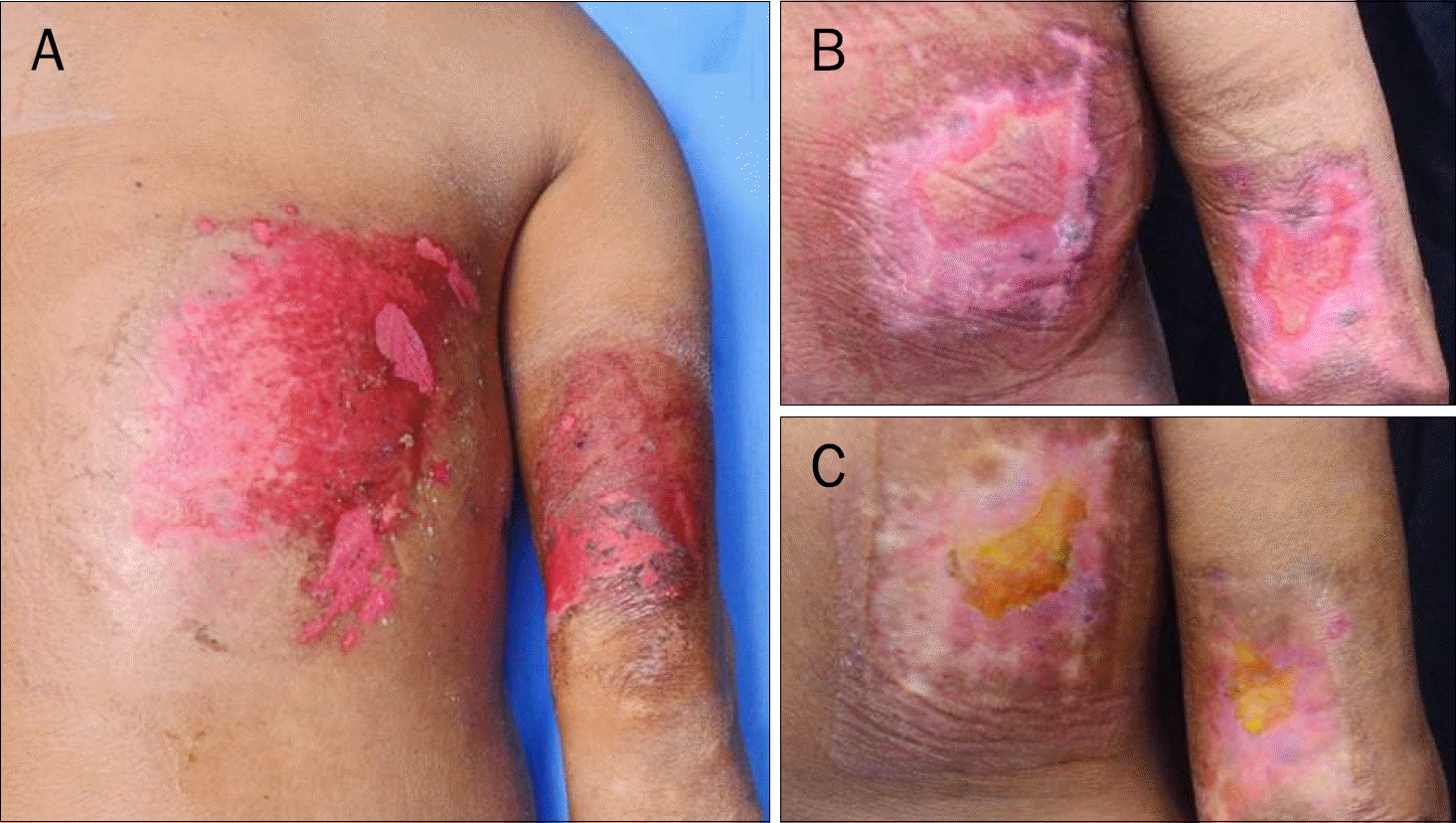
Fig. 1.
(A) One day after radiation therapy, the lesion is described as a relatively sharply demarcated, erythematous, square shaped patch with erosion and moist desquamation. (B) One month later, re-epithelialization is starting from the margin and a pinkish plaque is present. (C) Two month later, the entire lesion is almost healed and re-epithelialized.

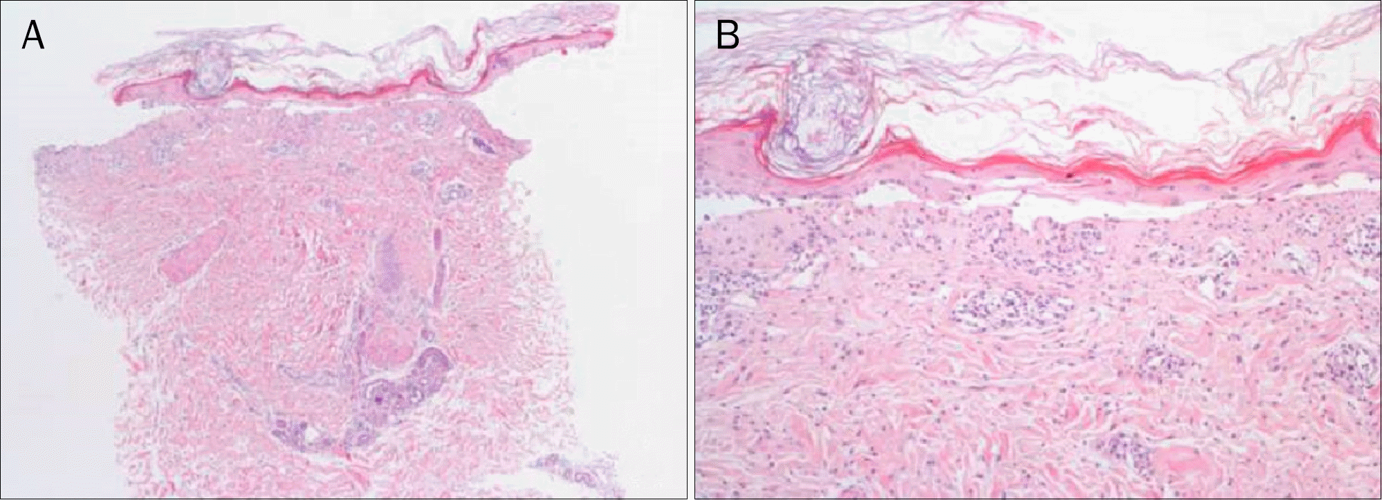
Fig. 2.
Microscopic findings (H&E). (A) A dermal-epidermal separation and a mild inflammatory infiltrate involving the epidermis, papillary dermis, and vessels is seen (×40). (B) High-power field showed a atrophic change of the epidermis with scattered atypical keratinocytes, hyperkeratosis, and spongiosis (×100).


 PDF
PDF ePub
ePub Citation
 Print
Print


 XML Download