

 PDF
PDF ePub
ePub Citation
 Print
Print


INTRODUCTION
Nicotinamide riboside (NR) is a recently discovered vitamin B3, which is mainly present in milk [1]. It mediates several physiological processes acting as an NAD+ precursor [23]. NR is converted to NAD+ via two pathways: NR kinase (NRK) pathway and nicotinamide (NAM) salvage pathway [23]. NR phosphorylation by NRK produces nicotinamide mononucleotide (NMN), which is adenylated to NAD+ by nicotinamide mononucleotide adenylyltransferase [3]. NR may break down to NAM by purine nucleoside phosphorylase [2]. Nicotinamide phosphoribosyltransferase catalyzes NAM to NMN, which is metabolized to NAD+.
Nicotinic acid (NA), which is one version of niacin, is used to treat dyslipidemia, especially by increasing high-density lipoprotein cholesterol as well as by decreasing triglyceride and low-density lipoprotein cholesterol in humans [4]. However, high doses (more than 1 g/day) of NA often induce flushing response, which limits the use of NA as a pharmacological agent [5]. NAM is a more effective NAD+ precursor than other types of niacin in stressed conditions including exposure to high-fat (HF) diet and hyperglycemia [6]. It improves systemic glucose control and hepatic NAD+-sirtuin pathway as well as mitochondrial biogenesis [6]. NAM preserves the mass and function of pancreatic cells [7]. However, its potential side effects include liver toxicity, oncogenicity, and growth inhibition in animals and humans [8]. In contrast, no side effects of NR have yet to be reported. In addition, oral intake of NR elevates hepatic NAD+ more effectively in mouse compared with NA or NAM [9]. However, NR is more expensive than other types of niacin. Both in vitro and in vivo studies have demonstrated the potential of NR to improve hearing loss, obesity, and mitochondrial myopathy. The mechanism of action involves increased NAD+ levels, activation of sirtuins (Sirt) and induction of mitochondrial biogenesis [101112]. NR supplementation prevented the development of diabetic neuropathy in a rodent model of type 2 diabetes [13].
NR elevates or maintains NAD+ levels in liver, which may affect the development of non-alcoholic fatty liver disease (NAFLD) by altering biological pathways including mitochondrial unfolded protein responses and NAD+-dependent Sirt pathway [1014]. Short-term NR treatment decreased hepatic nucleotide binding and oligomerization domain-like receptor family, pyrin domain containing 3 (NLRP3) inflammasome in obese/diabetic KK mice [15]. Increasing NAD+ by NA supplementation ameliorated hepatic steatosis and increased hepatic biogenesis [16].
NR is available from commercial reagent suppliers, and an oral supplement was also introduced under the brand name NIAGEN®. However, the evidence of health benefits, especially for liver, is still limited. Therefore, we hypothesized that NR ameliorates hepatic inflammation and increases levels of mitochondrial markers, and tested the hypothesis in an experimental model of hepatocyte steatosis.
MATERIALS AND METHODS
Cell culture and treatments
Mouse hepatocyte AML12 cells (American Type Culture Collection, Manassas, VA, USA) were cultured in DMEM/F12 medium (Invitrogen, Carlsbad, CA, USA) supplemented with 10% fetal bovine serum, penicillin-streptomycin (100 U/mL), a mixture of insulin, transferrin, and selenium and 0.1 mM dexamethasone at 37℃ in a humidified atmosphere enriched with 5% CO2. Steatosis was induced by treating AML12 cells with 250 µM palmitic acid (PA, Sigma-Aldrich, St. Louis, MO, USA) for 48 h. The cells were treated with phosphate-buffered saline (PBS) or NR (10 µM and 10 mM; BOC Sciences, Shirley, NY, USA) for 24 h. Morphological changes in the cells were captured using a Leica DM IL LED (Leica, Wetzlar, Germany) and TrueChrome Metrics (Fuzhou Tucsen Photonics Co., Fujian, China). Original magnifications of acquired images were 100 x and 200 x.
Cell viability
PrestoBlue® assay was performed according to the manufacturer's instructions. Each well contained the cells cultured medium removed. PrestoBlue® solution (Invitrogen) was added to each well, and the plates were incubated at 37℃ with 5% CO2 for a specified duration. After incubation, the absorbance was measured using an Epoch microplate spectrophotometer (BioTek Instruments, Winooski, VT, USA) at 570 nm.
Oil Red O staining
Cells were gently rinsed twice with PBS, fixed in a with 4% paraformaldehyde-PBS solution for 1 h at room temperature, stained with the 0.5% Oil Red O-isopropyl alcohol for 1 h, and then washed with distilled water. The cells were checked by a Leica DM IL LED (Leica, Wetzlar, Germany) and TrueChrome Metrics (Fuzhou Tucsen Photonics Co., Fujian, China). Original magnifications of acquired images were 40 x.
Sirt1 activity
Sirt1 activities in the cell were measured using a commercial Sirt1 activity assay kit (Abcam, Cambridge, UK), and the assay was conducted according to the manufacturer's instructions. ddH2O, Sirt1 assay buffer, fluoro-substrate peptide, NAD, and developer were mixed in microplate wells. Cell lysate was added to each well, and mixed at room temperature. Fluorescence intensity was read for 30 min to 1 h using SpectraMax M3 multiplate reader (Molecular Devices, Sunnyvale, CA, USA) with excitation/emission at 360/460 nm.
RNA extraction and quantitative reverse transcriptase polymerase chain reaction (qRT-PCR) analysis
Total RNA was isolated using PureLink RNA Mini kit (Invitrogen) according to the manufacturer's instructions. RNA concentration was measured on Nanodrop 2000 (Thermo Fisher Scientific, Wilmington, DE, USA). Reverse transcription was performed using High Capacity cDNA Reverse Transcription kit (Invitrogen). Power SYBR Green PCR Master Mix (Applied Biosystems, Forester city, CA, USA), cDNA and specific primers (Bioneer, Daejeon, Korea) were mixed. Real-time PCR was performed using StepOnePlus (Life Technologies, Carlsbad, CA, USA). Relative expression levels were determined based on the Ct values and normalized to Ct values for the 18S gene.
Western blot analysis
Cells were lysed using a cold lysis buffer containing protease and phosphatase inhibitors (Santa Cruz Biotechnology, Santa Cruz, CA, USA). The lysates were centrifuged, and the supernatants were collected. Western blotting analysis was performed by denaturing 25 mg of protein at 95℃ for 5 min in a Laemmli sample buffer and 2-mercaptoethanol. Proteins were separated by SDS-PAGE and transferred onto PVDF membranes. The membranes were blocked, and incubated with antibodies against Sirt1, Sirt6 (Abcam), NLRP3 (Santa Cruz Biotechnology), and beta-actin (Cell Signaling Technology, Danvers, MA, USA). Membranes were exposed to horseradish peroxidase-conjugated secondary antibody (Santa Cruz Biotechnology). Detection was performed using enhanced chemiluminescence (GE Healthcare, Piscataway, NJ, USA). The bands were scanned with LAS 4000 imager (Fujifilm, Duesseldorf, Germany). Densitometry analysis was performed using ImageJ software (NIH, Bethesda, MD, USA).
RESULTS
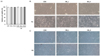
Cell viability, cell morphology, and fat accumulation
Cytotoxicity of NR was assessed by PrestoBlue® assay in AML12 mouse hepatocytes. Addition of NR did not affect cell viability (Fig. 1A). 10 µM and 10 mM of NR treatments did not alter cell morphology (Fig. 1B). The effects of NR on fat accumulation were examined by Oil Red O staining of AML12 hepatocytes. PA treatment tended to increase fat accumulation which was stained with pink or red colors (Fig. 1C). NR treatment tended to reduce fat accumulation, as indicated by decreased Oil Red O staining in AML12 hepatocytes.
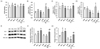
Effects of NR on gene expression and activity of sirtuins
To test whether NR treatment affected sirtuins, the expression levels of Sirt1, Sirt3, Sirt6 and Sirt1 activity were evaluated. Low-dose (10 µM) NR significantly increased Sirt1 activity and Sirt3 expression without altering the gene expression of Sirt1 and Sirt6 in PA-untreated hepatocytes (Fig. 2). Unexpectedly, high-dose (10 mM) NR had no effect on the gene expression and activity of sirtuins in PA-untreated hepatocytes. In a condition of hepatocyte steatosis, the increases in Sirt1 and Sirt6 expression following treatment with low-dose NR were noticeable, which was even higher than under high-dose NR (Fig. 2B and 2D). Sirt1 activity (Fig. 2A) was not affected by doubling the dose of NR treatment in PA-treated cells. Sirt3 expression was upregulated by high-dose NR, and not by low-dose NR in PA-treated hepatocytes (Fig. 2C). High-dose NR significantly increased Sirt1 protein levels in PA-untreated hepatocytes (Fig. 2F), however, the effects of two doses of NR on Sirt1 protein levels were not statistically significant in PA-treated hepatocytes even though the P-values were close to the significance levels (CON vs. NR_H, P = 0.052) in PA-treated hepatocytes (Fig. 2F). Sirt6 protein levels were not significantly altered by two doses of NR (Fig. 2G).
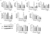
Alteration of inflammation-related markers by NR
To investigate the role of NR treatment in inflammation, the expression TNF-alpha, interleukin (IL)-6, NLRP3 inflammasome components, peroxisome proliferator-activated receptor (PPAR)-alpha, adiponectin, fetuin-A, and nuclear factor kappa B (NF-κB) was analyzed. High-dose NR significantly decreased TNF-alpha expression in PA-untreated and -treated hepatocytes (Fig. 3A). Two doses of NR significantly reduced protein concentration of TNF-alpha in PA-treated hepatocytes (Fig. 3J). Gene expression of IL-6 and caspase 1 as well as NLRP3 protein levels were not affected by NR in PA-untreated hepatocytes (Fig. 3B, 3C, 3H, and 3I). PA treatment induced hepatocyte steatosis, and significantly upregulated caspase 1 expression (Fig. 3C). Low-dose NR treatment significantly downregulated IL-6 and caspase 1 in PA-treated hepatocytes (Fig. 3B and 3C). High-dose NR treatment altered IL-6 expression, but not caspase1 in PA-treated hepatocytes (Fig. 3B and 3C). PPAR-alpha mRNA expression was increased by low-dose NR treatment in PA-untreated hepatocytes (Fig. 3D). PA treatment did not affect PPAR-alpha level. High-dose NR significantly increased PPAR-alpha expression, and not by low-dose NR in PA-treated hepatocytes (Fig. 3D). Adiponectin gene expression in PA-untreated hepatocyte was not affected by NR (Fig. 3E). Induction of hepatocyte steatosis by PA treatment significantly decreased adiponectin level, which was rescued by high-dose NR treatment (Fig. 3E). Gene expression of fetuin-A and NF-κB was not altered by doubling the dose of NR in PA-untreated and -treated hepatocytes (Fig. 3F and 3G). PA treatment significantly upregulated fetuin-A level, and low-dose NR tended to downregulate fetuin-A levels (P = 0.057) (Fig. 3F).
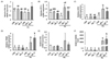
Effects of NR on mitochondria-related markers
To determine the effect of NR on mitochondrial markers, the gene expression of peroxisome proliferator-activated receptor gamma coactivator-1 alpha (PGC-1α), carnitine palmitoyltransferase 1 (CPT-1), uncoupling protein 2 (UCP2), transcription factor A, mitochondrial (TFAM) and nuclear respiratory factor 1 (Nrf1) as well as mitochondrial DNA (mtDNA) in AML12 hepatocytes was analyzed. NR treatment did not affect the expression of these mitochondrial markers in PA-untreated hepatocytes and Nrf1 in PA-untreated and -treated hepatocytes (Fig. 4). UCP2 gene expression was increased by low dose of NR in PA-treated hepatocytes (Fig. 4C). High-dose NR treatment significantly increased PGC-1α in PA-treated hepatocytes (Fig. 4A). Low and high doses of NR treatment significantly increased CPT-1, TFAM, and mtDNA in a condition of hepatocyte steatosis (Fig. 4B, 4D and 4F).
DISCUSSION
The present study investigated whether NR attenuated hepatic inflammation and altered mitochondrial markers in mouse hepatocytes. Administration of NR did not affect cell viability or morphology. No undesirable effects of NR in animals and humans have been reported until now [691115]. Further, findings from the current study supported the hypothesis that NR treatment attenuates hepatic inflammation and induces mitochondrial biogenesis in mouse hepatocytes.
NR prevented the development of HF diet-induced NAFLD in an aged mouse model [17]. In addition, NR administration attenuated hepatic metaflammation by modulating the NLRP3 inflammasome levels in a mouse model of type 2 diabetes [15]. NR delayed the progression of mitochondrial myopathy by boosting mitochondrial biogenesis and preventing mitochondrial DNA deletion in a mouse model of mitochondrial myopathy [12]. These results suggest the possibility of NR as a therapeutic agent for liver diseases by improving inflammation and mitochondrial biogenesis.
Sirtuins are a family of seven proteins that function as NAD+-dependent deacetylases. Among the seven sirtuins, which exhibit distinct functions and locations, Sirt1, 2, 3, 6, and 7 exert anti-inflammatory effects in fatty liver disease, metabolic syndrome and other chronic inflammatory diseases [18192021]. Moreover, Sirt3, mainly located in mitochondria, is involved in mitochondrial function and mitochondrial diseases [222324]. As expected, NR treatment significantly upregulated Sirt1 gene expression. Sirt3 gene expression was significantly upregulated by NR in hepatocytes (Fig. 2B and 2C). These alterations may suggest that these sirtuins play a role in NR-mediated effects on inflammation and mitochondrial function. Despite some interesting findings on NR and sirtuins in this study, our analyses had limitations; part of interpretation from the data was based on the alteration of mRNA expression, and there was discrepancy between mRNA expression and protein levels. The discrepancy may be due to post-transcriptional (e.g. mRNA stability), translational (e.g. initiation factor and trans-acting protein) or post-translational (e.g. proteolysis and phosphorylation) regulations. Transcription of Sirt was regulated by various transcription factors. For example, Forkhead box O1, PPAR-alpha/beta, and cAMP response element-binding are known to increase Sirt1 expression [25262728]; however, PPAR-gamma and carbohydrate response element-binding protein down-regulate Sirt1 expression [2629]. On the other hand, post-transcriptional or translational regulation of Sirt was not well investigated. Also, Sirt activity is the most direct factor regulating deacetylase activity of sirtuins, and is regulated at various levels. Two main types of post-translational modifications of Sirt are phosphorylation and sumoylation [30313233]. Sirt can be phosphorylated by cyclin B-cyclin-dependent kinase 1 complex, JUN Nterminal kinase, dual specificity Tyr-phosphorylated and regulated kinase 1 (DYRK1) and DYRK3 [303132]. Moreover, sumoylation of Sirt1 increases Sirt1 activity [33]. In addition to post-translational modifications, formation of complexes between Sirt and other positive (active regulator of Sirt1) and negative proteins (nuclear receptor co-repressor 1-silencing mediator of retinoid and thyroid hormone receptors complex and deleted in breast cancer 1) as well as NAD+ availability can alter Sirt activity [3435363738]. In our study, low dose NR increased Sirt1 activity in PA-untreated hepatocytes, but, the NR-mediated increases in Sirt1 activity were not shown in PA-treated hepatocytes (Fig. 2A). In addition to these limitations, lack of dose-dependent responses of NR on sirtuins made the clear interpretation difficult.
Inflammation plays a major role in the pathogenesis of NAFLD and obesity-induced insulin resistance [39]. A coordinated network of various pro-inflammatory and anti-inflammatory cytokines controls the inflammatory response. In the current study, NR ameliorated hepatic inflammation by reducing the levels of the pro-inflammatory cytokines, TNF-α and IL-6 (Fig. 3A, 3B, and 3J). The inflammatory response accompanies systemic activation of many signaling pathways. NF-κB induces the expression and activation of inflammatory cytokines [40]. Knockdown of NLRP3 reduced NF-κB activation in human monocyte cell line THP-1 [41]. In the present study, NR upregulated anti-inflammatory molecule adiponectin, and tended to downregulate hepatokine fetuin-A levels (Fig. 3E and 3F). High levels of fetuin-A have been reported in animal models of diet-induced obesity (DIO) and obese diabetic patients, whereas reduction of hepatic fat content is associated with the decline of fetuin-A in humans [4243]. Resveratrol lowered fetuin-A in a mouse model of DIO with direct interaction of adiponectin and fetuin-A [2]. NR appears to inversely regulate adiponectin and fetuin-A in mouse hepatocytes.
Accumulating evidence indicates that mitochondrial dysfunction contributes to the pathogenesis of liver diseases including NAFLD because it affects hepatic lipid homeostasis, and promotes reactive oxygen species production, lipid peroxidation, and cytokine release [44]. Impaired mitochondrial biogenesis occurs in Alzheimer's disease, cardiovascular disease, and aging [454647]. Various transcriptional networks regulate the mitochondrial metabolism [48]. At the molecular level, several transcription factors and cofactors mediate in the activation and regulation of mitochondrial biogenesis [49]. PGC-1α is a master regulator of mitochondrial biogenesis by integrating and coordinating the activity of multiple transcription factors such as Nrf-1, and -2, mitochondrial transcription factor A, and PPAR-alpha [50]. NR increased levels of mitochondrial biogenesis-related factors (PGC-1α, CPT-1, UCP2, and TFAM) as well as mtDNA (Fig. 4). The gene expression of PGC-1α is directly linked to mitochondrial biogenesis [51]. PGC-1α modulates Nrf1 and other mitochondrial biogenesis-related factors [52]. Fatty liver was associated with impaired activity of PGC-1α in mice [53]. Activation of PGC-1α leads to increased expression of genes related to gluconeogenesis, fatty acid oxidation, and lipid transport [5455]. Our findings from the current study demonstrated that NR induced PGC-1α, CPT-1, UCP2, and TFAM as well as mtDNA in AML12 mouse hepatocytes.
This study suggests the potential role of NR in inflammation regulation and mitochondrial biogenesis in a model of hepatocyte steatosis. As inflammation and mitochondrial dysfunction are involved in the pathophysiology in NAFLD, our study has potential clinical and public health implications if the findings are confirmed in human clinical trials. In conclusion, our findings demonstrate that NR treatment attenuates hepatic inflammation, and induces mitochondrial biogenesis in mouse hepatocytes. These findings suggest the therapeutic value of NR in hepatic inflammation and impaired mitochondrial biogenesis.
 XML Download