

 PDF
PDF ePub
ePub Citation
 Print
Print


Introduction
Type 2 diabetes is a hyperglycemic metabolic disease, a condition caused either by insufficient insulin secretion or insulin resistance (Berry et al., 2007; Sriplang et al., 2007). The number of diabetic patients is rapidly rising in most parts of the world. People with type 2 diabetes are often sedentary, obese, middle-aged adults with an increased risk of macrovascular disease, retinopathy, nephropathy, neuropathy, and hypertension (Berry et al., 2007; Goldberg, 1981). These health complications lead to increased morbidity and premature death (Hammoud et al., 2000; Reven, 1987). Specific goals of medical nutrition therapy for diabetic patients include achieving and maintaining near-normal blood glucose levels, achieving optimal serum lipid levels, consuming adequate calories to achieve a reasonable body weight, and improving overall health by maintaining a balanced intake of macro- and micro-nutrients (American Diabetes Association, 2006; Hammoud et al., 2000). In general, maintaining blood glucose concentrations near normal ranges in these patients is accomplished with oral hypoglycemic/antihyperglycemic agents and insulin (American Diabetes Association, 2006; Kilo, 1987; Saudek & Eder, 1979). However, these treatments have limited efficacy and are associated with undesirable side effects that have led to an increasing interest in the use of medicinal plants as an alternative management for type 2 diabetes (Betteridge, 1989). In fact, the American Diabetes Association (ADA) recommends a daily consumption of 20-35 g of total fiber from sources that include both soluble and insoluble fiber (Ness & Powles, 1997; Steinmetz & Potter, 1996). Reports indicate that consuming diets rich in soluble and insoluble fiber induces satiety, improves glycemic control, and reduces total energy intake, adiposity, and blood lipids (Qureshia et al., 2001; Qureshia et al., 2002).
Seaweeds are frequently consumed in Asia and occasionally consumed in the rest of the world. Edible seaweeds are rich in non-starch polysaccharides (dietary fiber), proteins, minerals, and vitamins (Jurkovic et al., 1995; Urbano & Goni, 2002). They have low lipid content and provide few calories. However, seaweed could interfere with the bioavailability of other dietary components (Lahaye, 1991; Wong et al., 1999). As seaweed polysaccharides cannot be entirely digested by human intestinal enzymes, they are considered to be a source of dietary fiber. Seaweed dietary fiber differs in composition, chemical structure, physico-chemical properties, and biological effects from the fiber of land plants. Therefore, seaweed consumption could increase the variety of dietary fiber (Lahaye, 1991; Lahaye & Kaeffer, 1997; Michel & MacFarlane, 1996).
There is great interest in the role of increased oxidative stress in the complications suffered by those with diabetes. Increased oxidative stress may result from an increase in free radical production. Seaweed is exposed to intense light and high oxygen concentrations, leading to the formation of free radicals and other strong oxidizing agents in it's environment (Lahaye & Kaeffer, 1997; Lahaye, 1991; Michel & MacFarlane, 1996; Wong et al., 1999). The absence of oxidative damage in their structural components (polyunsaturated fatty acids) and the stability during storage suggest that their cells possess protective antioxidative systems (Lahaye, 1991; Wong et al., 1999). As of yet, only a few reports on the antioxidant activity of seaweeds have been published. Since seaweeds are rich in polysaccharides, minerals, proteins, and vitamins, a documented antioxidant activity would elevate their value in the human diet and as food and pharmaceutical supplements (Albu et al., 2004; Berge et al., 2002; Burritt et al., 2002; Heo et al., 2005; Yuan & Walsh, 2006).
Subjects and Methods
Subject selection
Nine men and eleven women with type 2 diabetes were selected according to the following specific criteria: diabetes controlled by diet and or oral hypoglycemic agents, body mass index (kg/m2) < 35, fasting plasma glucose concentrations > 150 mg/dl (150-300 mg/dl), no consumption of lipid-lowering drugs, and being 40 to 70 years of age. Aside from diabetes, all subjects were in good general health and had no clinical or laboratory evidence of renal, hepatic, or cardiovascular disease.
Experimental design
This study was approved by the Ethics Committee and the Institutional Review Board for Human Subjects Research at Hanyang University Hospital, Seoul, Korea. Subjects were randomized into either a control group or a seaweed supplementation group. Pills with equal parts of dry powdered sea tangle and sea mustard were provided to the seaweed supplementation group three times a day for 4 weeks. Total daily consumption of seaweed was 48 g. The subjects continued normal daily activities and exercise patterns.
Dietary assessment
Food intake of each subject was quantitatively evaluated by 24 hour recall. The dietitian collected data using graduated models to estimate the size of food portions. Daily nutrient intake was calculated using the Computer Aided Nutritional Analysis program (Can-Pro) software (version 3.0; The Korean Nutrition Society) based on data from Korean food-composition tables.
Biochemical analysis
Blood samples were taken at study entry and at 4 weeks. We measured blood glucose, total cholesterol (T-C), high-density lipoprotein cholesterol (HDL-C), low-density lipoprotein cholesterol (LDL-C), and triglycerides (TG), hemoglobin A1c (HbA1c) after fasting, and 2-hour postprandial blood glucose levels (PP-2hr BG). The following determinations were made on the same day, according to standard protocol at the biochemical laboratory of Hanyang University Hospital, a certified clinical laboratory using HITACHI 7600-110 Auto Biochemistry Analyzer (Hitachi Electronics, Japan). Glucose was measured using the glucose oxidase (GOD)-peroxidase (POD) enzymatic colorimetric test. T-C was determined using the enzymatic cholesterol oxidase (COD)-peroxidase (POD) colorimetric test. TG was based on enzymatic method using lipoprotein lipase, glycerol kinase, glycerol phosphate oxidase and peroxidase (Fossati & Prencipe et al, 1982). HDL-C was determined enzymatically with dextran sulfate-magnesium method (Warnick et al., 1982). LDL-C was measured enzymatically (Friedewald et al., 1972) and HbA1C was analyzed using high performance liquid chromatography (HPLC) method (Jeppsson et al., 1986).
For erythrocyte lysates, blood was collected in tubes containing EDTA and then centrifuged at 1500 g for 10 minutes at 4℃. The supernatant containing the plasma and buffy coat was discarded. The red blood cell pellet was washed three times with cold saline and frozen immediately at -70℃ until analysis. Hemoglobin was separated from the blood cells by precipitation with an ethanol/chloroform mixture, followed by continuous shaking for 5 min and centrifugation at 2500 g for 20 min. The supernatants were used to determine enzyme activity.
The total amount of lipid peroxidation products was assayed with the thiobarbituric acid method, which quantifies thiobarbituric acid reactive substances (TBARS) at 532 nm. Superoxide dismutase (SOD, EC 1.15.1.1) activity was measured using pyrogallol (Marklund, 1984). Catalase (EC 1.11.1.6) activity was calculated by spectrophotometrically measuring the disappearance of H2O2 at 240 nm (Aebi, 1984, Claiborne, 1984). Glutathione peroxidase (GSH-Px, EC 1.4.1.9) activity was assayed according to the method of Flohe and Gunzler (1984)
The hemoglobin concentration of lysates was determined spectrophotometrically at 546 nm using the cyanmethaemoglobin method of Mahoney et al. (1993). All assays were carried out in triplicate using a spectrophotometer (Beckman-Coulter DU 400, Fullerton, CA, USA).
Statistical analysis
For statistical analyses, the SPSS/PC computer program (Statistical Package for Social Science 12.0) was used. Data was expressed as means ± S.E. The significance in differences between the two groups was assessed by independent t-tests. The paired t-test was used to compare means for experimental periods in each group.
Results
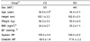
Table 1 shows the characteristics of the two groups at the beginning of the study. There were no significant differences between subjects in the two groups with respect to age, height, weight, BMI or blood pressure. The 10 subjects in the control group included 5 men and 5 women, aged 54.8 ± 2.5 yr, with BMI of 24.2 ± 0.7 kg/m2, systolic BP of 125.0 ± 3.3 mmHg, and diastolic BP of 80.0 ± 1.9 mmHg. The 10 subjects in the seaweed supplementation group included 4 men and 6 women, aged 54.4 ± 3.1 yr, with BMI of 23.2 ± 1.1 kg/m2, systolic BP of 120.0 ± 2.2 mmHg, and diastolic BP of 77.8 ± 2.2 mmHg.
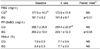
Nutrient intake of the two groups during the 4 weeks is presented in Table 2. Intake of the two groups was identical in regards to the proportion of carbohydrates, fat, and protein. 65% of the calories were derived from carbohydrate, 16% were from protein, and 19% were from fat. Throughout the 4 weeks, food intake did not differ appreciably between the two groups, except for fiber intake. As expected, the mean total dietary fiber intake in patients receiving seaweed supplementation was 30.1g/day, which was 2.5 times higher than the 12.3g/day in the control group (p<0.001).
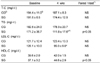
The mean change in fasting blood glucose (FBG) levels, PP-2hr BG, and HbA1C are shown in Table 3. There were no significant differences in FBG, PP-2hr BG, and HbA1C at baseline between the two groups. After seaweed supplementation, the concentrations of FBG and PP-2hr BG were decreased significantly in patients receiving seaweed supplementation, but there were no significant differences in controls. PP-2hr BG concentration in controls was 254.4 ± 22.8 mg/㎗ and significantly higher as compared to 203.1 ± 12.3 mg/㎗ in the seaweed supplementation group (p<0.05). In other words, increased dietary fiber intake resulting from supplementation had beneficial effects on FBG and PP-2hr BG. Little change was observed in the level of HbA1C in both the supplemented and control groups after 4 weeks and these changes were not statistically significant.
The mean change in blood lipid concentrations is shown in Table 4. There were no significant differences in T-C, TG, LDL-C and HDL-C concentration at baseline between the two groups. After seaweed supplementation, the concentration of TG was decreased and HDL-C was increased significantly (p<0.05). There were no significant differences in the control group for the same time period. Both groups had lower T-C level compared with baseline, but these differences were not significant. Seaweed supplemented group showed significantly lower TG and LDL-C levels compared with control group (p<0.05) after 4 weeks.
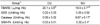
Our results on oxidants and antioxidants are presented in Table 5. Seaweed supplementation significantly lowered the level of TBARS in erythrocytes as compared to controls (p<0.05). Seaweed supplementation also increased catalase and GSH-Px activities (p<0.05). However, SOD activity was not affected by seaweed supplementation.
Discussion
We evaluated the effects of seaweed intake on blood glucose levels, lipid profile, and antioxidant enzyme activities in patients with type 2 diabetes.
Seaweed was provided in the form of pills that were made with powdered sea tangle and sea mustard. Sea tangle and sea mustard are frequently consumed in Asia and occasionally in the rest of the world. Seaweeds are classified into three groups: brown algae, red algae, and green algae, and each of these groups differ with regard to their reserve and cell-wall polysaccharides (Jimenez-Escrig & Sanchez-Muniz, 2000). Sea tangle and sea mustard are the most commonly used seaweeds in Korea (Heo et al., 2005), and both belong to the family of brown algae. Dietary fiber from brown algae is essentially derived of four families of polysaccharides: laminarans, alginates, fucans, and cellulose (Jimenez-Escrig & Sanchez-Muniz, 2000; Renn, 1990). Sea tangle and sea mustard, which are rich in indigestible polysaccharides, appear to be good sources of soluble dietary fiber. The total dietary fiber content of seaweeds ranges between 25-75% (on a dry weight basis), and 51-85% of which is water-soluble fiber (Renn, 1990).
In this study, the total dietary fiber intake of subjects given seaweed was 30.1g/day, which was 2.5 times higher than the 12.3 g/day of controls. Consistent with the increase in dietary fiber, the concentrations of FBG and PP-2hr BG were decreased significantly relative to baseline, which may result from a delay in glucose absorption by fiber in seaweed. The decreased FBG levels after seaweed supplementation agreed with the results of Simpson et al. (1979, 1981). In their studies, however, it was not clear whether the effects were due to the high-fiber diet, restricting the intake of fat, or both (Simpson et al., 1979). In a second study, the difference in daily fiber intake was extremely high and was achieved mainly by a leguminous diet (Simpson et al., 1981). In this study, the individual energy intake was kept constant and the ratio of protein, fat, and carbohydrates was maintained to isolate the effect of fiber.
Diabetic and insulin resistant individuals tend to have an atherogenic blood lipid profile with increased serum triglyceride, low HDL-cholesterol, and small, dense LDL-particles (Saudek & Eder, 1979; Kilo, 1987). Seaweed supplementation resulted in lower levels of TG and higher levels of HDL-C. Small changes were observed in the levels of T-C and LDL-C, however, these changes were not statistically significant. The results agree with earlier studies that reported decreased lipid levels with diets containing gel-forming fiber (Jurkovic et al., 1995; Qureshia et al., 2001, 2002; Unbano & Goni, 2002; Wursch & Pi-Sunyer, 1997)
The mechanism by which fiber lowers blood cholesterol remains undefined. Evidence suggests that some soluble fibers bind bile acids or cholesterol during the intraluminal formation of micelles (Schroder, 2007; Shafrir, 1996). The resulting reduction in the cholesterol content of liver cells leads to an up-regulation of the LDL receptors and thus to an increased clearance of LDL cholesterol. Other suggested mechanisms include inhibition of hepatic fatty acids synthesis by products of fermentation (production of short-chain fatty acids such as acetate, butyrate, propionate); changes in intestinal motility; fibers with high viscosity causing decreased absorption of macronutrients, leading to increased insulin sensitivity; and increased satiety, leading to lower overall energy intake (Schroder, 2007).
Seaweeds are exposed to a combination of light and high oxygen concentrations, which lead to the formation of free radicals and other strong oxidizing agents. However, they seldom suffer from serious photodynamic damage during metabolism. This fact implies that their cells have protective antioxidative mechanisms and compounds (Yuan & Walsh, 2006). Seaweeds are rich source of antioxidants. Recently, potential antioxidants have been identified as pigments (e.g. fucoxanthin, astaxanthin, carotenoid) and polyphenols (e.g. phenolic acid, flavonoid, tannins). These compounds are widely distributed in seaweeds and known to exhibit high antioxidative activities (Yuan et al., 2005a). These antioxidants act by scavenging reactive oxygen species and inhibiting lipid peroxidation (Yuan et al., 2005b).
TBARS are the most commonly used markers for oxidation. In this study, TBARS levels in erythrocytes of those given seaweed supplementation were significantly lower than in controls. Catalase and GSH-Px activities in the seaweed supplementation group were higher than in controls. However, SOD activity was not affected by seaweed supplementation. These results suggest that seaweed supplementation decreases oxidative stress in vivo due to a combination of improved antioxidant activities and decreased peroxidation processes.
We conclude that addition of seaweed influences glycemic control and may be effective in lowering blood lipids and improving antioxidant enzyme activities. Accordingly, such effects may reduce risk factors for cardiovascular disease in patients with type 2 diabetes. However, to confirm these effects and to make dietary recommendations for patients with type 2 diabetes, further studies are necessary.

 XML Download