

 PDF
PDF Citation
 Print
Print


Abstract
Tumor incidence in wild mammals is reportedly very low. Wild nutria, a large rodent, is known to carry many infectious diseases, but rarely exhibits neoplastic diseases. We necropsied a male wild nutria and found a large nodular mass in the left inguinal region, adjacent to the penis. Histopathologically, the mass was diagnosed as preputial gland adenoma. Spontaneous preputial gland adenomas are extremely rare in all animals. Moreover, reports of tumors in nutrias have been limited to adenocarcinomas of the lungs and uterus, as well as subcutaneous fibromas. Here, we describe preputial gland adenoma in a wild nutria.
References
1. Marchlewska-Koj A, Pochroń E, Sliwowska A. Salivary glands and preputial glands of males as source of estrus-stimulating pheromone in female mice. J Chem Ecol. 1990; 16:2817–2822.

2. Rudmann D, Cardiff R, Chouinard L, Goodman D, Küttler K, Marxfeld H, Molinolo A, Treumann S, Yoshizawa K. INHAND Mammary, Zymbal's, Preputial, and Clitoral Gland Organ Working Group. Proliferative and nonproliferative lesions of the rat and mouse mammary, Zymbal's, preputial, and clitoral glands. Toxicol Pathol. 2012; 40:7S–39S.
3. Coleman GL, Barthold W, Osbaldiston GW, Foster SJ, Jonas AM. Pathological changes during aging in barrier-reared Fischer 344 male rats. J Gerontol. 1977; 32:258–278.
4. Reznik G, Ward JM. Morphology of hyperplastic and neoplastic lesions in the clitoral and preputial gland of the F344 rat. Vet Pathol. 1981; 18:228–238.
5. Mitsumori K, Elwell MR. Proliferative lesions in the male reproductive system of F344 rats and B6C3F1 mice: incidence and classification. Environ Health Perspect. 1988; 77:11–21.
6. Hong IH, Kang SY, Kim JH, Seok SH, Lee SK, Hong SJ, Lee SY, Park SJ, Kong JY, Yeon SC. Histopathological findings in wild Nutrias (Myocastor coypus) with Capillaria hepatica infection. J Vet Med Sci. 2017; 78:1887–1891.
7. Kim HS, Kong JY, Kim JH, Yeon SC, Hong IH. A case of fascioliasis in a wild nutria, Myocastor coypus, in Republic of Korea. Korean J Parasitol. 2018; 56:375–378.
8. Gang DG, Sim CH, Lee TJ, Kong JY, Hong IH. Sebaceous cell differentiation in a canine oral papilloma. J Vet Diagn Invest. 2018; 30:569–571.
9. Turusov VS. Morphology and histogenesis of anal region and clitoral gland tumors induced in mice by 1,2-dimethylhydrazine. J Natl Cancer Inst. 1980; 64:1161–1167.
10. Ratcliffe HL. Incidence and nature of tumors in captive wild mammals and birds. Cancer Res. 1933; 17:116–135.
Fig. 1.
Necropsy of a male wild nutria. (A) The ventral aspect of the animal. The ventral skin has been peeled back, revealing a large muscle-covered mass (asterisk) lateral to the penis (arrow). (B) Cut surface of the mass. Yellowish exudates are observed.
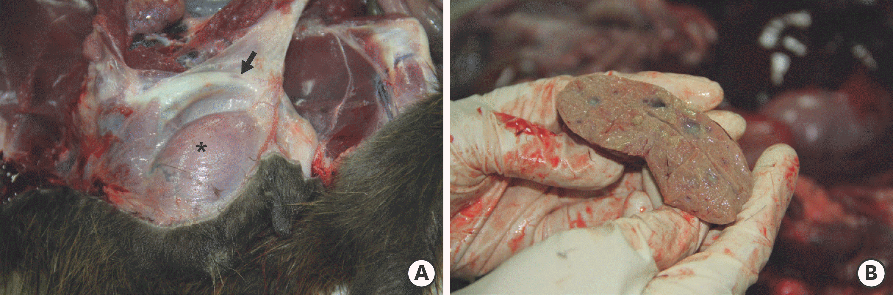
Fig. 2.
Microscopic examination of the mass in a male wild nutria. (A) The mass is composed of reserved basal cells and secretory cells containing oily and waxy matter, which constitute a type of sebaceous gland. The gland has a long excretory duct with a wide lumen lined by hyperplastic squamous epithelium. (B, C) The tumor cells have a central round nucleus with one or (rarely) 2 large nucleoli. The cytoplasm contains small to large vacuoles. (D) Adipophilin is expressed in intracytoplasmic lipid vacuoles in sebaceous cells. Hematoxylin and eosin (A-C); immunohistochemistry of adipophilin (D) (all scale bar = 100 µm).

 XML Download