

 PDF
PDF ePub
ePub Citation
 Print
Print


Introduction
Ovarian cysts (OCs) of clinical relevance are commonly found in domestic animals [16] in which they cause gynecopathies [21828] and hyperestrogenism [15]. OCs can originate from different ovarian structures and their development, frequency, and size varies across species [16]. Histology is used to distinguish different types of OCs [1015]. The most common types are follicular cysts (FCs), cysts of subsurface epithelial structure (SES), cystic rete ovarii (CRO), lutein cysts (LCs), and cystic corpora lutea (CCL) [10151629]. While OCs and related gynecopathies are well characterized in sows [28] and cattle [2], the association of OC morphology based on histology and immunohistochemistry in relation to the clinical manifestation of the patient has rarely been investigated in dogs [46]. In particular, it should be noted, that histologically evaluated LCs in dogs have not been fully documented in recent reports [4].
The present study investigated advanced histology features of canine OCs in relationship to corresponding clinical manifestations that were observed when the bitch was presented in our clinic. It was hypothesized that multiple cyst types occur simultaneously on a single ovary in bitches having clinically manifest gynecopathies, and that it is possible to determine LCs histologically in bitches with gynecopathies.
Materials and Methods
Ethical statement
The study is in accordance with German legal and ethical requirements for appropriate animal procedures. Good veterinary practice was applied to all procedures whenever animals were handled. The aims of this study were explained to all pet owners. All experimental procedures were approved by the Ethics Committee of Regierungspraesidium Gießen Germany (approval No. V 54-19c20 15h02Gl18/14kTV13/2017).
Bitches
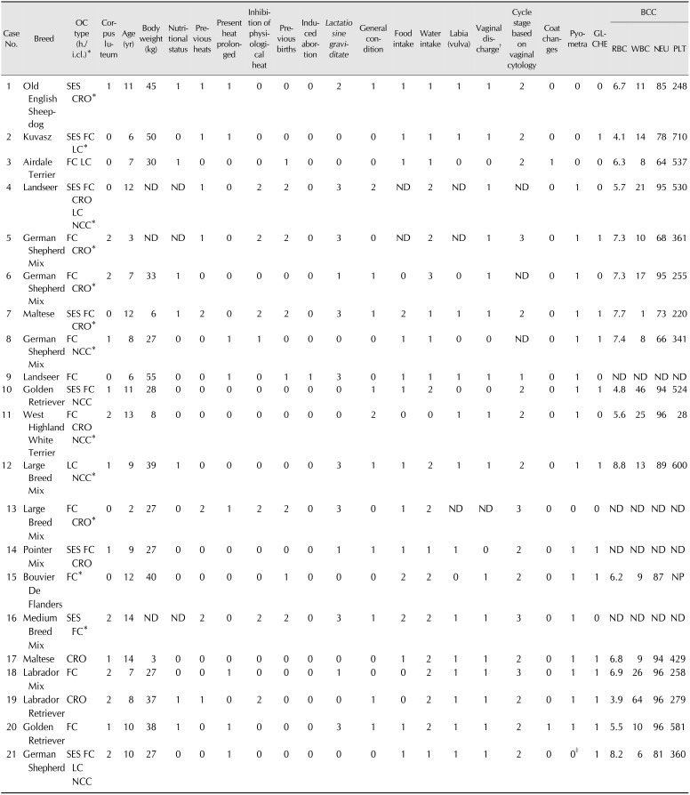
A total of 21 bitches of different ages and breeds were presented to the Clinic for Obstetrics, Gynecology and Andrology of Large and Small Animals, Justus-Liebig-University in Giessen, Germany due to different gynecological symptoms. In accordance with current reports, we defined an OC as a fluid-filled cavity [25] of any size present outside the physiological estrus cycle [15] and located within or on the ovary's surface. Diagnosis of an OC was based on medical and reproductive histories, as well as on a thorough clinical and gynecological examination. A standardized questionnaire and clinical and gynecological examination protocols were applied to all cases. Exfoliative vaginal cytology assessment followed a protocol published elsewhere [34]. Ultrasound (Sono Ace-9900 ultrasound machine with a 7.5-MHz convex transducer; Sonoace, Germany) was used to visualize cysts on the ovaries and to check the integrity of adjacent organs by following a routine procedure. Blood counts were performed using the Hematology System Abott Cell-Dyn 3500 (Abbott Laboratories, USA). In order to be included in the study, female dogs needed to have a medical history of prolonged estrus (> 28 days) and show pathological vaginal discharge, coat changes, pyometra, and/or disturbed general condition. Moreover, detection of cystic structures on the ovaries via ultrasound was an additional inclusion criterion. All bitches underwent ovariohysterectomy (OHE) following a protocol described elsewhere [13].
Histology
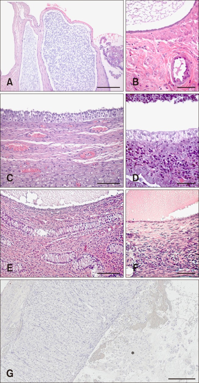
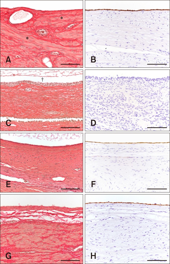
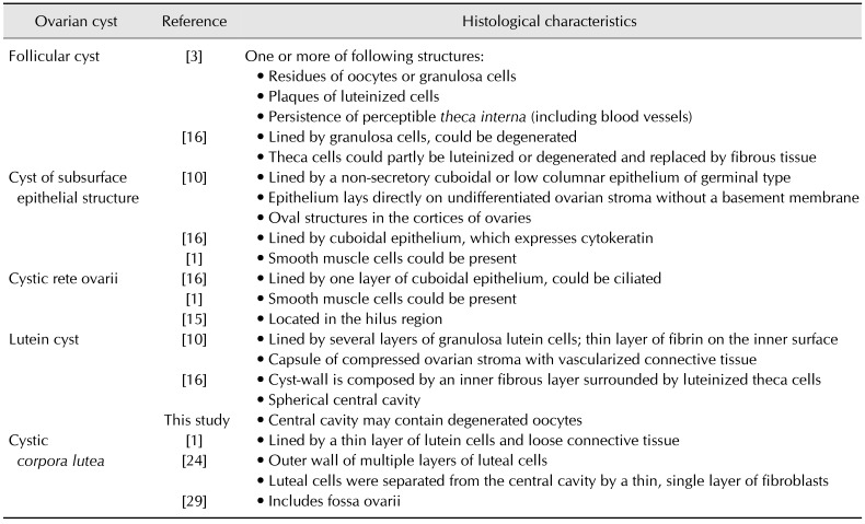
Surgically removed ovaries were fixed in toto in 10% neutral buffered formalin for a minimum of 72 h at 4℃. Specimens were trimmed and paraffin embedded using an automated embedding station (Microm Laborgeräte, Germany). Thin tissue layers (4 µm thick) were routinely stained with H&E in an automated process (Varistain Gemini; Thermo Fisher Scientific, Germany). Cystic structures of 42 ovaries were examined to determine the type of lining cells (granulosa, theca, or epithelium cells, as well as ciliated cells) and the form of these cells (cuboidal, palisade shaped, or flattened). The existence of plaques composed of luteinized cells and a luminal fibrinous layer was noted. Finally, ambient cells and tissue were described (smooth muscle cells, connective tissue, blood vessels). Histological evaluation criteria of canine OCs are summarized in Table 1. In addition, samples of consecutive sections were stained using the Elastica van Gieson staining kit (Merck, Germany) according to the manufacturer's protocol. A tissue section of a healthy bitch's uterus served as a positive control for smooth muscles, while a tissue section of dog's skeletal muscles served as a negative control. Each slide was examined under light microscopy (Leica Microsystems, Germany) by two independent investigators. Evaluation was carried out using the Leica Image Manager (Leica Microsystems).
Immunohistochemistry
In all cases in which classification was not possible on the basis of H&E and Elastica van Gieson staining, additional immunohistochemistry was performed to depict cytokeratin. Contiguous sections were prepared and placed on SuperFrost slides (Gerhard Menzel, Germany). Specimens were treated with mouse anti-cytokeratin antibody (keratin, pan Ab-3, clone Lu-5, horse anti-mouse IgG, ABC complex) according to the manufacturer's protocol. Epidermis tissue of a dog was used as a positive control. Examination of stained slide specimens followed the procedure described above. Cell classification derived from histological characteristics was performed as follows: cells of FC react negative to cytokeratin, whereas cells of cysts of SES were positive (25–75% of the cells react positively to cytokeratin) to strong positive (> 75% of the cells react positively to cytokeratin), while cells of CRO were weakly positive (< 25% of the cells react positively to cytokeratin) to positive (25–75% of the cells react positively to cytokeratin).
Statistical analysis
Data were evaluated using PRISM 7 for Macintosh (GraphPad Software, USA). Data were tested for the presence of a normal distribution by utilizing Shapiro-Wilk and D'Agostino & Pearson omnibus normality tests. In cases of Gaussian distribution, data are presented as mean ± SD. The interference of blood cell-concentration with the reference range of healthy dogs was analyzed by using the Wilcoxon signed rank test.
Results
Patient data and medical history
The age of the presented 20 female dogs ranged between 3 and 14 years (9.5 ± 3 years). The patient population was divided into four groups based on dog breeds and half-breeds with OCs (n = 21): (1) small, n = 3 (Maltese, West Highland White Terrier); (2) medium, n = 3 (Airedale-Terrier, Pointer-Mix); (3) large, n = 12 (German Shepherd, German Shepherd-Mix, Golden Retriever, Labrador-Retriever, Labrador-Mix, Old English Sheepdog, Bouvier de Flanders), and (4) huge, n = 3 (Kuvasz, Landseer). The study population included 38.9% (n = 7/18) of the bitches with an obese nutritional status, whereas 61.1% (n = 11/18) were normal weighted. Individual information on body weight (in kg) and nutritional status (normal/obese) at the date of presentation is provided in Table 2. In 16 of 21 cases, pet owners provided information on previous heats in their dog. Based on this, 4 of 16 bitches (25.0%) showed prolonged or shortened inter-estrus intervals in past heats. In 75.0% (12 cases) physiological heats were regular recurrent. One bitch (n = 1/15) received regular proligestone (Delvosteron) treatment to suppress the physiological estrus cycle. Furthermore, 3 of 15 dogs (20.0%) had given birth to one or more litters. Most of the female dogs with OCs were nulliparous (n = 12/15, 80.0%), including one animal with induced abortion. Four of 13 patients developed regular (3) or irregular (1) lactatio sine graviditate. Data are summarized in Table 2.
Clinical examination and additional macroscopic findings during OHE
On the date of presentation in the clinic, general condition was good in 10/21 dogs (47.6%), moderate in 42.9% (n = 9/21), and poor in 2/21 patients. Food intake (inappetent/good/moderate) and water intake (no/normal/increased/multiplied) per individual is summarized in Table 2. Edema of the vulva was present in 72.2% (n = 13/18) of the bitches. Pathological vaginal discharge was detected in 16 of 20 patients (80.0%). Based on the exfoliative vaginal cytology, bitches were assigned to the following cycle stages: proestrus (n = 1/18), estrus (n = 13/18, 72.2%), metestrus (n = 4/18), anestrus (n = 0/18). Two animals displayed fur changes (n = 2/21). Pyometra was diagnosed in 16 of 21 cases (76.2%). Glandular cystic hyperplasia of the endometrium (GLCHE) was macroscopically observed in 13 of 21 bitches (61.9%). Corpora lutea were recorded macroscopically in 66.7% (n = 14/21) of the dogs. Bitches with OC and pyometra had a significantly higher leucocyte concentration than the reference range of 13 healthy dogs (6–12 G/L, Wilcoxon signed rank test, p = 0.02), whereas concentrations of thrombocytes were not significantly different between both groups. Erythropenia was shown in three patients (< 5.5 T/L). In 6 cases, thrombocytopenia was present (< 280 G/L), and it was especially pronounced in one bitch (28 G/L). A combination of erythropenia and thrombocytopenia was observed in one case. The blood cell counts (red blood cells, white blood cells, neutrophils, platelets) per individual are provided in Table 2.
Histology and Immunohistochemistry
There were a total of 193 cystic structures observed and they were identified as cysts of SES (n = 72; panels A and B in Figs. 1 and 2, respectively), FCs (n = 61; panels C and D in Figs. 1 and 2, respectively), CRO (n = 38; panels E and F in Figs. 1 and 2, respectively), LCs (n = 13; panel G in Fig. 1), and non-classifiable cysts (n = 9; panels G and H in Fig. 2). The allocation of the characterized OC type per individual case is included in Table 2. The observations resulted in the differentiation of two groups: Group 1, only endocrine inactive OCs including case No. 17 and 19, and Group 2, endocrine active and inactive OCs including all other case numbers.
Discussion
Frequently, more than one type of cyst is present on a single ovary [126]. The multiple cellular differentiation processes that are involved in follicular growth and maturation may explain the differences in the prevalence of the different types of cysts. This is consistent with our findings in bitches with clinical signs of gynecopathies. We divided our cases into two groups: Group 1, only endocrine inactive OCs and Group 2, endocrine active and inactive OCs. In Group 1, we confirmed only two cases. In that group, only CROs were present (case No. 17 and 19) and were associated with clinical signs of gynecopathies. However, CROs do usually not produce hormones [26], although they may replace the surrounding physiological ovarian tissue [23], which could result in a functional loss. However, in the preliminary stages of our study, hyperestrogenism could be present due to delayed ovulation. Theoretically, the endocrine active OCs could have disappeared due to atresia or spontaneous ovulation, as has been described in cattle [17]. Other clinical observations in these two CRO cases, like edema of the vulva, the estrus cycle stage based on vaginal cytology, pyometra, and GLCHE would also relate to the prolonged presence of estrogen or a hormonal disturbance. Overall, CRO was the third most common type of OC detected in this study, although cells of the rete ovarii were present in the follicular stroma in a higher number than epithelial cells and granulosa cells. A possible explanation for this is, that rete ovarii tissue is physiologically uninvolved into sex-hormone-regulated differentiation and maturation processes [32]. Group 2 included the other 19 of 21 cases and was dominated by FCs (present in n = 17 bitches, 81.0%); FCs were the second most common cystic structures in this study. The transition of a mature physiologic follicle into a pathological FC is fluent and in general is based on an imbalance of the underlying endocrine regulatory mechanisms during estrus, on the local and superior levels [7]. In terms of clinical importance, hormone-producing FCs are the most often described cystic structure in relation to clinically persistent estrus or anestrus [525] as well as to infertility [1231]. In four cases of Group 2, FCs were present together with LCs. Clinical symptoms of FCs and LCs overlap and the bitches in this study had an estrogen-characterized clinical picture. LCs occur more frequently in sows and cows than in bitches [1629]. Hormone-producing canine LCs were described histologically by Dow [10] in 10% of 90 animals with cystic structures of ovaries that were submitted to routine post-mortem examination. In that study, clinical data of the bitches and histological pictures of LCs were not included, and evidence of oocyte residues in LCs was not a criterion in the histological evaluation. However, there are a few histological descriptions, but no histological figures, of canine LCs [1629]. In our study, we were able to detect more than twice the percentage of LCs reported by Dow [10] in dogs (n = 5/21, 23.8%) with medical histories of gynecopathies. The underlying pathological mechanism of LC, an insufficient LH-secretion of the adenohypophysis [23], is similar to the one described for FCs. A possible explanation for the difference in prevalences of LCs and FCs may be related to the sequence of cellular processes involved in follicular growth and maturation. The development of an LC ultimately requires luteinization of granulosa cells and therefore involves an additional cell differentiation step. Residues of oocytes can be found in FCs [3] and, as we have shown in our study, in LCs. We, therefore, propose to include this characteristic in the histological evaluation criteria for canine OCs as a contribution to clearly defining LCs in bitches (Table 1; “central cavity may contain degenerated oocytes” of LC). Moreover, this characteristic could help to distinguish histologically LC from CCL. The latter is characterized by a fossa ovarii [29] as a sign of ovulation. However, the possible misinterpretation of luteinized anovulatory follicles as LCs should be considered. Since only bitches which fulfill the inclusion criteria for OCs were included in this study, we are confident about our results. With regard to clinical presentation, we note that in cases of conservative therapy measures of cycle aberrations, such as the application of human chorion gonadotropin (hCG, e.g., Ovogest) or gonadotropin-releasing hormone analog (GnRHa, e.g., Receptal) [19], the simultaneous occurrence of LCs and FCs has to be considered, which could explain failures of conservative treatment.
In addition to OCs, corpora lutea (CL) occurred in 66.7% (n = 14/21) of our cases. This is consistent with previous findings [6]. Conservative treatment of pyometra with an antigestagen (Aglepristone) is often not effective [35] since hormonally active CL and OCs can have an enduring effect of the endometrium [35]. Regardless, hormonally active LCs may also contribute to conservative treatment failure.
In contrast to previous findings, in which FCs were the most cystic ovarian structures in bitches [1020], the number of cysts of SES dominated all other types of OCs in this study. The development of cysts of SES may be induced by accidental scattering of ovarian serosa that was introduced to the ovarian tissue during ovulation [37]. This process can potentially occur during any physiological heat of the bitch. Gulliver and Hurst [14] reported that estradiol exposure can result in ovarian surface epithelium hypertrophy and hyperplasia in mouse ovaries. On that basis, Chuffa et al. [9] analyzed the expression of sex steroid receptors in polycystic ovaries of dogs and noted moderate expression levels in unspecified OCs. It is likely that canine cysts of SES also express estrogen receptors, which could explain their frequent occurrence in bitches with high estrogen levels (case No. 1, 2, 4, 7, 10, 14, 16, 21). Overall, it should be critically noted, that only OCs ≤ 1 cm in diameter were sampled for histological examination, and OCs > 1 cm in diameter (which were present in 12 of the 21 bitches in this study; Table 2), were sampled for the presence of corresponding cyst fluid. Results of the estradiol-17ß and progesterone concentrations for these bitches have been published elsewhere [18]. Case No. 1 had to be excluded from Group 1 and transferred into Group 2 due to additional 15 OCs with a diameter > 1 cm that were not histologically evaluated, but contained increased estradiol-17ß and progesterone concentrations in cyst fluid published elsewhere [18]. Moreover, for case No. 21 we considered that at least one more hormonally active OC was present in addition to the histologically evaluated LC. Nevertheless, the clinical symptoms did not differ, as shown in Table 2.
Based on previous reports, the age of female dogs with OCs varies between 1 and 18 years [10112231]. This was confirmed by our results in which bitches had an average lifetime of 9.5 ± 3 years. However, we were unable to confirm that the prevalence of OCs is in direct proportion to dog age, as reported by Marino et al. [22]. Since life expectancy of different dog breeds varies, there is a tendency of increasing OC prevalence in dogs over the age of six. A relationship between obesity and OC incidence has been reported in human [21], mouse [27], and rat [36], but investigations into that association in dogs are lacking. Our study population included 38.9% (n = 7/18) overweight bitches, whereas 61.1% (n = 11/18) were normal weighted. A nutritional assessment was carried out during the dogs' first clinical examination and followed generally accepted protocols. However, based on our results we can only speculate that, with an increase in adipose tissue, the risk of developing OCs also increases in dogs; further investigations are needed. Most female dogs with OCs were nulliparous (n = 12/15, 80.0%), which supports findings reported elsewhere [610].
In Germany and other prosperous societies, most dogs are well integrated into households and are not allowed to breed. Under the assumption that most female dogs in Germany are nulliparous, it is not surprising that a similar situation was reflected in our OC study population. Studies that include free-ranging stray bitches may derive different results. Referring to the medical and reproductive histories of our bitches, there was no correlation between previous treatment of bitches with hormones to suppress their physiological estrus cycle or induce abortion and the prevalence of OCs. But, under the assumption that treatment with hormones can promote OC formation [5811], we recommend further investigations, including studies with standardized control and patient groups. Prolonged or shortened inter-estrus intervals of the past heats seem to be relatively low-risk factors for the development of OCs. Pet owners reported irregular past heats in 25% of the study animals, which did not reveal any gynecopathies. This could be due to the spontaneous healing of endocrine active OCs, as has been described in cattle [17].
The leading symptom in the bitches with OCs in our study was prolonged vaginal discharge (16 of 20 examined cases, 80.0%), which was consistent with a previous report on bitches with OC syndrome [6]. The most common OC-accompanying diagnosis was pyometra in 16 of 21 cases (76.2%). In this study population, the endometrium of 11 bitches (68.8%) also showed GLCHE. Another dog had a GLCHE accompanied by a mucometra. Overall, 13 bitches with OCs had GLCHE, which is indicative of longer-term exposure of the endometrium to estrogen and progesterone. Due to the association of OCs with pyometra in the majority of animals in this study (n = 16/21, 76.2%), we qualified the statement of an estrogen-induced leukocytosis with neutrophilia. Blood parameters of bitches with OCs should be separately evaluated from those in bitches with OCs and pyometra due to in utero inflammatory processes obscuring the changes. Our results indicated leukocytosis without accompanying pyometra in one of four dogs with OC. OCs may trigger hyperestrogenism through the non-regulated release of steroid sex hormones. However, dogs are estrogen sensitive [29]. An estrogen-induced myelotoxic effect on the hematopoietic system can lead to anemia and a loss of condition [253033]. In our study, we found one bitch with a thrombocyte level clearly below the physiological range (28 G/L), and that level in five further animals was in the lower part of the normal range (280–200 G/L). Only in one case (No. 19), was this linked with an erythropenia (3.9 T/L) and a loss of condition. Despite the low number of animals in this study, we suggest, that hemograms are not useful as a proxy for the presence of hyperestrogenism. It should, however, be emphasized that a thorough clinical examination must include a hemogram in order to exclude life-threatening conditions in bitches with gynecopathies and to determine the most appropriate treatment approach (conservative or surgical).
Supporting our hypothesis that multiple cyst types occur simultaneously on a single ovary in bitches having clinically manifest gynecopathies, we observed more than one type of OC on single ovaries isolated from bitches with clinical signs of gynecopathies. Despite the relatively small study population (n = 21) we identified four of five OC types with the occurrence of LCs being higher than that reported previously. Because we surprisingly could not detect a clinical difference between dogs with endocrine active and endocrine inactive OCs at the time of histological classification, our results indicate that every OC-affected bitch should be considered as an individual case. Therefore, further investigations are needed to identify the primary factors that trigger ovarian dysfunction and to better understand the complex pathophysiology of OC development.
 XML Download