

 PDF
PDF ePub
ePub Citation
 Print
Print


Introduction
Thoracic radiography in adult horses is particularly challenging because of the size of the equine chest. In addition, the muscles of the forelimb overlay the cranial thorax and increase the tissue depth in this area. For thoracic radiography, the thorax can be divided into three overlapping areas: craniodorsal, caudodorsal, cranioventral, and caudoventral [56]. For routine diagnostics of chronic respiratory disease, the caudodorsal and caudoventral view are sufficient in most cases, as the caudodorsal projection provides the largest unobstructed view of the lungs and the caudoventral projection allows evaluation of a small triangle of lung bounded by the caudal vena cava, caudal border of the heart, and cranioventral aspect of the diaphragm [18]. Nevertheless, if the diagnostic workup necessitates the exclusion of differentials, such as thoracic masses, the projections cranial to the heart may be beneficial to the clinician.
Indications for thoracic radiography include clinical signs like dyspnea, tachypnea, dysphagia, cough, nasal discharge, or exercise intolerance. Radiography is the diagnostic method of choice for evaluating the pulmonary parenchyma, especially in diseases affecting the deep lung and the mediastinum [12]. While a low correlation between the severity of exercise-induced pulmonary hemorrhage and its radiographic appearance has been reported, significant differences were found in horses with different stages of equine asthma [2223]. In contrast to this, radiographs have been shown to be of no predictive value in mild-moderate asthma (formerly known as inflammatory airway disease, IAD) [17]. Regardless, radiographic observations must always be interpreted in the light of clinical findings and clinical pathology results.
Lower airway disease is common in adult horses. Dixon et al. [4] reported a greater than 50% prevalence of equine asthma in horses referred for respiratory problems. Nevertheless, other differentials need to be excluded from the diagnostic workup. In addition to a clinical examination, endoscopy, blood gas analysis, pulmonary function testing, bronchoalveolar lavage (BAL) fluid cytology, and radiographic examination of the thorax are often performed. In most cases, radiography serves to exclude differentials other than equine asthma and may also help to stage the disease as an increased interstitial pattern, as well as bronchial radio-opacity and thickening, have been reported to be significantly correlated to disease severity and progression [23]. BAL fluid cytology is considered a reference test for diagnosing equine IAD [220]. Collection of BAL fluid can be performed either blindly or by using an endoscope [1314]. To our knowledge, there are no previous reports on whether BAL, which is performed routinely in the workup of chronic respiratory disease, influences the interpretation of thoracic radiographs and, therefore, whether it should be performed after a radiographic examination.
We hypothesized that BAL may cause an interstitial pattern on thoracic radiographs, which may be misinterpreted as inflammatory or congestive edema formation or fibrosis. As a consequence, a standardized BAL procedure would increase the interstitial opacity in various chronic pneumopathies and healthy controls, which should allow observers to identify, whether the radiograph was taken before or after the BAL procedure.
Materials and Methods
Horses
Fifty-three horses, which had either no history of respiratory disease or were presented to the clinic for examination of the lower airways, were examined for this study. The horses were of mixed breed, age, and gender and had been dewormed on a regular basis. No corticosteroids, anti-histaminics, or bronchodilators were given to any of the horses for a minimum of two weeks before being examined. Horses without a history or clinical finding of respiratory disease were included as controls, and horses with a BAL in their diagnostic workup and that had been diagnosed with severe equine asthma (formerly known as recurrent airway obstruction, RAO) as defined by Robinson [20] or with mild-moderate equine asthma as defined by Couëtil et al. [2] were included as chronic pneumopathies.
The evaluation of horses affected by respiratory disease was not classified as an animal experiment by the State Office of Health and Social Affairs Berlin, and performance of similar evaluations of the control horses was approved (No. L0294/13). The owners gave permission to involve their horses in the study.
Pre-participation examination
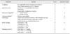
All horses were examined by two clinicians experienced in the diagnostics of equine respiratory disease. The horses were assigned to three different groups according to their history of chronic respiratory disease and their respiratory tract clinical examination, arterial blood gas analysis, tracheal secretion endoscopic scoring, and BAL fluid cytological results. These results were incorporated into a validated and internationally accepted scoring system [81920] shown in Table 1. Controls (Group 1) had no history or clinical signs of airway disease, Group 2 horses had severe equine asthma and Group 3 had mild-moderate equine asthma.
Bronchoalveolar lavage fluid cytology
Local anesthesia was performed by the application of 20 mL lidocaine (lidocain 2%; Bela-Pharm, Germany) prior to the procedure. Phosphate buffered saline (500 mL; Lonza, Belgium) at 37℃ were infused through a 300 cm balloon catheter (Silicone Bronchoalveolar Lavage Catheter; Smiths Medical, USA) as described elsewhere [20]. The entire amount of re-aspirated fluid was documented and macroscopically evaluated for color, turbidity, and amounts of foam and mucus. For cytological examination, the samples were centrifuged at 250 × g for 10 min; then, a direct smear was taken and stained using the May-Grünwald Giemsa staining method. Five hundred cells of each smear underwent magnification (500× and 1,000×) examination to evaluate the relative percentages of macrophages, lymphocytes, neutrophils, eosinophils, and mast cells.
Thoracic radiography
For radiographs of the thorax, a Gierth HF 400 ML generator (Gierth X-Ray International, Germany) and needle imaging plates (35 cm × 43 cm; CR MD4.0 General Cassette; Agfa Healthcare, Belgium) were used with a focus-film distance of 1.5 m. Caudoventral and caudodorsal images were obtained from all horses before and within 30 min after BAL and were developed with a developing device (Curvix 60; Agfa, Germany). Exposure factors were in the range of 102 to 117 kV and 25 to 32 mA for caudoventral and caudodorsal fields as described by Mair and Gibbs [15]. For individual horses, exposures were the same before and after BAL. Additionally, the images were adjusted to achieve the best comparable opacity of the ventral borders of the thoracic vertebrae between different horses before grading the pulmonary structures.
The radiographs were taken, under sedation administered for respiratory endoscopy, at the end of inspiration.
Interpretation of radiographs
Radiographs were scored by two independent clinicians; observer A was a boarded specialist in equine internal medicine and B a board-qualified specialist in large animal diagnostic imaging. The observers were blinded to the four randomized radiographs of each horse, so that horse history, ancillary test results, diagnosis, and timing of the radiographic examination (before or after BAL) were not available. The radiographs were classified as described by Tilley et al. [23]. Single parameters included interstitial opacity, bronchial opacity, tracheal thickening, bronchial thickening, and expansion of the ventral curvature of the lung field, as well, air bronchograms and peribronchial infiltrations were scored from 0 (unremarkable) to 3 (severe findings). Observers had the option to exclude a radiograph from scoring of a single parameter, if, in their opinion, the image was insufficient for evaluation.
Statistical analysis
After describing all variables by using standard statistical procedures, several analyses were performed. Numerical correlation between scores assigned by the two observers was assessed by using Spearman rank correlation coefficients. Score agreement was assessed by using Kendall's tau-B (agreement measure corrected for ties in score values), and systematic shift in scores was assessed by calculating the difference in scores between the two observers (A – B) and applying a non-parametric one-sample test on the differences (Wilcoxon signed-rank test for difference in medians). The proportion of images considered of poor quality was compared between observers by using cross-classification and Chi-squared/Fisher's exact tests. After describing all parameters using standard measures of central tendency and variability, a multivariable analysis of variance (ANOVA) model was used to compare the median scores given by the two observers between disease, time, and projection categories. A multivariable mixed logistic regression model was used to assess the influence of a predefined set of factors on the chance of misclassification of the projection timing (dependent variable, correct time identified = 0, wrong time identified = 1). The individual horse was included in the model as a random effect to account for the hierarchical level of the data (multiple measurements within each horse). Independent factors assessed in the model were: (a) disease category (3 classes), (b) radiograph projection (caudoventral; caudodorsal), (c) amount of recovered BALF (> 200 mL; ≤ 200 mL), and (d) difference in before and after BAL interstitial pattern scores (0, < 0, > 0). All factors were first assessed individually and, then, analyzed together in the multivariable mixed model to obtain adjusted odds ratio (OR) values with 95% confidence intervals. Data were stored in MS Excel 2016 (Microsoft, USA) and analyzed in IBM SPSS (ver. 22; IBM, USA) and STATA (ver. 12; Stata, USA). The alpha level of significance (p) was set to 0.05.
Results
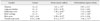
Based on the clinical examination, blood gas analysis, respiratory endoscopy, BAL cytology, and thoracic radiography results, 45 of initially 53 horses were included in the study as either healthy control (n = 12), severe equine asthma (n = 12), or mild-moderate equine asthma (n = 21) horses. Six horses were excluded, as they were diagnosed with different disease. The radiographs of two horses (No. 9 and 26) were excluded from the study due to poor image quality, which made it impossible to use the scoring system for these radiographs. The results of the clinical examinations are summarized in Table 2.
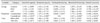
A high correlation was detected between observers A and B, as Spearman correlation and Kendall's tau (corrected for ties) were above 0.7 for all parameters, although a significant systemic deviation was evident. Data for the inter-observer analysis are presented in detail in Table 3.
The BAL fluid return volume ranged between 215 and 258 mL with no significant difference between diagnosis groups. Significant differences between caudoventral and caudodorsal projections were found in interstitial and bronchial opacity, as well as in bronchial thickening, while significant differences between groups were only detected in the expansion of the ventral curvature. Radiographic scores and the differences between categories of the three variables (a) disease group, (b) projection, and (c) time are shown in Table 4.
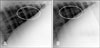
In addition, we assessed the question of which parameters were associated with the observers' ability to classify the correct radiographic timing (taken before or after BAL) by using a multivariable logistic regression model. Our underlying hypothesis was that the BAL procedure would increase interstitial opacity. The ability of the observers to identify the time of the radiograph correctly was significantly influenced by diagnosis and projection. In radiographs of horses diagnosed with severe equine asthma, the chance (adjusted OR) of misinterpretation of the correct BAL timing was about 5 times higher than in healthy controls (OR = 5.373, p = 0.028). No significant differences were detected for mild-moderate equine asthma horses (p = 0.0157). The chance of misinterpretation of the correct time of performing BAL was about 4 times lower in caudodorsal projections than in caudoventral projections (OR = 0.241, p = 0.004). The amount of re-aspirated fluid did not have a significant influence (p = 0.205). The ability of observers to correctly identify whether the radiograph was taken before or after BAL, was highly correlated with an increase in interstitial opacity, which increased the chance of correct identification by a factor of 10 (OR = 9.976, p < 0.0001). Fig. 1 shows two caudodorsal projections taken before and after BAL in a healthy control horse.
Discussion
Our hypothesis that BAL increases interstitial opacity in radiographs of the equine thorax was confirmed by the results of our study, as this parameter was shown to increase the chance of correctly determining whether the radiograph was taken before or after BAL, by a factor of almost 10; however, this effect was only true for the caudodorsal projection. The influence of BAL on opacity was shown by the positive effect of projection timing on the ability of observers to correctly identify the timing of the radiograph, which was more than 4 times higher in caudodorsal than caudoventral projections. It must be assumed that blind placement of the balloon catheter also takes place in the caudodorsal lung field comparable to the endoscope under visual control, as several studies have shown no difference in cytologic results obtained by either technique [20]; moreover, it seems mechanically improbable that a balloon catheter would leave the main bronchus to be redirected into a ventral bronchus displayed on a caudoventral projection.
In the present study, the chance of misinterpretation of the correct radiograph timing was about 5 times higher in severe equine asthma horses than in the healthy controls, which may be explained by the positive correlation coefficient for the radiographic interstitial pattern in the course of asthma, as described by Tilley et al. [23]. If the initial interstitial opacity is higher in severe equine asthma horses than in healthy horses, the influence of BAL may be harder to see. In contrast, a lower grade of inflammation, as in mild-moderate equine asthma horses, did not affect the ability of observers to identify the timing of the radiograph correctly.
A problem in diagnosing interstitial radiographic patterns is that as the quality of the image improves as the amount of perceived interstitial lung pattern decreases [24]. This is particularly true when using digital systems in which contrast and brightness can be adjusted individually. Therefore, we adjusted the obtained images to achieve comparable opacity of the ventral borders of the thoracic vertebrae among the observed horses and, then, graded the pulmonary structures. Nevertheless, using a bony structure as a reference to evaluate soft tissue is not ideal; an intrathoracic structure would be preferable but, unfortunately, is not possible in lateral radiographs. Moreover, in this study, computed tomography and magnetic resonance images were not available.
In a previous study evaluating the correlation of interstitial radiographic patterns and exercise-induced pulmonary hemorrhage in racehorses, interstitial and bronchointerstitial radiographic results were influenced by various factors, and a negative correlation between interstitial opacity and perceived diagnostic quality of the images was reported [24]. Interstitial fluid accumulation produces a typical radiographic appearance, as is often seen in patients suffering from acute respiratory distress syndrome in concert with lung edema. Even in horses free of respiratory disease, re-aspiration of the full amount of fluid given during the BAL procedure is unlikely. The fluid return volume in horses is negatively correlated with BAL, neutrophil percentage, and severity of respiratory disease and it is associated with bronchial collapse during re-aspiration of fluid, a phenomenon reported in human medicine [10]. Asthmatic patients with moderate to severe airway obstruction usually have poor BAL fluid return [716]; therefore, a higher amount of BAL fluid is likely to remain in the airways of more severely affected horses, which has been reported in severe equine asthma by several authors [39]. This fluid retention may affect radiographic interpretation in such patients, but whether increased interstitial opacity is caused by the primary disease or the remaining fluid in the lung has not been elucidated.
Thorax radiography is routinely performed in equine medicine to diagnose different forms of chronic pneumopathy or to rule out substantial pathologies like abscesses or fluid accumulation in the thoracic cavity [21]. Increased diffuse interstitial opacity and bronchial pattern have been described in severe equine asthma [1]; the latter being better described in smaller bronchi than in trachea and main bronchi [11]. Several radiographic parameters can be shown to correlate significantly with disease severity [23]. In contrast, radiographs have been shown to have no predictive value in mild-moderate equine asthma [17]. Overall, no significant differences were found in all parameters examined in our study, neither in severe nor mild-moderate equine asthma horses. Among the severe equine asthma horses, this could be explained by the relatively low mean clinical scores of the horses presented for participation in this study. Our results might have been different if more horses suffering from long-lasting severe equine asthma had been available.
With regard to the inter-observer agreement results, our analysis showed that the evaluation of thoracic radiographs was highly reliable, as shown by the high Spearman and Kendall's tau correlations, in observers A and B, both of whom were experienced in the interpretation of diagnostic imaging of the equine chest.
The clinical relevance of this study lies in our recommendation to undertake BAL after radiographic examination of the thorax during a diagnostic workup of chronic respiratory disease, as the BAL procedure can affect radiographic interpretation, at least in early respiratory disease; that is, before the occurrence of interstitial fibrosis. As the interpretation of results and the classification of disease severity are usually the last steps in clinical routines and take place after all standard procedures, including BAL and thoracic radiography, have been completed, the severity of disease is unclear when radiographs are obtained. Thus, the possible influence on a diagnosis of undertaking BAL prior to radiography can be avoided.

 XML Download