

 PDF
PDF ePub
ePub Citation
 Print
Print


Introduction
Hepatic diseases are of great economic importance in bovine medicine [1127]; however, they receive little attention because they may be unnoticed due to their nonspecific clinical signs [11]. Abattoir-based survey studies are commonly conducted for detection of liver afflictions [8] due to difficulties reaching an accurate diagnosis in clinic-based situations. Additionally, laboratory-based diagnosis of hepatobiliary diseases, such as that from hepatospecific enzyme tests, is insufficient and time consuming [28].
The negative effects and economic losses of hepatobiliary diseases on animal's productivity have been documented; moreover, affected livers are condemned at slaughter, and adhesions to the surrounding organs or diaphragm may necessitate carcass trimming [27].
Recent advances in diagnostic tools and the introduction of ultrasonic imaging have facilitated screening of different diseases of internal organs, including hepatic diseases in buffalo under field conditions [35]. A complete ultrasonographic examination of the liver can provide detailed information about the nature of an affliction, the parenchymal pattern of the liver, the size and position of the gallbladder (GB), and the intra- and extrahepatic bile ducts in cattle [11] and buffalo [18].
Both focal and diffuse liver afflictions including abscesses, neoplasia, cyst, and cirrhosis, as well as hepatobiliary diseases such as cholangitis and cholelithiasis, have been recorded separately in various animal species [1]. These afflictions can develop as primary diseases or as sequelae to other diseases such as traumatic reticuloperitonitis (TRP) [567]. They are commonly reported in feedlot and dairy cattle being fed rations that predispose to rumenitis [26].
In recent years, ultrasonography has become an increasingly important tool for diagnosis of various abdominal disorders in bovine practice [9]. Several studies concerned with ultrasonography of digestive system disorders have been carried out in buffalo, particularly studies into TRP [241922] and intestinal disorders [2021], and cattle [2429]. However, there are few studies describing ultrasonographic results associated with hepatobiliary diseases in buffalo [1]. Therefore, this research was undertaken to document ultrasonographic observations of naturally developed hepatobiliary diseases in buffalo and relate those results to results from clinical and biochemical assessments.
Materials and Methods
Animals
The diseased group included 30 Egyptian buffalo (Bubalus bubalis; aged 7 months to 8 years) of both sexes were admitted to the Veterinary Teaching Hospital (VTH) at Assiut University (Egypt) with histories of weight loss or poor growth rate, as well as reduction of milk yield in female animals. Some cases had a history of long-standing diarrhea. A control group (n = 20) was selected from a group of healthy, non-pregnant female buffalo in a herd at the VTH.
Ethical guidelines
All animal procedures performed in this study were conducted in accordance with Institutional Animal Care and Use Committee guidelines of Assiut University which basically conform to the Guide for the Care and Use of Laboratory Animals of the National Institutes of Health in the USA (NIH publication No. 86-23, revised 1996).
Clinical examination
All animals underwent a thorough clinical examination as described by Cockcroft [16].
Radiographic examination
Left lateral recumbent plain radiographs of the cranial, abdominal, and caudal thoracic regions were obtained from both control and hepatobiliary diseased buffalo. The examination was carried out according to the method reported by Braun et al. [12] by using a ceiling-suspended X-ray apparatus (45–65 Kv and 45–55 mA/s). The following features were recorded: foreign body nature and location (reticular, diaphragmatic, or pericardial position), diaphragm status, and heart visualization (good versus bad line of demarcation).
Ultrasonographic examination
Hepatic ultrasonography was performed according to the methods reported by Braun [11] and Khalphallah et al. [18] by using 3.5 MHz sector transducer connected with the ultrasound device (FF Sonic UF-400; Fukuda, Japan). In preparation for ultrasonography, the right thorax and abdomen areas were clipped and shaved, and a coupling gel was applied. Ultrasonographic examination of the liver was performed on the right side of the abdomen in the region of the 7th to 12th intercostal spaces (ICSs). The circumferences of the liver and GB were determined. The nature of hepatic lesions, focal or diffuse, and observed disorders in the biliary system were recorded. The left thorax area and the area behind the xiphoid process were also prepared and screened for TRP according to the method reported by Braun and Götz [13].
Laboratory examination
Whole blood and serum samples were collected, and all precautions for sample collection and preparation to ensure accurate evaluation of hematological and biochemical indices, as described by Coles [17], were taken into consideration.
A fully automated blood cell counter machine (CA620 Vet Hematology Analyzed; Medonic, Sweden) was used to determine various hematological parameters. The differential leukocytic count was determined by using the four-field meander method.
Serum concentrations of aspartate aminotransferase (AST), γ-glutamyltransferase (GGT), alkaline phosphatase (ALK), total protein, albumin, cholesterol, and triglyceride (TG) were assessed by using the Optizen32220 UV/Visible spectrophotometer (Mecasys, Korea). Serum globulin was determined by subtracting albumin level from total protein level.
All kits and reagents were obtained from Spectrum Reagents (Egyptian Company for Biotechnology, Egypt).
Statistical analysis
Data were analyzed by using the SPSS statistical software packaged program for Windows (ver. 10.0.1; SPSS, USA). Data are presented as mean ± SD values. Analysis of the variance of the obtained data was performed by using Student's t-test and significance level of results was set at p ≤ 0.05.
Results
Clinical observations
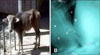
Most of the hepatobiliary diseased buffalo (21/30) had a poor body condition score (BSC < 2) (panel A in Fig. 1) and a history of inappetence. Some cases (9/30) experienced long-standing diarrhea. Body temperature and respiratory rate were normal in most diseased buffalo (28/30); however, ruminal movement was greatly reduced. The visible mucous membranes, including the conjunctiva, were pale and rarely (2/30) icteric. The episcleral blood capillaries were empty in most diseased buffalo. Six buffalo with hepatobiliary afflictions associated with TRP and/or traumatic pericarditis (TP) showed anorexia, tucked up appearance, tensed abdomen, and recurrent tympany.
Radiographic observations
In both the control buffalo and most of the hepatobiliary diseased buffaloes (24/30), radiography showed an absence of any reticular metal objects. The diaphragm was imaged clearly between two radio-opaque structures; reticulum and heart. The heart appeared as radio-opaque with clear margins, normal size, and a characteristic shape (cone shape with base of the heart directed dorsally and apex directed ventrally).
Radiography of buffalo with hepatobiliary diseases associated with TRP and/or TP (n = 6) showed different radio-opaque metallic foreign bodies (needles, hair-pins, and wires) within the reticulum in buffalo with normal heart (n = 4) or affected heart (n = 2; panel B in Fig. 1).
Ultrasonographic results
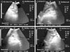
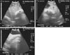
Five types of hepatobiliary afflictions were successfully imaged in the hepatobiliary diseased buffalo (Table 1). Based on ultrasonographic results, these afflictions included focal parenchymal lesions including liver abscess and cyst in 18 of the 30 diseased buffalo. Hepatic abscess, recorded in 12 (40.0%) of the diseased buffalo, imaged as a well-demarcated focal mass with an echogenic capsule and hypoechoic or anechoic contents producing distal acoustic shadowing (Fig. 2). Sometimes (2/12), hepatic abscess was associated with visualization of an echogenic clear tract within the hepatic tissue extending from the abscess and with accumulation of thick echogenic deposits around the GB, abscess, cystic duct, and common hepatic duct (i.e., pericholecystic hyperechogenic material).
Buffalo with hepatic abscess associated with TRP and/or TP (n = 6) showed a thick wall reticulum with uneven contour, reduced reticular contractions with displacement of reticulum away from diaphragm, peritoneal effusions, reticular abscess, and spleen involvement. Reticular abscess was imaged as a well-distinguished echogenic capsule with hypoechoic content. The involved spleen was completely covered with a fibrinous echogenic deposit and was surrounded with hypoechoic fluid associated with echogenic fibrinous deposits leading to adhesions between spleen and the abdominal wall, as well as between spleen and reticulum and/or the craniodorsal blind sac of the rumen. In cases with TP (n = 2), cardiomegaly with loss of the characteristic cardiac shape, thickened cardiac wall, and accumulation of pericardial hypoechoic fluids interspersed with echogenic deposits was also imaged.
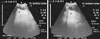
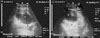
Liver cyst was recorded in 6 buffalo representing 20% of the total diseased buffalo. Liver cyst appeared as a pear-shaped anechoic sac with a well-defined thin bright echogenic wall and a peripheral refractive zone situated ventromedially to the liver causing distal acoustic enhancement (Fig. 3).
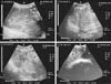
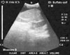
Diffuse parenchymal lesion (hepatobiliary fibrosis or liver cirrhosis) was diagnosed in 5 buffalo representing 16.7% of the 30 diseased buffalo. The liver showed echogenic linear bands with less distinct imaging of the portal vasculature. Multiple echogenic nodules and anechoic foci on the liver were imaged, giving the liver a heterogeneous nature. Fibrinous echogenic deposits were visualized around the caudal vena cava, resulting in reduction of its lumen (Fig. 4). Liver size was reduced, and liver could not be imaged from the right 12th ICS. Ultrasonography also was used to diagnose complete hepatobiliary cirrhosis with a dilated loop of the small intestine. The dilated loop of the cranial part of the duodenum (6.2–6.5 cm) was intertangled medially to the GB and liver (Fig. 5).
Obstruction of hepatobiliary passages was diagnosed in 7 buffalo representing 23.3% of the total diseased buffalo. The obstructions included cholestasis (n = 4) and hepatocholelithiasis (n = 3).
Cholestasis was recorded in 4 buffalo representing 13.3% of the total diseased buffalo. Dilatation of GB with thickening in its wall, dilatation of the bile duct and cystic duct, alterations in GB content (homogeneous or heterogeneous), and hepatomegaly were the most common ultrasonographic observations in buffalo with cholestasis (Fig. 6).
Hepatocholelithiasis was reported in 3 buffalo representing 10% of the total diseased buffalo. It appeared as an echoic structure inside the hepatic parenchyma or inside and around the GB and bile duct. In addition, there was greater echogenicity of the hepatic parenchyma than that of normal parenchyma (Fig. 7). Distal acoustic shadowing was also observed.
Laboratory results
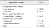
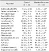
Results of blood and serum biochemical analyses of buffalo with confirmed hepatobiliary diseases are summarized in Table 2. Compared to the control buffalo, the diseased animals had a significantly decreased (p < 0.05) RBC count, lower packed cell volume, and lower hemoglobin concentration with neutrophilic leucocytosis. Blood results in buffalo with hepatobiliary diseases associated with TRP and/or TP showed lymphocytic leucocytosis. Serum biochemical analysis of buffalo with either hepatobiliary disease only or hepatobiliary disease associated with TRP and/or TP showed a significant (p < 0.05) decrease in serum total protein (hypoproteinemia) and albumin (hypoalbuminemia) from levels in control buffalo. Significant (p < 0.05) elevations of blood serum levels of GGT, ALK, and AST were present in hepatobiliary diseased buffalo; however, serum levels of TG and cholesterol were not significantly different between the groups.
Discussion
The clinical signs associated with hepatobiliary diseases in cattle and buffalo are too vague to allow specific diagnosis, and hence, there are no reports of successful treatment. Cattle may carry hundreds of small abscesses or several large abscesses with no clinical signs, and the abscesses are detected only at the time of slaughter [1141523]. Therefore, the present study was designed to describe the most common ultrasonographic observations in hepatobiliary diseased buffalo. The present results show that liver ultrasonography is an efficient tool for diagnosis of five hepatobiliary diseases: hepatic abscess and cyst, liver cirrhosis, cholestasis, and hepatocholelithiasis.
In this study, buffalo with hepatobiliary diseases showed no specific clinical signs, such as poor BSC, long-standing diarrhea, pale and rarely icteric mucous membranes, or empty episcleral blood capillaries. These results are in agreement with results of previous studies in cattle [123].
Blood composition and chemistry of the hepatobiliary diseased buffalo showed the presence of neutrophilic leucocytosis, hypoproteinemia, and significant elevation of the serum enzymes GGT, ALK, and AST. Although these indices may suggest liver dysfunction, they are nonspecific for diagnosis of hepatobiliary diseases. Similar observations have been reported previously [1].
Ultrasonographic examination of the liver is recommended when buffalo have poor production and/or exhibit weight loss in the absence of specific clinical signs because such examination is rapid, noninvasive, and accurate for the diagnosis of several abdominal disorders [25]. Moreover, radiographic examination of the caudal thoracic and cranial abdominal regions is also recommended whenever TRP and/or TP are suspected.
Liver abscess images showed well-demarcated focal masses within the normal hepatic parenchymal tissue and revealed echogenic capsule with contents of various echogenicity according to the consistency of the pus. Similar observations have been reported in cattle [15]. In this study, ultrasonography of liver abscess with TRP and spleen involvement revealed complete reduction of reticular contraction with displacement of reticulum away from the diaphragm along with imaging of peritoneal effusions and reticular abscess. A similar description of TRP with spleen involvement was reported in cows [12].
Ultrasonographic observation showed that liver cysts appear as black pear-shaped sacs with a bright echogenic margin and a well-defined thin wall within normal hepatic parenchymal tissue producing a peripheral refractive zone. Khalphallah et al. [23] reported similar observations in cattle.
Hepatobiliary cirrhosis was easily diagnosed by ultrasound in buffalo. In such cases, the liver did not extend caudal to the last rib, indicating a reduction of liver size. The cirrhotic liver showed linear echogenic bands and indistinct imaging of the portal vasculature. These results are similar to those reported for cattle [23]. Failure of visualization of portal vasculature is due to the heterogeneous nature of hepatic parenchyma with accumulation of echogenic fibrinous deposits and hepatic parenchymal bands. A complicated case of liver cirrhosis with proximal ileus was diagnosed in the present study. The characteristic ultrasonographic observation, in that case, was the presence of dilated small intestine loops related to liver and GB. Similar observations have been previously recorded in cattle [14] and buffalo [20].
Hepatobiliary obstruction in buffalo, including cholestasis and hepatocholelithiasis, was also diagnosed by ultrasonography in this study. Buffalo with cholestasis showed GB dilatation with thickened GB wall and altered contents (homogeneous or heterogeneous). Cholestasis may be associated with hepatitis or hepatic degeneration. In the absence of liver changes, the main cause of cholestasis is mechanical biliary obstruction by calculi, biliary cirrhosis, cholangitis, and cholecystitis, as has been reported previously in cows [1423].
Hepatocholelithiasis presented as echoic structures within the hepatic parenchyma, or within and around GB and bile duct, with increased echogenicity of the hepatic parenchyma than that observed in normal parenchyma. Dilatations of the bile duct, cystic duct, and GB, which had a thickened wall and altered contents, were also observed. These results are essentially similar to those reported in previous studies of cattle with hepatocholelithiasis [1023].
This study has several limitations, including its small sample size and difficulties in performing follow up of diseased buffalo after slaughter; consequently, there was a lack of both gross and histopathological examination results to confirm the diagnosed hepatobiliary diseases. Regardless, we conclude that ultrasonography can be used effectively as a rapid noninvasive tool for screening of common hepatobiliary diseases in buffalo under field conditions.
 XML Download