

 PDF
PDF ePub
ePub Citation
 Print
Print


Introduction
Mesenchymal stem cells (MSCs) obtained from bone marrow stroma possess the capacity for self-renewal and the potential to differentiate into other cell types of embryonic lineage, including adipocytes [28], muscle cells [12], neurons [36], and placenta [39], depending on the surrounding microenvironment. These characteristics make MSCs potentially useful in various kinds of cell therapy and regenerative medicine [427]. In particular, some studies have demonstrated that MSCs can be isolated from adipose tissue, and this has rapidly increased interest in the developmental and therapeutic potential of MSCs isolated from this tissue [1840]. Because adipose tissue is a readily accessible and abundant source of adult stem cells, MSCs isolated from this source could be ideal candidates for cell therapy and tissue engineering. Several studies have provided evidence that adipose tissue-derived stem cells (ASCs) are multipotent, differentiating in vitro along the adipocyte, chondrocyte, myocyte, and osteoblast lineages, thus representing a promising source of multipotent stem cell for regenerative medicine [51140].
Recently, many studies have demonstrated potential therapeutic effects of ASCs. It was reported that ASCs attenuated pulmonary hypertension by improving pulmonary blood flow and also elevated expression of an anti-inflammatory marker, interleukin-10 (IL-10), in a mouse pulmonary fibrosis model [31]. Another study suggested that systemic infusion of human ASCs alleviated the severe symptom of colitis by regulating inflammatory and autoimmune responses in mice [15]. In addition, it was demonstrated that human ASCs secrete multiple proangiogenic growth factors including vascular endothelial growth factor (VEGF) and hepatocyte growth factor (HGF), which have improved blood perfusion in nude mice [32], while administration of porcine ASCs moderately ameliorated acute myocardial infarction in a porcine model by promoting angiogenic processes in ischemic tissue[14].
However, each of these interventions has revealed some issues because the effects and adverse reactions arising from such stem cell therapies have not been fully characterized. It has been reported that adverse responses were detected following allogenic or xenogenic stem cell transplantations: pulmonary parenchymal hemorrhage in dogs [20], pulmonary sequestration and embolism in mice [13], and collapse and sepsis in humans [17]. Accordingly, as stem cell therapies are being used more frequently in regenerative medicine, the need to control the processing of stem cells continues to grow in order to prevent a high risk of complications and adverse effects in the recipient. Therefore, the aim of study was to assess the safety, based on physical and blood examinations, of intravenous transplantation of human ASCs into dogs, which share similar physiological characteristics with humans. In addition, further investigation of ASCs and their angiogenic, pro- and anti-inflammatory effects after transplantation was performed to elucidate the beneficial effects of human ASCs on dogs.
To the best of our knowledge, few investigations have reported physiological assessment of human ASCs posttransplantation into intact animals, particularly whether ASCs exhibit angiogenic and inflammatory characteristics, which are some of the main functions of ASCs. In one previous report, three different concentrations (5.0 × 10⁶, 3.5 × 10⁷, or 2.5 × 10⁸ cells/kg) of human MSCs were injected into mice using different injection rates, and no adverse events were observed except at the highest dose with rapid injection [30]. Therefore, based on that previous research outcome, in the present study, we transplanted human ASCs intravenously at a specific dose (7.5 × 10⁶ cells/mL, total 10 mL), infusion rate (3 mL/min), and bolus rate (60 mL/min) into intact dogs. Following transplantation of ASCs, we performed clinical evaluations based on physical assessment and blood analysis. In addition, we measured the concentration of matrix metalloproteinase 9 (MMP9), VEGF, basic fibroblast growth factor (bFGF), HGF, tumor necrosis factor-α(TNF-α), and IL-10 in the treated dogs to investigate the effect of ASCs on secretions of various angiogenic and pro-inflammatory/anti-inflammatory factors.
Materials and Methods
Ethics in animal experiments
Twelve healthy, 4-year-old, intact male beagle dogs without clinical signs of disease were recruited for this research. All dogs were continuously monitored throughout the research period and fed a consistent amount of commercial adult dry food (Natural Balance; Natural Balance Pet Food, USA) and water daily. Also, all dogs were housed in an identical manner since their birth in individual cages. Protocols for the ethical use of animals were followed during the course of this research. All experiments and studies were conducted in accordance with recommendations described in “The Guide for the Care and Use of Laboratory Animals” published by the Institutional Animal Care and Use Committee of Seoul National University (approval No. SNU-150331-4). In this regard, dog care facilities and the procedures performed met or exceeded the standards established by the Committee for Accreditation of Laboratory Animal Care at Seoul National University.
Isolation and culture of ASCs
The procedure for preparation of human ASCs was performed under Good Manufacturing Practices conditions at the R Bio Stem Cell Research Center (Korea) with approval from the Life Ethics Committee of the Biostar Stem Cell Institute (RBIO 2015-12-001; Korea). The ASCs were isolated from disposed human adipose tissues obtained from the lower abdomen of patients, with their agreement, and were primary cultured as previously described [30]. In detail, human subcutaneous adipose tissues were obtained by simple liposuction from abdominal subcutaneous fat with informed consent and digested with collagenase I (1 mg/mL) under gentle agitation for 60 min at 37℃. The digested tissues were then filtered through a 100-µm nylon sieve. The tissues were centrifuged at 470 × g for 5 min and resuspended in a Dulbecco's modified Eagle's medium (DMEM; Invitrogen, USA)-based medium containing 0.2 mM ascorbic acid and 10% fetal bovine serum (FBS). After re-centrifuging at 470 × g for 5 min, the supernatant was discarded and the cell pellet collected. The obtained cells were cultured overnight at 37℃ under 5% CO2 in DMEM-based medium containing 0.2 mM ascorbic acid and 10% FBS. The cell medium was changed to Keratinocyte-SFM (Invitrogen)-based medium containing 0.2 mM ascorbic acid, 0.09 mM calcium, 5 ng/mL recombinant epidermal growth factor (rEGF), and 5% FBS. The cells were maintained for 4 to 5 days until confluent (passage 0). When the cells reached 90% confluency, they were subculture-expanded in Keratinocyte-SFM-based medium containing 0.2 mM ascorbic acid, 0.09 mM calcium, 5 ng/mL rEGF, and 5% FBS and cryopreserved in 10% dimethyl sulfoxide containing a freezing solution. Cryopreserved ASC were thawed and cultured until 90% confluency, and then trypsinized for use in ASC transplantation.
Transplantation of ASCs
The 12 healthy male dogs were divided into four groups: (i) 10 mL saline infusion (n = 3, injection rate 3 mL/min); (ii) 10 mL saline bolus (n = 3, 60 mL/min); (iii) 10 mL ASC infusion (n = 3, 7.5 × 10⁶ cells/mL, injection rate 3 mL/min); and (iv) 10 mL ASC bolus (n = 3, 7.5 × 10⁶ cells/mL, injection rate 60 mL/min). The saline and prepared ASCs were injected into the cephalic vein through a catheter.
Clinical assessment
Physical examinations such as determining body temperature, respiratory rate, and pulse rate were performed before and every 5 min after transplantation for up to 30 min. In addition, for monitoring the occurrence of adverse responses, electrocardiography (ECG) was performed and neurological symptoms, swelling of the four limbs, and heart murmur were assessed before injection and at 30 min after injection. Lead II ECG tracings were recorded from each dog. For evaluation of neurological symptoms, cranial nerve reflexes (olfaction, menace, pupillary light reflex, cotton ball, strabismus, nystagmus, palpebral reflex, hearing, and gag reflex) and postural reflexes (proprioception, hemi-standing/walking, wheelbarrowing, placing reaction-tactile/visual, and withdrawal reflex) were examined.
Blood analysis
Complete blood counts (CBC) and serum chemistry panel tests were performed (IDEXX Laboratories, Korea) on days 1, 7, 14, 21, and 28 post-transplantation of saline or ASCs for assessment of adverse responses to transplantation. A total of 8 parameters for CBC tests were measured: red blood cell (RBC), white blood cell (WBC), packed cell volume (PCV), hemoglobin (HB), and platelet (PLT), neutrophil (NEUT), basophil (BASO), and eosinophil (EOS) counts. In addition, a serum chemistry panel test was performed including measurement of sodium (Na), potassium (K), chlorine (Cl), alkaline phosphatase (ALP), alanine aminotransferase (ALT), aspartate aminotransferase (AST), blood urea nitrogen/urea (BUN/UREA), creatinine (CREA), phosphorus (P) and calcium (Ca) levels.
Enzyme-linked immunosorbent assay analysis (ELISA)
For ELISA analysis, blood samples were collected, taking aseptic precautions, and allowed to clot to obtain serum, then centrifuged at 800 g for 10 min to separate serum. Sera were stored at −80℃ until use. The concentrations of TNF-α (R&D Systems, USA), IL-10 (R&D Systems), VEGF (R&D Systems), HGF (Biorbyt, UK), MMP9 (USCN Life Science, China) and bFGF (MyBioSource, USA) in serum from each dog were analyzed by ELISA. The assays were performed following the manufacturer's instructions. Briefly, standard solutions and samples were added to each well of the ELISA plates and incubated for 1 to 2 h at room temperature. To remove unbound antigen, wash buffer using a squirt bottle was applied five times to each well. After the last wash, conjugates were added to each well and samples were again incubated for 1 to 2 h at room temperature and washed five times with wash buffer. Substrate solutions were then added to each well followed by incubation for 30 min at room temperature with protection from light. Lastly, stop solutions were added to each well, and gentle tapping was performed to ensure thorough mixing. The spectroscopic absorbance of each well was determined immediately by using a microplate reader (Tecan Sunrise, USA) at 450 nm excitation/590 nm emission.
Statistical analysis
Statistical analysis was performed by using GraphPad Prism 5.0 (GraphPad, USA). Differences among the groups were examined with two-way ANOVA followed by the Bonferroni post-test. Differences associated with p < 0.05 were considered significant, and all data are presented as mean ± SEM values.
Results
Clinical assessment
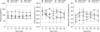
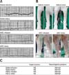
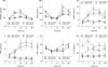
There were no remarkable clinical results for any of the dogs. Also, there were no significant differences in vital signs such as heart rate, respiratory rate, and body temperature (p > 0.05), and all vital signs were within normal ranges for all of the dogs after transplantation (Fig. 1). In addition, an ECG test was performed before and after transplantation in order to evaluate adverse circulatory responses such as arrhythmia and coronary artery anomalies. All groups showed normal patterns of wave interval and height (P width, P height, PR interval, QRS width, R height and QT interval) before and after saline or ASC transplantation (panel A in Fig. 2). Auscultation of heart murmur, neurological symptoms, and signs of swelling in the limbs were recorded for 30 min (panels B and C in Fig. 2); no group showed adverse responses such as heart murmur or limb swelling, and all dogs showed normal neurological responses for cranial nerve reflexes and postural reflexes.
Blood analysis
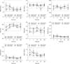
We analyzed CBC and serum chemistry results on days 1, 7, 14, 21, and 28 post-transplantation of saline or ASCs to survey for possible adverse effects in all dogs (Figs. 3 and 4). All serial test results for CBC after transplantation were within normal ranges and showed no significant differences among the four groups (reference intervals: WBC, 5.2–17.0 K/µL; RBC, 5.7–8.8 M/µL; PLT, 143–400 K/µL; PCV, 37.1%–57.0%; HB, 12.9–18.4 g/dL; BASO, 0.0–0.5 K/µL; EOS, 0.0–1.82 K/µL; NEUT, 3.2–10.9 K/µL). Moreover, the serum chemistry results post-transplantation were within normal ranges for all groups (reference intervals: Na, 145–155 mmol/L; K, 2.7–5.5 mmol/L; Cl, 96–122 mmol/L; ALP, 0.0–97.9 U/L; ALT, 5.8–83.3 U/L; AST, 11.7–42.5 U/L; CREA, 0.4–1.3 mg/dL; albumin, 2.6–4.4 g/dL; BUN/UREA, 6.0–24.0 mg/dL; total protein, 5.4–7.5 g/dL; Ca, 8.0–11.9 mg/dL; P, 1.3–6.3 mg/dL). However, there were significant differences in serial test results for BUN/UREA and P. The BUN/UREA results revealed significant differences among groups on days 7 and 21, whereas the P results showed a significant difference only on day 21 following transplantation of ASCs. There was significantly increased BUN/UREA in the ASC bolus group (17.7 ± 0.3 mg/dL) compared with levels in other groups on day 7 post-transplantation (p < 0.05). Also, on day 21 post-transplantation, the saline infusion and saline bolus groups (19.0 ± 2.0 and 22.0 ± 2.1 mg/dL, respectively) showed significantly higher concentrations of BUN/UREA than those of the ASC infusion and ASC bolus groups (10.0 ± 0.6 and 10.7 ± 0.9 mg/dL, respectively). In addition, there was significantly increased P in the saline infusion group (5.5 ± 0.0 mg/dL) compared with the levels in the other three groups on day 21 following transplantation (p < 0.05).
ELISA analysis
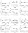
The concentrations of MMP9, VEGF, bFGF, HGF, TNF-α, and IL-10 in serum of each group on days 1, 7, 14, 21, and 28 post-transplantation of saline or ASCs were analyzed (Fig. 5). There was no significant difference in serum TNF-αconcentrations among the four groups, whereas the concentration of MMP9 was significantly high in both ASC groups on days 7 and 14 following transplantation (p < 0.05): ASCs infusion group 76.9 ± 5.8 and 89.1 ± 7.6 ng/mL, respectively; ASC bolus group 78.7 ± 5.3 and 74.0 ± 4.0 mg/mL; saline infusion group 48.5 ± 3.6 and 57.0 ± 2.2 ng/mL; saline bolus group 51.1 ± 1.6 and 53.2 ± 1.7 ng/mL.
The serum concentrations of VEGF were significantly increased in the saline bolus and ASC bolus groups on day 1 post-transplantation: 114.1 ± 7.3 and 114.1 ± 2.7 pg/mL, respectively, compared with those of the infusion groups. On day 21 post-transplantation, VEGF was significantly upregulated in the ASC infusion group (150.4 ± 18.1 pg/mL) compared with the other groups, and, on day 28 post-transplantation, VEGF was significantly increased in the ASCs infusion group (124.4 ± 9.3 pg/mL) compared to that in the saline bolus group.
The bFGF concentrations in both ASC groups were significantly increased on days 14 and 21 (ASC infusion group 13.9 ± 2.9 and 10.9 ± 3.0 pg/mL, respectively; ASC bolus group 9.8 ± 1.2 and 10.1 ± 2.0 pg/mL) compared with those in the saline groups. On day 28 post-transplantation, bFGF was significantly increased in the ASC infusion group (9.2 ± 3.5 pg/mL) compared to those in the saline groups.
The concentration of HGF was significantly upregulated in both ASC groups on days 7, 14, 21, and 28 following transplantation (ASC infusion group 1.4 ± 0.1, 2.9 ± 0.4, 3.0 ± 0.2, and 2.8 ± 0.2 pg/mL, respectively; ASC bolus group 1.7 ± 0.1, 2.3 ± 0.2, 2.5 ± 0.2, and 2.3 ± 0.2 pg/mL) compared with the saline groups.
The serum concentration of IL-10 in the ASC infusion group was significantly upregulated on days 1, 7, 14, 21, and 28 post-transplantation (36.8 ± 4.7, 48.7 ± 7.1, 33.7 ± 4.4, 38.1 ± 6.1, and 35.6 ± 4.8, respectively) compared to those in the other groups.
Discussion
Use of MSCs from tissues, bone marrow, or cord blood is a promising technology that can overcome the limitations of current medical technologies. These cells can be applied to various incurable diseases, including critical limb ischemia and arthritis, which cannot be treated with current medical technology [1622]. In clinical applications, MSCs have favorable properties, such as having immunomodulatory characteristics and protective effects on immunological disorders [212938] as well as angiogenic effects [37]. However, those properties may also have adverse effects. Some studies have demonstrated that transplanting MSCs into mouse brain elicits an inflammatory response and rapid rejection [89]. Moreover, it was reported that the immunosuppressive capacity of MSCs may, in some cases, favor the growth of tumor cells [10], and ASCs can promote growth of breast tumors and metastasis [24]. These contradictory results indicate that investigations into the characteristics of ASCs are far from complete, and further studies and consensual research to clinically assess transplantation of ASCs are necessary to fully elucidate their effects because there are no reports assessing the deleterious aspects of ASC transplantation into intact animals. To address these issues, we investigated clinical alterations after human ASC transplantation into healthy dogs to investigate possible applications in human clinical therapies. Furthermore, to determine whether transplanted human ASCs can maintain their positive aspects, including pro-inflammatory/anti-inflammatory and angiogenic properties as mentioned above, ELISA analyses were carried out to evaluate these characteristics.
In the present study, our results showed that after intravenous injection of human ASCs with two different injection rates (infusion and bolus), there was no incidence of adverse reactions in laboratory studies including physical examination (heart rate, respiratory rate, body temperature, heart murmur, ECG, neurological symptoms, swelling of four limbs). A previous study reported that MSCs transplanted into mice (15.7–24.3 g body weight) via a high dose (2.0 × 10⁸ cells/kg) bolus injection died soon afterward, while no adverse events were observed when the high dose was injected slowly [30]. In the present study, we obtained no abnormal clinical assessment results related to injection rates. In addition, blood analysis results showed that all parameters were within normal ranges post-transplantation. Also, there were no significant differences among groups, except for P and BUN/UREA levels which did show significant differences among groups after transplantation. A previous study reported that the level of BUN could vary depending on the measurement methods [7]. In support of our results, there was no adverse response based on the BUN results, and we suggest that significant differences in these factors (P and BUN/UREA) might be caused by variations in measurement methods.
In the present study, we measured the concentration of angiogenic and pro-inflammatory/anti-inflammatory factors in serum following ASC transplantation. We found that various angiogenic factors including MMP9, VEGF, bFGF, and HGF were increased, which could lead to angiogenesis.
MMPs produced by stromal cells and adipocytes are capable of degrading the extracellular matrix (ECM) to facilitate invading endothelial cells [3]. Also, they are prerequisite factors for releasing ECM-sequestered proangiogenic factors including VEGFs and FGFs that have been clearly implicated in angiogenesis [2]. In support of those results, our study showed that the concentration of MMP9 was significantly increased in the ASC groups on days 7 and 14 following transplantation along with increased levels of VEGF and bFGF. Therefore, the results indicate that MMP9 might have facilitated the release and increase in the levels of VEGF and bFGF on days 21 and 14, respectively, post-transplantation.
ASCs make a considerable contribution to angiogenesis by producing VEGF, HGF, and bFGF [2635]. In particular, it has been determined that VEGF and HGF act in a synergistic manner, which makes them valuable in angiogenesis therapy [34]. Therefore, ASCs could contribute to angiogenesis by secreting angiogenic molecules and supplying vascular cells to blood vessels. For instance, increased expression of VEGF mRNA and protein has led to rapid and dramatic increases in myocardial capillary diameter, capillary density, and blood flow in anemic fetal sheep [23]. Another study found that when human ASCs were cultured in vitro, consistent augmentation of HGF secretion was observed by ELISA [25]. It was also reported that MSC implantation into ischemic limbs and myocardium markedly improved vessel formation, blood perfusion, and capillary density associated with release of large amounts of VEGF and bFGF [1933]. In our study, transplantation of ASCs into intact animals induced high levels of VEGF, bFGF, and HGF. The results indicate that transplantation of ASCs into intact animals might invoke angiogenesis by markedly increasing the concentrations of VEGF, bFGF, and HGF.
A previous study proposed that MSCs mediate their immunomodulatory effects by controlling various kinds of cytokines including TNF-α and IL-10, which are pro-inflammatory and anti-inflammatory cytokines, respectively [6]. Another report demonstrated the immunomodulatory functions of human MSCs by co-culturing them with immune cells and suggested that human MSCs changed the secretion of cytokines in immune cells by inducing secretion of IL-10 and decreasing that of TNF-α [1]. That study proposed that when human MSCs are present in an inflammatory environment they could change the on-going immune response, thereby causing a shift from a pro-inflammatory situation to an anti-inflammatory one. Thus, they demonstrated a close interaction between human MSCs and immunomodulatory cytokines. Our results showed that the level of IL-10 was significantly increased in the ASC infusion group compared with that in other groups after transplantation, while there were no significant differences in TNF-α concentrations. Therefore, we assume that ASCs might mediate the high expression of IL-10 in intact animals.
In conclusion, transplantation of human ASCs intravenously into intact dogs elicited no adverse responses based on our physical assessment and blood analysis results. Thus, for the first time, we have demonstrated that human ASCs can be safely transplanted into dogs for clinical use. Furthermore, transplantation of human ASCs could induce a markedly high concentration of angiogenic factors (MMP9, VEGF, bFGF, HGF) and the anti-inflammatory factor IL-10, which suggests that human ASCs might represent a novel therapeutic tool for stimulating anti-inflammatory and angiogenic factors in animals with ischemic vascular diseases.
 XML Download