

 PDF
PDF ePub
ePub Citation
 Print
Print


Introduction
As the field of minimally invasive surgery has been expanding in veterinary medicine, one-lung ventilation (1LV) is often accompanied in thoracoscopic lung surgery to facilitate motionless surgical approach with a benefit of optimal hilar visualization. During 1LV, however, collapse of the operated lung area blocks its ventilation completely, and there is a potential risk for considerable intrapulmonary right to left shunt of deoxygenated blood through the non-ventilated lung [1516]. This theoretical ventilation-perfusion (V/Q) mismatch may result in hypoxemia and decreased oxygen saturation [2151618283031].
Hypoxic pulmonary vasoconstriction (HPV), a critical physiologic homeostatic mechanism, is activated in hypoxic condition, and pulmonary blood flow is diverted away from collapsed, hypoxic areas to better-oxygenated areas of the pulmonary vascular bed to match regional ventilation and perfusion [181931]. This phenomenon decreases shunt fraction and accordingly improves patients' oxygenation [2527]. In clinic, most indications of lung lobectomy are in pathologic status of impaired oxygenation inducing poor alveolar distension-contraction such as pulmonary abscess, bullous emphysema, consolidation, neoplasia, or lung lobe torsion [22]. Associated hypoventilation, V/Q mismatching, shunt fraction, and dead space may deteriorate hypoxemia and hemoglobin desaturation. Moreover, 1LV in these patients might induce further risk potential with an artificially increased shunt fraction [15]. Under such conditions, attenuation of HPV could result in fatal intraoperative hypoxemia [2361827].
In general, it has been accepted that volatile anesthetics are known to inhibit HPV with a vasodilative effect counteracting the protective mechanism of HPV, while intravenous anesthetics do not depress HPV [72527]. However, various authors have reported a variety of results, and there is no consensus currently [51032]. According to previous reports, in vitro study in rat lung showed a negative effect with a median effective dose (ED50) of 0.6 minimum alveolar concentration (MAC) isoflurane inhalation [20], and in vivo study in chloralose-based intravenous anesthetized rabbit with 1LV reported increased regional blood flow to non-ventilated lung fields after conversion to 1.5% (0.75 MAC) isoflurane inhalation [7]. In human medicine, propofol improved oxygenation and shunt fraction during 1LV in an open chest condition for esophageal surgery compared with isoflurane and sevoflurane [1], and a three-fold greater shunt fraction with isoflurane compared with propofol was also reported [12]. On the other hand, a neutral to weak inhibitive action on HPV was observed with modern inhalants of isoflurane, sevoflurane, and desflurane in human medicine [4182731], whereas a total intravenous anesthesia (TIVA) of propofol-alfentanil combination in another human study did not decrease the risk of hypoxemia compared with 1 MAC isoflurane during 30-minute 1LV [26].
These results are inconsistent and depend on conditions of the study such as method of induction of hypoxic condition (hypoxic gas inhalation vs. 1LV), assessment of HPV (arterial blood gas analysis, pulmonary arterial catheterization, colored microsphere technique), type and dose of anesthetics, experimental animals, or in vivo vs. in vitro study [51032]. Moreover, some studies excluded surgical maneuver and evaluated only under 1LV [31417]. Consequently, it is inappropriate to compare results among studies, thus effects under real surgical condition remain unpredictable in dogs.
Considering these aspects, this study investigated the effects of two anesthetic regimens, isoflurane inhalation and propofol-remifentanil combined TIVA, on HPV and oxygenation during thoracoscopic lung lobectomy with 1LV in Beagle dogs. The aims of this study were (1) to evaluate the effect of each anesthetic encountered during the overall steps of thoracoscopic lung lobectomy with 1LV, and (2) to determine differences between the two anesthetic groups.
Materials and Methods
Experimental animals
This study was performed under the guidance of the Chungnam National University Institutional Animal Care and Use Committees (CNU-00413). Ten 3-year-old, purpose-bred intact male Beagle dogs weighing 8.88 ± 0.75 kg (range, 8.0–10.0 kg; body condition score, 4/9–6/9) were included. All dogs were clinically healthy based on physical examination, complete blood count, serum biochemistry, electrolytes panel, and thoracic radiography. The dogs were randomly allocated into one of two groups of anesthetic regimens (isoflurane inhalation, ISO group; propofol-remifentanil combined TIVA, TIVA group).
Anesthetic regimen
All dogs were oxygenated for 5 min prior to anesthetic induction. Normal saline was administered at 10 mL/kg/h. Glycopyrrolate (Mobinul, 0.011 mg/kg intravenous [IV]; Myungmoon Pharm, Korea) and diazepam (Diazepam, 0.1 mg/kg IV; Samjin Pharm, Korea) were used in the premedication of each dog. Anesthesia in five dogs (ISO group) was induced with propofol (Provive, 4 mg/kg IV; Myungmoon Pharm) and maintained with isoflurane (up to 2%, 1.5 MAC) after endotracheal intubation. In the other five dogs (TIVA group), propofol and remifentanil (Ultiva; GlaxoSmithKline, UK) were administered for induction (4 mg/kg IV, 4 µg/kg IV) and infused for maintenance (up to 0.6 mg/kg/min constant rate infusion [CRI] and 6–20 µg/kg/h CRI) after endotracheal intubation. Rates of inhalation and infusion were controlled in response to patient monitoring parameters and recorded. Meloxicam (Metacam, 0.2 mg/kg IV; Boehringer Ingelheim Vetmedica, Germany) was administered immediately prior to anesthetic induction, and pure oxygen (FiO2 = 1) was used throughout the procedure.
Anesthetic phases
Total anesthetic duration was divided into the following five phases according to the surgical process. (A) Pre: after anesthetic induction, two-lung ventilation with a closed chest, 10 min; (B) O2LV: after portal placements, two-lung ventilation with open chest, 20 min; (C) O1LV: after bronchial block, 1LV with open chest, 20 min; (D) LOBEC: thoracoscopic right middle lung lobectomy under 1LV, 10 min; and (E) REVENT: re-ventilation and expansion of the collapsed lung, 5 min. Each dog served as its own control. The durations of total anesthesia and 1LV (phase O1LV and LOBEC) were 65 min and 30 min, respectively.
In phase Pre, all dogs were placed in left lateral recumbency, and the right hemithorax was surgically prepped. The dorsal pedal artery was catheterized for systemic blood pressure monitoring and arterial blood sampling. A pulmonary artery catheter (Swan Ganz Catheter, 132F5, 110 cm; Edwards Lifesciences, USA) was introduced through the right external jugular vein using the Seldinger technique with characteristic monitoring pressure tracking to measure the pulmonary arterial pressure and cardiac output (CO) and to obtain mixed venous blood sample. Atracurium besylate (Atra, 0.2 mg/kg IV; Hana Pharm, Korea) was administrated and the lungs were mechanically ventilated with a volume- and time-controlled ventilator after spontaneous respiration disappeared; Royal 77 (Royal Medical, Korea), tidal volume = 15 mL/kg, respiratory rate = 20 breaths/min, I/E ratio = 1:2, peak inspiratory pressure limit = 20 cmH2O.
During phase O2LV, an open chest condition was obtained by taking an 8-6-5 intercostal approach with three soft threaded trocars (Thoracoport; Covidien, USA) as described previously [23]. Three openings were created for the 5 mm telescope, 12 mm stapler and specimen retrieval pouch, and 5 mm grasper, respectively.
Phase O1LV followed. 1LV was initiated with an endobronchial blocker (Arndt Endobronchial Blocker Set, 5 Fr; Cook Critical Care, USA) occluding a main bronchial opening to the entire right lung field with guidance via a fiber-optic bronchoscope (EPK-700; Pentax Medical, Japan). Tidal volume was reduced by half of baseline during the O1LV and LOBEC phases. Each dog was vigilantly monitored, and the atelectasis of the right lung field was confirmed via thoracoscopy. Dislodging of the endobronchial blocker was monitored also.
In the LOBEC phase, thoracoscopic right middle lung lobectomy was performed with an endoscopic stapler and the resected lung lobe was retrieved with a specimen retrieval pouch. The surgical field was re-explored for hemorrhage, air leakage, or pedicle tearing.
In the REVENT phase, the collapsed lung lobes were re-expanded 10 times after the deflation of the endobronchial blocker by applying 5 cmH2O positive end-expiratory pressure ventilation with 20 cmH2O peak inspiratory pressure. The tidal volume restored to baseline, and ventilation was continued for additional 5 min.
After termination of all phases, dogs were humanely euthanatized under anesthesia.
Evaluation
Patient monitoring included electrocardiogram, heart rate (HR), respiratory rate, capnogram, end-tidal carbon dioxide concentration, tidal volume, rectal temperature, core temperature, arterial blood pressure (systolic [SAP], mean [MAP], and diastolic), and pulse oximetry (SpO2). These parameters were monitored and recorded by using a Datex S/5 system (Datex Ohmeda, Finland).
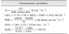
Hemodynamic changes, pulmonary arterial pressures (systolic, mean [MPAP], diastolic, and wedge [PAWP]) and CO with a thermodilution technique were measured and recorded at the following times of each phase: Pre-10 min (PRE), O2LV-20 min, O1LV-10 min, O1LV-20 min, LOBEC-10 min (LOBEC), and REVENT-5 min (REVENT). The mean value of three consecutive CO measurements was used for each datum. Arterial and mixed venous blood samples were collected anaerobically and analyzed (i-STAT Portable Clinical Analyzer; Heska, USA) simultaneously. Data were calculated using following standard formulae (Table 1); cardiac index (CI), shunt fraction (Qs/Qt), arterial oxygen content (CaO2), oxygen delivery index (DO2I), pulmonary vascular resistance index (PVRI).
Statistical analysis
All continuous variable data are presented as mean ± SD values. Normality of data distribution was examined by the Shapiro-Wilk test, and non-parametric methods were selected for non-normal distributions. For a comparison among all phases in each group, data were analyzed using Friedman's test which is related to non-parametric ANOVA with repeated measures, and further multiple comparisons were performed with a Wilcoxon signed rank test when any significant difference was identified (p < 0.05). For differences between two independent groups in the same phase, data were compared by using the Mann-Whitney test. All analyses were performed with IBM SPSS Statistics (ver. 21.0; IBM, USA) and p < 0.05 was considered to indicate a significant difference.
Results
The anesthetic concentrations for maintenance were lower than that of the pre-planned protocol. In the ISO group, isoflurane was inhaled up to 1.5%, which was equivalent to approximately 1.125 MAC in dogs. In the TIVA group, the range of infusion rates of propofol and remifentanil were 0.2 to 0.4 mg/kg/min and 6 to 11 µg/kg/h, respectively.
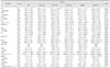
Intraoperative blood gas analysis and cardiopulmonary variable data at every assessment time in each group are shown in Table 2. HR in the ISO group significantly decreased after 1LV, while there was no significant difference among phases in the TIVA group or between the two groups. SAP and MAP increased with open chest and 1LV but it was not significantly different among phases in each group. In the REVENT phase, SAP (p = 0.032) and MAP (p = 0.016) in the ISO group were lower than that of the TIVA group.
CI levels did not differ between groups or among the phases in the ISO group; however, it was significantly higher in phases of O1LV-20 and LOBEC than that of PRE and O2LV-20 phases in TIVA. During 1LV, Qs/Qt increased significantly in each group, but there was no significant difference between groups. As for PaCO2 and pH, PaCO2 increased while pH decreased with 1LV significantly, but they restored in the REVENT phase. PaCO2 was not different between groups, but a significantly lower pH level was identified in the ISO group than that of the TIVA group in O1LV-10 phase (p = 0.008).
Regarding intraoperative oxygenation, SpO2, arterial oxygen pressure (PaO2), arterial oxygen saturation (SaO2), and CaO2 in both groups decreased significantly during 1LV but restored in the REVENT phase to the level of PRE (SpO2, SaO2), O2LV-20 (PaO2, CaO2 in ISO), and O1LV-10 (CaO2 in TIVA). DO2I in the ISO group showed no significant change among phases although there was a tendency of decrease during 1LV. In the TIVA group, on the other hand, significantly higher DO2I were identified in O1LV-20 and LOBEC phases than that in the PRE phase, and the level decreased in the REVENT phase. None of these 5 parameters showed a significant difference between the two groups.
There were significant increases of MPAP and PAWP in the ISO group and MPAP in the TIVA group during 1LV, but there was no difference between groups. PVRI values were not significantly different in either group and also between groups except in the REVENT phase (p = 0.032).
Discussion
This study attempted to answer the question of how we can reduce burden on canine patients for lung lobectomy while mitigating the risk of hypoxia. In lateral recumbency, an intercostal thoracotomy enables over-inflation of the non-dependent lung as the thoracic wall does not limit its expansion, while ventilation of the dependent lung is impaired because the loss of negative intra-thoracic pressure induces compression of the lower lung via the weight of the heart, mediastinum, and abdominal organs. However, lung perfusion is more concentrated on the dependent side due to the gravity [1318], and these complicated conditions contribute to V/Q mismatch.
Contrary to the approach in thoracotomy, the thoracic wall is intact during thoracoscopy and it prevents the over-inflation of the non-dependent lung. Therefore, a higher proportion of ventilation occurs in the dependent lung, resulting in a smaller Qs/Qt and a lower reduction in PaO2, SaO2, and CaO2. Previously, a 19% reduction in PaO2 with unaffected SaO2 was reported during thoracoscopic cannulae placement, whereas a 29% to 39% reduction in PaO2 accompanied by a significant decrease in SaO2 was induced by intercostal thoracotomy [13].
Thus, we selected a thoracoscopic procedure for lung lobectomy and included 1LV, which could be decisive in some cases. With 1LV, the non-dependent lung is excluded from the ventilatory circuit completely, thus it becomes a true shunt [18]. In this scenario, ventilation is restricted to the dependent lung, although Qs/Qt increases. At this moment, a concentrated blood flow to the region where the gas exchange occurs is key to improve patients' oxygenation; this is why HPV should be optimized with 1LV. HPV can reduce shunt flow through the operative lung by 40% approximately [18].
The mechanism of HPV has not been fully described. It is thought of as a local constriction of vascular smooth muscle in the pulmonary circulation in response to regional hypoxia; an initial decrease in alveolar oxygen pressure (PAO2) followed by a mixed venous oxygen pressure (PvO2) [181933]. HPV occurs within a few seconds and reaches an initial peak in 15 to 20 min (phase 1) and can be prolonged for 2 to 4 h (phase 2) as a peak effect [1819]. HPV may be assessed with oxygenation and PVRI indirectly or via a microsphere technique to measure the direct regional blood flow [31718]. The net effect of the hemodynamic and respiratory changes of any treatment can be assessed through the evaluation of DO2I [14]. The incidence of hypoxemia associated with 1LV was recently reported as 5%; that rate has decreased from 25% in the 1970s owing to improvement of lung isolation technique, anesthetic agents, and better comprehension of the physiology of 1LV [19].
HPV is influenced by several factors such as general anesthesia, surgical manipulation, trauma, size of the hypoxic segment, intensity of the hypoxic stimulus, or temperature and age [2361827]. Whereas surgical manipulation in lateral recumbency reduces blood flow to the non-dependent lung with gravity passively [18], the anesthetic agents, muscle paralysis and mechanical ventilation, lateral recumbency, and atelectasis can deteriorate Qs/Qt, V/Q mismatch, and arterial hypoxemia [13]. However, these changes might be inevitable during practical surgery, and there is a need to evaluate anesthetic effects under the actual surgical conditions. This study was conducted under the most extreme hypoxic condition of left-sided 1LV, which excludes the right lung area that occupies 58% of the total lung volume [8]. Efforts were focused on maintaining anesthetic techniques and surgical procedures relevant to real clinical practice. The experimental phases followed the same courses as those of thoracoscopic lung lobectomy, and anesthetic depth was controlled to be just suitable for surgery.
In this study, obvious impairments in gas exchange were induced during 30 min 1LV in both anesthetic groups. There were significant decreases in PaO2, SaO2, and CaO2, whereas PaCO2 increased and respiratory acidosis was intensified. However, neither hypoxemia nor desaturation that were unacceptable (i.e., PaO2 < 60 mmHg, SpO2 < 90, SaO2 < 91% [929]) did not develop in either group. In addition, despite CaO2 decreasing numerically during 1LV, it was relatively well maintained in all phases and in both groups within or over the normal range (16–20 mL/dL) [9]. Moreover, none of the assessment times showed significantly lower CaO2 compared to that of the PRE phase.
When we consider the insignificant change in PVRI despite significant increases in MPAP or PAWP, as well as the unaffected oxygen delivery regardless of significantly increased shunt fractions, neither anesthetic regimen in this study appeared sufficient to impair oxygenation. Qs/Qt increased significantly after initiation of 1LV in both groups but did not show a significant decrease until end of 30 min 1LV. Oxygen delivery was maintained regardless of any of open thorax, 1LV, or surgical maneuver, and it was not different between groups. This might be attributed to the high fraction of inspired oxygen (FiO2 = 1.0) or low anesthetic concentration, and the latter could be a reflection of the characteristics of minimally invasive surgery; a smaller incision and less pain. It is also possible that the overall effects of complicated in vivo compensation mechanisms avoided hypoxemia in advance of HPV. The effects of HPV seem to be maximized in conditions of low PAO2 from 25 to 50 mmHg, low V/Q with hypoxic lung area between 30% and 70%, and hyperthermia [19].
In several reports, HPV was reported to be decreased by volatile anesthetics under in vitro and in vivo conditions. The attenuation appeared in a dose-dependent manner, especially with old agents [5], and some authors suggested that the clinical relevance of this effect is negligible [18]. Suppressive effects of volatile anesthetics are more potent when the pulmonary vascular region of HPV is exposed through the alveolus rather than via mixed venous blood [19]. If 1LV is stable, the agent is delivered by mixed venous blood. The anesthetic concentration in this study was even lower than that in a previous study that reported non-affected DO2I by 1LV under 1.5 MAC isoflurane inhalation; in that study, oxygenation was evaluated in the same manner as in present study, but it was not accompanied by surgical procedure [14]. Although respiratory acidosis was more severe in the ISO group than in the TIVA group after initiation of 1LV, it might be less influential on HPV. As mentioned above, HPV is a response to the oxygen pressure (hypoxia) rather than that of carbon dioxide. Indeed, HPV is less associated with hypercapnia or respiratory acidosis but is attenuated by hypocapnia, instead [19].
During 1LV, CI increased significantly in the TIVA group, which is counter to the general characteristic of propofol anesthesia presenting a decrease in CO. It seemed to be associated with an increase in HR for a decrease in PaO2, although that change was insignificant. It is followed by an increase in DO2I because DO2I is determined by both CO and CaO2. Even though CaO2 declined, the degree of change in CO was relatively greater than that of CaO2 at the end of 1LV. Nevertheless, it did not result in a significant change in PVRI. On the other hand, in the ISO group, there was a significant decrease of HR after initiation of 1LV, which was accompanied by a non-significant decrease in CI and DO2I. With significant increases in MPAP or both MPAP and PAWP associated with 1LV in both groups, PVRI values during the phases with 1LV were higher than those of the other phases in both groups, but the differences were not significant. Similarly, there was a tendency to higher values in the TIVA group than in the ISO group in every phase, but there was no significant difference between two groups other than in the REVENT phase.
Previous canine studies with 1LV usually did not include surgical procedures [3101432]. Recent retrospective research in human medicine investigated during thoracic surgeries with 1LV but evaluated mainly only PaO2, blood pressure, or HR [5242729]. Our results are supported by these human studies, which concluded there were no significant differences in PaO2, MAP, or HR between propofol-based TIVA and volatile anesthetics using isoflurane [2629] or sevoflurane [24]. Regarding thoracoscopic lung lobectomy in human medicine, a propofol-remifentanil combination was suitable and did not alter PaO2 [527], and one study reported that combination provided better oxygenation than a desflurane-remifentanil combination [5]. A previous study on thoracoscopic subphrenic pericardiectomy with 1LV was performed under isoflurane inhalation (up to 2.0%) in normal dogs. The authors monitored only PaO2, PaCO2, and SpO2 and reported relatively acceptable ventilation and oxygenation, although they did report a significant decrease in PaO2 during 1LV [21]. In another case report of thoracoscopic pericardial fenestration in a dog with a heart base tumor showed slight respiratory acidosis and considerable impairment in oxygenation parameters on arterial blood analysis; minimal PaO2 was 96 mmHg at 170 min [2]. That study was performed under sevoflurane inhalation with 1LV excluding the right lung field as in the present study.
When interpreting our results, the following limitations should be considered. First, this study included a small number of healthy dogs, and there was a lack of data after anesthetic recovery when patients might be in unstable cardiovascular/hemodynamic status with reduced ability of compensation and impaired immunity. Further study of a large number of clinical patients with impaired gas exchange with long-term tracking after recovery from anesthesia is warranted. Second, the experimental conditions might be not controlled completely among phases with serial intervention of a surgical procedure. In addition, the concentration of anesthetics was not fixed, which was because the study was performed to act as a replication of real surgery. Third, the duration of 30 min 1LV could be too short to monitor the further effects of HPV, although it might be enough for the surgical procedure. According to a previous review, PaO2 level reaches its lowest 20 to 30 min after 1LV and then increases gradually over the next 1 to 2 h [19]. Fourth, anesthesia and measurements were not carried out in a blind manner throughout the study, and the net effect of oxygenation was evaluated via an indirect method. Although a direct determination of blood flow with colored microsphere technique in pulmonary artery could provide further information on alteration in the distribution of blood flow at the level of capillaries in each phase, it was impractical to perform this technique in our clinic.
In summary, both 1.5% (1.125 MAC) isoflurane inhalation anesthesia and propofol-remifentanil combined TIVA did not affect patients' overall oxygenation throughout the thoracoscopic lung lobectomy procedure with 30-minute left-sided 1LV in dogs. Therefore, both anesthetic regimens can be used according to the surgeon's preference for thoracoscopic procedures with ILV. However, observations of 1) severe hypercapnia and respiratory acidosis with 1LV in ISO group than TIVA group, 2) an increase in DO2I with CI during 1LV in TIVA group, and 3) a highly maintained SAP and MAP in the TIVA group before end of the study should be considered during case selection. TIVA could be safer in patients with cardiovascular instability concerns. This study recommends vigilant anesthetic monitoring in patients with cardiopulmonary disease as significant alterations in gas exchange associated with ILV were induced regardless of the anesthetic regimen. Normocapnia and normothermia should be maintained during surgery, as HPV could be attenuated by hypocapnia and hypothermia [19]. Life-threatening hypoxemia during 1LV should be prevented by checking for the location of endobronchial blockers or double lumen tubes, increasing FiO2, applying 5 cmH2O positive end-expiratory pressure, re-expansion of the non-ventilated lung, or by administration of respiratory stimulants such as almitrine [111518].
 XML Download