

 PDF
PDF ePub
ePub Citation
 Print
Print


Introduction
Due to the presence of feeding materials and feces, the environment inside swine facilities is very dusty [730]. A study on Korean swine farms reported that 60% to 70% of the total dust in such facilities comes from feeding materials, which rises to approximately 90% during feeding time [19]. The dust is a biologically active aerosol because it contains microorganisms such as bacteria, viruses, and fungi and their organic compounds, including endotoxins [22].
Endotoxins are one of the most ubiquitous organic compounds in agricultural settings. This is partly due to their heat resistance, which allows them to persist in the environment even under extreme conditions [21]. Airborne endotoxin levels can be estimated by using the limulus amebocyte lysate (LAL) assay, which measures endotoxin levels as functional (bioactive) endotoxin units (EUs) per cubic meter of air (EU/m3) [3335]. There is still no internationally accepted endotoxin exposure threshold that indicates safe levels of exposure, even in humans. Several studies have proposed that the following thresholds may be useful for humans: a no-observed-adverse-effect level of 17 EU/m3 [25] or < 100 EU/m3 [20], and a lowest observed effect level of 30 to 75 EU/m3 [20]. However, Park et al. [28] suggested that indoor airborne endotoxin levels in homes as low as 0.02 to 19.8 EU/m3 could have a role in sick-building syndrome.
An endotoxin is a potent immunogenic stimulant, and even small amounts can non-specifically stimulate the immune system [212838]. Our previous study showed that swine-farm workers who were exposed to high endotoxin levels had type 2-helper (TH2)-skewed immune profiles; their peripheral-blood mononuclear cells (PBMCs) stimulated with phorbol 12-myristate 13-acetate and ionomycin produced lower levels of the proinflammatory cytokines interferon-gamma (IFNγ) and tumor necrosis factor-alpha (TNFα) and higher levels of the anti-inflammatory cytokines interleukin (IL)-4 and IL-13 than a control group of office workers. This suggests that endotoxin exposure may promote allergic responses [16]. Similarly, when healthy human subjects [1] and mice [40] inhaled low endotoxin doses, they exhibited TH2-skewed immune profiles. While the effect of endotoxin exposure on the immune status of livestock animals has not yet been extensively investigated, Kullik et al. [18] showed that intravenous injection of pigs with lipopolysaccharide (LPS) significantly increased their plasma TNFα and IL-6 levels. In addition, when normal porcine monocytes are differentiated into macrophages in the presence of organic dust extract from swine husbandry facilities, their differentiation and responses to LPS are impaired [29]. Similarly, pig barn dust extract treatment in vitro negatively affects porcine macrophage function [17].
Recently, we reported that indoor dust endotoxin levels in swine-confinement facilities do not associate significantly with the isolation of Gram-negative bacteria from the air in these buildings [33]. This suggests that when assessing the effect of endotoxins on the health of animals or workers, indoor endotoxin levels may be a more useful measure of endotoxin exposure than isolation of Gram-negative bacteria. Hence, to test the hypothesis that exposure to endotoxins in natural husbandry settings can alter the immune responses of pigs, we measured endotoxin levels in several swine farms and assessed the relationship of endotoxin level with the immunological profiles of the swine in these farms.
Materials and Methods
Pig farms and blood collection
Ten pig farms in six counties in Korea participated in this study. They were selected from a list that was prepared by a swine farmers' cooperative and were included in the study if the farmers allowed us to enter the pig-confinement buildings. The buildings were visited between July and September in 2012, 2014, or 2015. All pig farms employed open-type housing in which ventilation was provided by large purpose-built windows with a curtain winching system. In all farms, the stocking density ranged from 0.8 to 1.7 m2/pig, which is within the recommended stocking density (0.8 m2/pig for fattening pigs) of the Korean government [26]. In each farm, three to 10 pigs with no apparent clinical or pathological abnormalities (age ranged from 90 to 150 days) were randomly chosen for blood collection irrespective of sex. Blood samples were collected by local veterinarians. A maximum of 10 mL of blood was drawn aseptically from the jugular vein of each pig and placed into EDTA-coated vacutainers [18]. All animal handling, blood collection, and experimental procedures were approved by the Institutional Animal Care and Use Committee of Daegu Catholic University (IACUC No. CUD IACUC-2012-10).
Dust collection and endotoxin measurement
Several reports show that the dust and endotoxin concentrations are highest in the daytime since this is when the animals are fed and are most active [19]. Therefore, we measured the total indoor dust levels in the pig farms for 8 h from 9 AM until 5 PM. For this, we used polyvinyl chloride (PVC) membrane filters (SKC, USA) with a 2-stage cassette at a flow rate of 2.0 L/min. We also measured the concentration of respirable dust, particulate matter less than or equal to 10 µm (PM10) by using a PVC membrane filter with a 10 mm Dorr-Oliver nylon cyclone at a flow rate of 1.7 L/min for 8 h [163435]. Both total and respirable dust samples were collected at two different locations in each farm; namely, at one- and two-thirds of the total distance from the entrance gate. Blank and sample filters were weighed at least three times by using an electronic micro-balance (Quintix 125D; Sartorius, Germany).
Endotoxin concentrations in dust were measured as described previously [323335]. Briefly, endotoxin was extracted from the filters by adding 3 mL of endotoxin-free LAL water (Lonza, USA) with 0.5% Tween 20 followed by shaking for 1 h at 350 r/min. The endotoxin concentrations in the collected supernatants were measured by using a LAL Kinetic QCL kit (Lonza) according to the manufacturers' instructions. Given the proposed threshold endotoxin levels described in the Introduction, we decided that 30 EU/m3 in total dust would serve as the threshold distinguishing high and low endotoxin levels in swine farms. Consequently, the pig farms with ≤ 30 and > 30 EU/m3 in total dust were considered to be low and high endotoxin exposure farms, respectively.
Determination of porcine hematological values
Total and differential white blood cell (WBC), red blood cell (RBC), and platelet counts of each pig were determined by an ADVIA 2120 (Siemens, Germany) automated hematology analyzer. Veterinary software adapted for pigs was used to analyze the data.
PBMC collection and lymphocyte phenotyping
PBMCs were isolated by Ficoll-Hypaque density-gradient centrifugation (Ficoll-Paque Plus; GE Healthcare Life Sciences, USA) [163435]. Lymphocyte subpopulations were analyzed by using three-color flow cytometry (FACScan; BD, USA). Porcine B and T cells were identified by using mouse anti-pig CD1-FITC Ab and mouse anti-pig CD3ε antibodies (Southern Biotech, USA), respectively. Porcine cytotoxic T cells, helper T cells, and CD4+CD8+ T cells were identified by using mouse anti-pig CD8α-FITC Ab and mouse anti-pig CD4α-FITC antibodies (Southern Biotech). R-phycoerythrin- or FITC-conjugated isotype controls served to control for non-specific background fluorescence.
Enzyme-linked immunosorbent assay quantitation of plasma antibodies
Porcine serum IgG and IgA (Bethyl Laboratories, USA) and IgE (Elabscience, USA) levels were measured by using commercial enzyme-linked immunosorbent assay (ELISA) kits according to the manufacturers' instructions. Optical density was measured at 450 nm (Epoch; Bio-Tek, USA).
T-cell activation and measurement of secreted cytokine levels
Porcine PBMCs (106 cells/mL) were resuspended in complete RPMI medium (1 mM nonessential amino acids, 1 mM sodium pyruvate, 1% sodium bicarbonate, 2 mM glutamine, 50 M 2-mercaptoethanol, and 10% heat-inactivated fetal bovine serum) and activated with 5 µg concanavalin A and 10 IU recombinant human IL-2 for 48 h at 37℃ in a 5% CO2 incubator. IL-4 secretion into the medium was determined by using a mouse anti-swine IL-4 monoclonal antibody (mAb) (capture antibody), recombinant swine IL-4 (standard), and a biotin-conjugated mouse anti-swine IL-4 mAb (detection antibody) (Invitrogen, USA). IFNγ secretion was determined by using a mouse anti-swine IFNγ mAb (capture antibody) (Invitrogen), recombinant swine IFNγ (standard) (BioSource, USA), and a biotin-conjugated mouse anti-swine IFNγ mAb (detection antibody) (Invitrogen). IL-12/IL-23 p40 and TNFα were quantitated by using Duoset ELISA kits (R&D Systems, USA).
Statistical analyses
All statistical analyses were performed by using SigmaStat 3.5 (Systat Software, USA). Depending on the normality of the data, the high and low endotoxin groups were compared by using Student's t-test or the Mann-Whitney rank-sum test. Differences were considered significant when p was < 0.05. Correlations between husbandry and immunological variables were analyzed by determining Pearson's correlation coefficients.
Results
Endotoxin levels in the pig farms
The high (n = 5) and low (n = 5) endotoxin exposure farms had similar general husbandry conditions; they had similar numbers of pigs in the confinement area, stocking densities, average pig ages, and ventilation modes with curtain winching systems (Table 1). However, the high endotoxin exposure farms tended to have higher total dust levels (mean, 0.87 mg/m3; range, 0.10–1.54 mg/m3) than that in the low-exposure farms (mean, 0.58 mg/m3; range, 0.13–1.06 mg/m3; p= 0.0536). They also had significantly higher respirable dust levels (mean, 0.67 mg/m3; range, 0.28–0.83 mg/m3) than that in the low-exposure farms (mean, 0.32 mg/m3; range, 0.01–0.96 mg/m3; p= 0.0001).
The high endotoxin exposure farms had significantly higher endotoxin levels in the total dust (mean, 443.18 EU/m3; range, 47.1–1,198.8 EU/m3) than that in the low endotoxin farms (mean, 13.05 EU/m3; range, 0–29.6 EU/m3; p= 0.0003). In addition, the high endotoxin farms had significantly higher endotoxin concentrations in the respirable dust (mean, 7.22 EU/m3; range, 0.55–14.48 EU/m3) than that in the low endotoxin farms (mean, 1.03 EU/m3; range, 0.01–2.76 EU/m3; p= 0.0000) (Table 1).
Hematological cell and lymphocyte subpopulation counts in the blood
Blood was collected from 28 pigs on the high endotoxin exposure farms (mean, 5.6±2.6 pigs/farm) and 19 pigs on the low endotoxin exposure farms (mean, 3.8±1.1 pigs/farm). The pigs from the high endotoxin exposure farms had significantly higher total WBC counts than that in the pigs from the low endotoxin farms (p= 0.0024). Since the two groups did not differ significantly in monocyte, eosinophil, and basophil counts, but did differ significantly in neutrophil and lymphocyte counts (p= 0.0187 and 0.0172, respectively), the higher total WBC counts reflect the higher neutrophil and lymphocyte counts in the highly exposed pigs. The pigs with high and low endotoxin exposure did not differ in RBC and platelet counts. The highly exposed pigs tended to have higher CD1+ B-cell counts than that in the pigs with low endotoxin exposure (p= 0.0546) but did not differ in other lymphocyte subsets (Table 2) [924].
Cytokine secretion by in vitro-stimulated PBMCs and plasma immunoglobulin levels
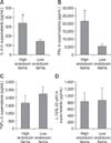
PBMC secretion of cytokines that have key roles in cell-mediated immunity was examined. The PBMCs from highly exposed pigs secreted significantly more IFNγ and IL-4 than that in the PBMCs from the pigs with low exposure (p= 0.034 and 0.037, respectively) (Fig. 1). Since the IFNγ:IL-4 ratio of non-specifically activated PBMCs can indicate an immune system skewing toward TH1 or TH2 immune responses, we calculated the IFNγ:IL-4 ratio by dividing the IFNγ concentration by the IL-4 concentration in the same culture supernatant and then multiplied the result by 10. High endotoxin exposure was associated with high IFNγ:IL-4 ratios(5.8±1.4 vs. 3.9±0.9). The two exposure groups did not differ in terms of PBMC production of IL-12/IL-23 p40 and TNFα (p= 0.951 and 0.513, respectively).
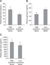
The highly endotoxin-exposed pigs tended to have higher plasma IgG and IgE levels than those with low endotoxin exposure, but the differences were without statistical significance (p= 0.213 and 0.574, respectively). However, high-exposure pigs had significantly lower plasma IgA levels than that in low-exposure pigs (p= 0.009) (Fig. 2).
Relationship between swine husbandry conditions and immunological variables
Correlations between endotoxin level and other husbandry factors with swine immunological variables were analyzed. Table 3 presents only the significant results. Pig age correlated positively with in vitro IL-4 (r = 0.398) and IFNγ production (r = 0.622) and negatively with platelet levels (r = −0.679). Total dust endotoxin levels correlated positively with total WBC (r = 0.496), neutrophil (r = 0.420), lymphocyte (r = 0.351), and monocyte (r = 0.505) counts in peripheral blood and in vitro TNFα(r = 0.706) and IL-12/IL-23 p40 production (r = 0.663). Respirable dust endotoxin levels correlated positively with in vitro IL-4 (r = 0.579) and IFNγ production (r = 0.697) and negatively with in vitro TNFα (r = −0.744) and IL-12/IL-23 p40 production (r = −0.563). Thus, total dust endotoxin and respirable dust endotoxin levels had opposite effects on TNFα and IL-12/IL-23 p40 production. Respirable dust endotoxin levels significantly correlated with platelet (r = −0.361) and CD1+ B-cell counts (r = 0.555).
Discussion
The present study showed that, on average, the ten pig farms that were included in this study had 269.3 EU/m3 in total and 5.0 EU/m3 in respirable dust. These levels were particularly pronounced in the high endotoxin exposure farms (defined as > 30 EU/m3); on average, those farms had 443.18 EU/m3 in total and 7.22 EU/m3 in respirable dust. These results are comparable to those in another study of swine barns [3], which reported that swine barns in Lithuania had on average 1,360 ±500 EU/m3 in total dust. Schierl et al. [36] reported that swine barns in Southern Bavaria had median endotoxin levels of 668.7 EU/m3 in total and 23.1 EU/m3 in respirable dust. Several surveys have shown that airborne dusts and endotoxins have detrimental effects on the health of humans working in swine facilities [131621]. However, the adverse effects of endotoxin exposure on animal health and productivity in the natural husbandry environment remain incompletely researched at present. To address this, we assessed the relationships between endotoxin exposure and immunological profiles of intensively reared pigs in Korea.
When endotoxins interact with host cells, it induces them to release proinflammatory cytokines and affects phagocyte differentiation and function [2129]. Our study showed that pigs living on farms with high endotoxin levels had higher peripheral blood total WBC, neutrophil, and lymphocyte counts than pigs with low exposure to endotoxins. This is consistent with reports showing that animals (including pigs) develop neutrophilia or lymphocytosis when they are experimentally infected with Gram-negative bacteria or are challenged with LPS [4612].
During infection, one of the first cytokines released by macrophages is the proinflammatory cytokine TNFα. Many studies have shown that when pigs are acutely exposed to LPS under experimental settings, their TNFα production is upregulated [182731]. In addition, other studies have shown that repetitive endotoxin exposure can lead to endotoxin tolerance, as indicated by decreased TNFα expression. Wysocka et al. [39] showed that when mice are injected with LPS twice in 26 h, or their splenic cells are treated twice with LPS in 20 h, their serum and supernatant TNFα levels decrease. Moreover, Castegren et al. [5] reported that when pigs are infused for 24 h with endotoxin, and their blood is subsequently treated with endotoxin in vitro, their TNFα production decreases. The latter studies are consistent with our observations in which nonspecificallyactivated PBMCs from pigs with high endotoxin exposure tended to produce less TNFα than that from the PBMCs of pigs with low endotoxin exposure. Moreover, PBMC production of TNFα correlated negatively with the endotoxin levels in respirable dust. Concerning the positive or negative correlation of TNFα and IL-12/23p40 production from PBMCs with the endotoxin level in total dust and respirable dust, respectively (Table 3), we can offer no clear explanation of this result at the moment. Assuming endotoxin in respirable dust could deeply penetrate into the lungs due to its small diameter (4 µm) compared to total dust (100 µm) [2], endotoxin in respirable dust could have a greater chance of interacting with the immunologic microenvironment in pulmonary alveoli, which may result in a different immune alteration than that from a total dust-mediated immune disturbance. Moreover, in addition to endotoxins, quantitative or qualitative differences in other hazardous components, including microbiological agents or odorous chemicals, may contribute to the different immune modulations mentioned above.
IFNγ and IL-4 typically promote TH1- and TH2-mediated immune responses, respectively, and they are mutually antagonistic [14]. Our study showed that the in vitro-stimulated PBMCs from pigs with high endotoxin exposure produced significantly more of both of these cytokines than that produced by PBMCs from pigs with low exposure. We also found that PBMC production of IFNγ and IL-4 both associated positively with endotoxin levels in respirable dust. However, high endotoxin exposure was associated with moderate PBMC, skewing toward TH1-mediated immunity, as indicated by the higher IFNγ:IL-4 ratios in the high-exposure group. This is inconsistent with studies showing that endotoxin exposure promotes TH2-mediated immunity in broiler chickens and beef cattle [3435]. This discrepancy may be due to differences in the husbandry environments between our study and those of Roque et al. [3435]. First, the cattle-confinement buildings studied by Roque et al. [34] had a lower endotoxin concentration (101 EU/m3) than that in our swine facilities (443 EU/m3). Second, while our pigs and the broiler chickens of Roque et al. [35] were exposed to similar levels of airborne endotoxin, the broiler chickens were housed under this condition for only 1 month as opposed to approximately 5 months for our fattening pigs. The inconsistent results could be explained by the suggestion that the helper T cell-mediated immunity of intensively reared animals may transit from TH2 to TH1 skewness when endotoxin concentrations are particularly high and/or when the animals are exposed to endotoxins for long durations. These possibilities are supported by several studies. First, while humans and rodents exposed to low endotoxin doses exhibit a TH2 phenotype [11123], exposure to high endotoxin doses appeared to convert TH2 skewing to TH1 skewing in skin allergic disease. Second, while TH2 reactivity predominates in the acute phase of atopic dermatitis, the chronic phase associates with upregulation of TH1-mediated responses [1037].
In the present study, the highly endotoxin-exposed pigs tended to have higher plasma IgG and IgE levels than those in pigs with low endotoxin exposure, although no statistical significance was detected. This may reflect a tendency for highly exposed pigs to have higher peripheral CD1+ B-cell counts than that in pigs with lower exposure. This is supported by the positive correlation between CD1+ B-cell counts and the respirable dust endotoxin levels. Moreover, high endotoxin exposure markedly decreased the plasma IgA levels in pigs. The lower IgA levels may reflect the prolonged exposure to endotoxin: Iqbal et al. [15] showed that prolonged endotoxin exposure induces mucosal IgA responses, thereby causing serum IgA molecules to transit from the blood to the mucosal membranes. Notably, Daiwen et al. [8] reported that when weanling pigs are experimentally injected with LPS, their serum IgG, IgM, and IgA levels are unchanged. This may indicate that these pigs were only acutely exposed to endotoxin.
Our study showed that when fattening pigs are exposed to prolonged high endotoxin levels (> 30 EU/m3), it can disrupt their immune homeostasis. Specifically, it skewed the TH1-TH2 balance, distorted the peripheral frequency of several immune cells, and dysregulated immunoglobulin production. These observations, especially the altered production of IL-4, IFNγ, and TNFα being correlated significantly with endotoxin levels in respirable dust, suggest that endotoxins may create an immune environment in which animals cannot adequately resist infection by pathogens. Since only 10 pig farms were investigated in our study, generalizations from the results reported herein should be limited. Therefore, systemic investigations should be conducted to elucidate further the molecular mechanisms by which endotoxins in dust can influence the immune profile of pigs. In addition, evaluations of husbandry conditions contributing to indoor endotoxin levels, including humidity/temperature, ad libitum feeding system, floor dampness, and slatted floor coverage, are necessary for pig farm managerial purposes.


 XML Download