

 PDF
PDF ePub
ePub Citation
 Print
Print


Hepatitis E virus (HEV) is a member of the family Hepeviridae, which contains two genera, Orthohepevirus and Piscihepevirus [14]. There are currently four species classified in the genus Orthohepevirus, including Orthohepevirus A, which is further divided into seven genotypes (HEV-1 to HEV-7). Infections with HEV-1 and HEV-2 appear to be limited to humans and induce endemic or epidemic hepatitis E outbreaks in developing countries. However, HEV-3 and HEV-4 infections have been identified in both humans and several animal species, including domestic pigs [210]. Zoonotic cases of hepatitis E worldwide are sporadically caused by consumption of raw or undercooked animal meats and meat products containing infectious HEV-3 and HEV-4 strains [10]. HEV-5 and HEV-6 have been found in wild boars [14], and HEV-7 was recently isolated from camels and from humans that consumed camel meat and milk [9]. Ever since swine HEV was first reported in the USA, multiple strains of HEV have been isolated from pigs [11]. Swine HEV-3 has been identified in many countries worldwide, whereas swine HEV-4 has been mainly reported in Asian countries, including China and Japan [13]. In Korea, several studies have confirmed HEV infections in domestic pigs [37], although only HEV-3 isolates, and no HEV-4 isolates, were identified in the reported cases. However, it has been suggested that sporadic cases of acute HEV infection in humans might be related to infection with HEV-4 originating from animals, including domestic pigs [6]. The objectives of this study were to identify the HEV genotypes circulating in domestic swine populations in Korea and to determine a plausible role of swine HEV in human infections.
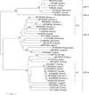
A total of 148 fecal samples were collected from weaners at 14 pig farms located in five different provinces in Korea. One gram of fecal sample suspended in 10 mL of phosphate-buffered saline was centrifuged at 1,200 × g for 30 min. The supernatant taken from the sample was then centrifuged at 16,000 × g for 10 min, and the resulting supernatant was collected and stored at −70℃ until analysis. Viral RNA was extracted from 150 µL of a porcine fecal sample by using the Patho Gene-spin DNA/RNA kit (iNtRON Biotechnology, Korea) according to the manufacturer's instructions. The extracted RNA samples were used for cDNA synthesis or were stored at −70℃. The cDNA was synthesized with the Superscript III First-Strand Synthesis for reverse transcriptase polymerase chain reaction (RT-PCR) kit (Invitrogen, USA) according to the manufacturer's instructions. The synthesis of the cDNA and RT-PCR processes was conducted as reported previously [6]. In brief, the initial PCR was designed to produce 531-bp nucleotide sequences specifically representing both HEV-3 and HEV-4 DNAs. The nested PCR was designed to produce 358-bp and 152-bp nucleotide sequences for HEV-3 and HEV-4, respectively, as described previously [6]. Phylogenetic analysis was conducted with 490-bp nucleotide sequences without the primer sequences from the 531-bp PCR products by using MEGA software (ver. 6.06; Kumar, Stecher, and Tamura 2015) as previously described [6]. The following HEV sequences acquired from GenBank (National Center for Biotechnology Information, USA) were included in the phylogenetic analysis: KX265101 (Korea), KX265100 (Korea), KX265099 (Korea), KX426271 (Korea), KX426270 (Korea), FJ426404 (Korea), FJ426403 (Korea), FJ763142 (Korea), M73218 (USA), D11092 (Japan), X98292 (India), AY230202 (Morocco), AY204877 (Chad), JF443721 (India), M74506 (Mexico), AF082843 (USA), FJ705359 (Germany), AB248521 (Japan), AB369687 (Japan), AF455784 (Kyrgyzstan), JQ013794 (France), FJ998008 (Germany), AY115488 (Canada), AB197673 (China), DQ279091 (China), AJ272108 (China), AY723745 (India), AB220974 (Japan), AB108537 (China), GU119961 (China), AB220972 (Japan), and AB291963 (Japan).
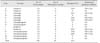
The nested-PCR protocol employed in this study produced 358-bp and 152-bp nucleotide sequences for HEV-3 and HEV-4, respectively, from the fecal samples of the pigs. HEV RNA was detected in seven of the 14 pig farms (50.0%) and in 30 of the 148 fecal samples (20.3%) collected from the 14 pig farms (Table 1). Among the seven HEV-infected farms, five farms contained only HEV-3, whereas the other two farms had HEV-4 exclusively. No farms were infected with both HEV-3 and HEV-4. Nucleotide sequence analysis of the 30 HEV strains showed that strains detected from a single farm were closely related (data not shown). Therefore, only one representative strain was selected from each HEV-infected farm for inclusion in the subsequent phylogenetic analysis. The 490-bp fragments (after removing primer sequences) of the seven HEV sequences were analyzed along with HEV-3 and HEV-4 reference strains isolated from pigs and humans. Phylogenetic analysis demonstrated that all five strains of swine HEV-3 determined in this study (GenBank accession Nos. MF095679, MF095680, MF095681, MF095682, and MF095683) were grouped into subtype 3a along with three reference swine HEV strains (AF082843, FJ426403, and FJ426404), showing 93.4% to 97.3% nucleotide sequence identities (data not shown). AF082843 is the reference strain of HEV subtype 3a reported from the USA, whereas strains FJ426403 and FJ426404 were previously reported in Korea (Fig. 1). The five pig farms with HEV subtype 3a were located in four different provinces of Korea. Therefore, these results might indicate that HEV subtype 3a is circulating over relatively broad areas in Korea. The two HEV-4 strains determined in this study (MF095684 and MF095685) were classified into subtype 4c along with recently reported human (KX265099, KX265100, and KX265101) and swine (KX426270 and KX426271) HEV-4 strains in Korea, but they were distant from a human strain (FJ763142) previously reported from a Korean hepatitis patient (Fig. 1). The two HEV-4 strains showed 98.9% to 99.5%, 98.7% to 99.3%, and 92.4% nucleotide sequence identities with those of the three human (KX265099, KX265100, and KX265101), two swine (KX426270 and KX426271), and the other human (FJ763142) HEV-4 strains isolated in Korea (data not shown). The two pig farms containing the HEV-4c strains were located in the same province, Gyeonggi. These results imply that circulation of HEV subtype 4c seems to be restricted to a relatively smaller area than that of HEV-3 in Korea.
In this study, we determined the HEV infection status and HEV genotypes circulating in domestic pigs in Korea. Among the seven HEV genotypes in the species Orthohepevirus A, both HEV-3 and HEV-4 are reported to cause most of the zoonotic transmissions to humans. Several studies have detected HEV in the feces, serum, and liver tissue of pigs in Korea. However, all of the previously isolated swine HEVs in Korea were HEV-3, with no report of HEV-4 [37], indicating that HEV-3 might be infecting the majority of pigs in Korea. Serological studies have demonstrated that approximately 20% of the Korean population produce antibodies to HEV [3], suggesting that the incidence of subclinical HEV infections might be substantial in Korea. Interestingly, all of the HEV strains identified from serum samples of Korean blood donors were also the HEV-3 strain [1], and the human HEV-3 strains were genetically close to swine HEV-3 strains isolated in Korea. Therefore, it was speculated that swine HEV-3 might be the causative virus inducing subclinical infections in Korean human populations. All the HEV-3 strains isolated from swine across Korea in the present study were classified into subtype 3a. We reported two complete genome sequences of swine HEV-3a subtype in a previous study [15]. The newly determined swine HEV-3 strains were still grouped into subtype 3a together with the two swine strains (FJ426403 and FJ426404) previously reported in Korea [15]. These results might indicate that the HEV-3 strains circulating in pigs in Korea are subtype 3a with absolutely no or very limited introduction of new HEV-3 subtypes. Although certain controversies persist, HEV-4 has been suggested to cause more severe cases of hepatitis than those from HEV-3 [12]. Sporadic cases of HEV infection have been reported in Korea, although the infection sources have largely not been determined except for a few cases [5]. Two of these exceptions were reported to be caused by the consumption of raw bile juice of wild boar or raw meat of roe deer infected with HEV-4 [48], and both HEV-4 strains were considered to have originated from China [13]. We recently learned that a considerable number of cases of cryptogenic hepatitis might have been caused by infection with HEV-4 strains that are closely related to the HEV strains originating from domestic pigs in Korea and from Japanese human strains [6]. Based on this background, we anticipated that substantial numbers of pigs in Korea might be infected with HEV-4. As expected, several HEV-4 strains were detected in pigs from two farms in the same province. Genetic sequencing helped to classify these strains, along with previously reported swine and human strains, into subtype 4c [6]. Therefore, HEV-4c is confirmed to be circulating in pigs in Korea; however, this result appears to be specific to only certain areas of Korea. Although this study was limited by the relatively small number of samples collected, the results indicate that domestic pigs are reservoirs for HEV-4 in some restricted areas in Korea. The swine HEV-4 strains detected in this study show a very close genetic relationship (98.9%–99.5% identity) with recently identified human HEV-4 strains. Therefore, swine HEV-4 seems to have zoonotic potential in causing acute hepatitis E in Korea.
In conclusion, we identified that both HEV-3 and HEV-4 are currently present in Korean pig farms. The swine HEV-3 and HEV-4 strains can induce cross-species transmission to humans, thereby inducing subclinical or clinical acute hepatitis E in Korea.
 XML Download