

 PDF
PDF Citation
 Print
Print


Introduction
Meniscal injuries are most often observed in association with cranial cruciate ligament (CrCL) insufficiency, with reported injury rates of 49% to76% in dogs with CrCL insufficiency [2,5]. The medial meniscus is more commonly affected than the lateral meniscus [2,9,13,14,15,16]. Medial meniscal injury is the most frequent complication of the surgical management of CrCL insufficiency, although it is sometimes undiagnosed when concurrent with a CrCL injury. In other cases, medial meniscal injury occurs subsequent to CrCL surgery [4,9,15,16]. Although medial meniscal release is often performed to prevent its injury during CrCL stabilization, injuries to the medial meniscus can still occur [4,16]. It is important to determine the association between meniscal injury and CrCL insufficiency because meniscal injury following CrCL corrective surgery leads to osteoarthritis, pain, and lameness.
CrCL insufficiency associated with meniscal injury has predominantly been reported in large-breed dogs, and a variety of surgical management techniques have been proposed [2,6,9,10]. However, meniscal injury has been considered rare in small-breed dogs with CrCL insufficiency, and conservative management of CrCL insufficiency works surprisingly well in this group [17]. However, up to 15% of dogs have been shown to not improve or to have worsened during 6 months of conservative management for CrCL insufficiency, resulting in the need for surgical stabilization and medial meniscectomy [17]. Although the prevalence of meniscal pathologies in large- and small-breed dogs may differ, body mass is not a factor that correlates with the presence of meniscal pathologies [11].
Arthrotomy and arthroscopy have been the gold standards for the definitive diagnosis of CrCL insufficiency and meniscal injury in veterinary orthopedics. Arthrotomy is relatively simple and does not require the use of specific devices or extensive training. However, it is invasive and can lead to postoperative morbidities, such as pain and lameness [5]. Additionally, the ability to visualize the caudal horn of the medial meniscus is limited [5]. Arthroscopy is less invasive and provides exceptional visualization of all intra-articular structures. The magnification and illumination provided by arthroscopy allow the surgeon to evaluate the caudal horn of the medial meniscus, which is otherwise difficult to see, and to classify all types of medial meniscal injuries [12]. However, arthroscopy can be difficult to use in small-breed dogs because of their narrow joint space, which may require smaller instrumentation [11]. Recently, joint distractors have been used to improve visualization and treatment of the meniscus during arthroscopic examination [3,7]. The device allows for unassisted stifle arthroscopy because it can induce cranial tibial thrust, and it removes the need for debridement of the fat pad. Therefore, it facilitates arthroscopic examination [3,7].
To the best of our knowledge, the sensitivity and specificity of arthroscopy for the diagnosis of medial meniscal pathology in small-breed dogs have not been reported. Furthermore, the use of a joint distractor during arthroscopy for the diagnosis of medial meniscal pathology has been investigated only in large-breed dogs [3,7]. We hypothesized that arthroscopy would provide high sensitivity and specificity for the diagnosis of medial meniscal pathologies, and that the use of a joint distractor would enhance the sensitivity and specificity of arthroscopic examination. Thus, the goals of present study were: first, to determine the sensitivity, specificity, and classification accuracy of arthroscopy with and without a joint distractor and the second, to determine whether the joint distractor increased the overall diagnostic accuracy of arthroscopy, as assessed by odds ratio analysis.
Materials and Methods
Specimen and design
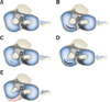
Sixty stifle joints were used in this study. They were collected from thirty mature dog cadavers, which were euthanized for reasons unrelated to this study. Dogs were not selected from specific breeds, but they were similar in size, with an average mass of 2.90 ± 1.21 kg (mean ± SD; range 1.32–5.89 kg). Radiographic images of the pelvic limbs and evaluations of the stifle joint confirmed that the specimens had no apparent orthopedic abnormalities. The cadavers were stored at −40℃ until used for the present study. By using computer software (Microsoft Excel; Microsoft, USA), The cadavers were randomly assigned to one of five groups: Group 1, no medial meniscal injury; Group 2, full-thickness vertical longitudinal tear; Group 3, partial-thickness vertical longitudinal tear; Group 4, multiple vertical longitudinal tears, and Group 5, peripheral detachment of the medial meniscus (Fig. 1). Twelve stifle joints were included in each test group, and no distinction was made between the left and the right joints.
Cadaver preparation
Pelvic limbs were thawed for 24 h at room temperature. Each limb was placed in a dorsally recumbent position. The hind limb of each dog was clipped with electric clippers. Self-adherent wrap was wrapped around the tails and feet, and the bodies were covered from waist to head with opaque surgical wrap (KimGuard; Kimberly-Clark, USA). The surgical site was kept moist during the surgery by spraying with saline solution. Two skin incisions (3–5 cm long) were made proximal to the tibial tuberosity and medial to the patellar ligament. Along the line between these incisions, subcutaneous tissue, medial retinaculum, and the joint capsule were cut to expose the joint. With the stifle joint flexed and the patella laterally luxated, the CrCL was identified, and desmotomy of the CrCL was performed. To minimize iatrogenic damage to the meniscus or other joint structures, no instruments were used for leverage. After the caudal horn of the medial meniscus was exposed and based on the experimental group to which the cadaver had been assigned, the simulated injury was created at the medial meniscus by using a No. 12 scalpel blade (Paragon Carbon Sterile Steel Blade; Medicom, UK). For the group with no medial meniscal injury, no injury was created. In the full-thickness vertical longitudinal tear group, a 3-mm-long injury that completely penetrated from the dorsal aspect to the ventral aspect of the meniscus was created. In the partial-thickness vertical longitudinal tear group, a 2-mm-long injury was created on the ventral aspect of the medial meniscus. In the multiple vertical longitudinal tear group, both a full-thickness vertical longitudinal tear and a partial-thickness vertical longitudinal tear were created. In the peripheral detachment of the medial meniscus group, the coronal ligament attaching the caudal horn of the medial meniscus to the joint capsule was transected with a No. 11 scalpel blade (Paragon Carbon Sterile Steel Blade; Medicom). After creating the meniscal injury, each layer of the surgical incision was closed.
Arthroscopic evaluation of the cadaver study
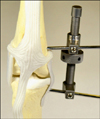
By using computer software (Microsoft Excel; Microsoft), arthroscopic evaluation of each stifle joint was randomly assigned to one of the following two treatment groups: arthroscopy alone or arthroscopy with the aid of an external joint distractor (Extra Articular Stifle Distractor; Veterinary Instrument, UK) (Fig. 2). All evaluations were performed by a single surgeon (HB Lee) with seven years of experiences in arthroscopy. The surgeon was blinded to the status of the medial meniscus of each stifle joint, and arthroscopic evaluation was performed on each specimen in a random order. After arthroscopic evaluation was performed for all specimens, a second arthroscopic evaluation was performed, but with the specimens presented in a different order.
In both groups, the medial meniscus was evaluated by using 2.5 mm and 1.8 mm meniscal probes (Meniscal Probe MPL/CCL Probe; BS.Corem, Korea). Arthroscopy of the stifle joint was performed by using a beveled-lens arthroscope (58 mm long, 1.9 mm diameter, 30° angle) with a 1080p HD three-chip camera (SynergyHD3; Arthrex, USA). Each cadaver was positioned in a dorsally recumbent position. The limb to be operated on was held in a vertical position with a brace. When a joint distractor was used, pins were inserted at the level of the medial fabella in the distal femur and at the level of the tibial tuberosity in the proximal tibia. A proximal percutaneous pin (1.6 mm diameter) was inserted into the distal femur at the level of the medical fabella. A distal percutaneous pin (1.6 mm diameter) was inserted into the proximal tibia at the level of the tibial tuberosity, as described by Böttcher et al. [3]. The distractor was installed to force the two pins apart and enlarge the viewing space. The joint was infused with lactated Ringer's solution with a 24-gauge needle until there was moderate pressure on the syringe. The estimated amount of fluid injected was 2 to 3 mL. After removal of the needle, a stab incision was made through the skin lateral to the patellar tendon and between the patella and the tibia for use as the primary arthroscopic portal. An arthroscope cannula with a blunt obturator was inserted into the joint. Then, the obturator was removed, and the arthroscope was inserted into the cannula. The fluid pressure was 35 mmHg and was regulated by a fluid pump (Continuous Wave III Arthroscopy Pump; Arthrex), but an egress portal was not created. The instrument portal was created on the side opposite the arthroscopic portal and was medial to the patellar tendon. The arthroscope was inserted proximal to the fat pad to avoid obstruction. However, if the fat pad blocked the arthroscopic view, a minimal amount of the pad was removed with a burr-type power shaver. During arthroscopy without a joint distractor, the assistant attempted to enlarge the space around the meniscus by forcing the joint into valgus and pulling the proximal end of the tibia in a cranial direction.
Statistical analysis
The statistical analyses used in our study have been described previously [11]. The body masses of the dogs in the 5 medial meniscal groups were checked for distribution normality with a Kolmogorov-Smirnov test and were compared with a one-way ANOVA. The threshold for statistical significance was set at p < 0.05. The diagnoses were classified as true positive, false positive, false negative, or true negative, and 2 × 2 contingency tables were constructed for each of the 5 groups. The sensitivity (Sn), specificity (Sp), positive predictive value (PPV), negative predictive value (NPV), and correct classification rate (CCR) for each method and the odds ratio (OR) comparing the two methods, along with the respective 95% confidence intervals (CIs), were calculated. The 2 × 2 contingency tables were used to compare the two arthroscopic methods. The 95% CIs were calculated by using Fisher's exact test. All analyses were performed with GraphPad Prism 5 (GraphPad Software, USA).
Results
Assessment of canine medial meniscal injury model
All stifle joints were positive for cranial drawer signs due to the desmotomy of the CrCL. Medial patellar luxation was present in 4 stifle joints (6.7%), but it could be reduced manually. No limbs were excluded because of an intact CrCL, medial patellar luxation, or altered state of the medial meniscus.
Sensitivity, specificity, positive predictive value, negative predictive value, correct classification rate, and odds ratio
The results of the statistical analyses are summarized in Table 1. The Sn, Sp, PPV, NPV, and CC were calculated separately for each diagnostic method, and an OR was calculated to compare the two methods. The use of a joint distractor increased the Sn and Sp. The Sn and Sp of arthroscopy using a joint distractor were 85% (95% CI, 73–93) and 96% (95% CI, 93–98), respectively, whereas the Sn and Sp of arthroscopy alone were 60% (95% CI, 47–72) and 92% (95% CI, 88–95), respectively. The difference between the methods was statistically significant for Sn (p = 0.0038), but not for Sp. The joint distractor increased the PPV and NPV from 65% (95% CI, 51–78) to 85% (95% CI, 73–93) and from 90% (95% CI, 86–94) to 96% (95% CI, 93–98), respectively. The difference between the methods was significant in both cases (p = 0.0175 and p = 0.0107, respectively). The use of the joint distractor also significantly increased the CCR (p = 0.0010) from 86% (95% CI, 80–90) to 94% (95% CI, 89–97). When the results from all stifle joints were pooled, the use of the joint distractor resulted in a 2.99-fold increase in the likelihood of accurately diagnosing the state of the medial meniscus (95% CI, 1.22–7.36) (Fig. 3).
Discussion
Because of the narrow space within the stifle joint in small-breed dogs, it has taken longer for arthroscopy to become accepted as a diagnostic method for this group than for large-breed dogs. This study examined whether arthroscopy can be used to diagnose medial meniscal injuries in small-breed dogs. It also examined the effect of using a joint distractor during arthroscopy. To determine the effectiveness of arthroscopy, cadaveric stifle joints from small-breed dogs were given surgical meniscal injuries via craniomedial incision. A previous cadaveric study of meniscal injuries in large-breed dogs used a craniocaudal incision on the medial aspect of the stifle joint [12], which may have some advantages over a craniomedial incision in terms of seeing the medial meniscus and avoiding coincidence with the arthroscopic instrument portal. However, a transverse incision in the collateral ligament and the joint capsule can unexpectedly force the joint into valgus or varus. Therefore, a craniomedial longitudinal incision was made in this study, despite its potential to interfere with the arthroscopic instrument portal.
Before performing the injury-model surgery, all stifle joints were confirmed to be free of apparent orthopedic abnormalities, such as patellar luxation. However, medial patellar luxation was detected in 4 stifle joints after surgery, which could have been due to the excessive tension generated by the suturing procedure. Although the influence of patellar luxation on arthroscopy used for the diagnosis of medial meniscal injury was not evaluated objectively in this study, the time taken for the arthroscopic examination was increased because the assistant had to expend considerable effort to replace the patella to its normal position.
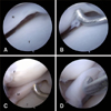
The meniscal state was defined as torn or intact based on what was visible via arthroscopy when moving the stifle joint through its range-of-motion or by probing the meniscus [11]. In a previous cadaveric study, arthroscopy combined with probing of the meniscus was shown to be more sensitive and specific than arthroscopic observation alone, and probing resulted in an 8.0-fold increase in the likelihood of an accurate diagnosis [12]. However, compared to large-breed dogs, a greater ability to manipulate a probe and smaller instrumentation may be required for the arthroscopic examination of stifle joints in small-breed dogs [11]. In this study, arthroscopy was attempted with two different probes (2.5 mm and 1.8 mm). The 2.5 mm probe was too large to easily manipulate in the narrow joint space, and the probe did not reach the meniscus, even with the aid of an assistant or the joint distractor. The 1.8 mm probe made evaluation of the state of the meniscus possible. Given that the menisci and joint spaces in small-breed dogs are smaller than those of large-breed dogs, this study noted limitations when performing arthroscopic examination alone.
Previously reported arthroscopic accuracy metrics (83% Sn, 96% Sp, and 93% CCR) for the diagnosis of meniscal lesions in large-breed dogs were higher than those in the present study (60% Sn, 92% Sp, and 86% CCR) [11]. However, the accuracy of arthroscopy with the use of a joint distractor in the present study (85% Sn, 96% Sp, and 94% CCR) was superior or equal to that found in arthroscopy on large-breed dogs. In this study, the joint distractor enhanced the Sn, PPV, NPV, and CCR of the arthroscopic examination and made an accurate diagnosis 2.99 times more likely than with the use of arthroscopy alone. The Sp also increased with the use of the joint distractor, but the increase was not statistically significant (p = 0.078). In this study, arthroscopy with probing and a joint distractor had a high Sn and PPV and may, therefore, provide reliable information about the state of the meniscus. However, the present study has some limitations. First, arthroscopic evaluation might be more difficult in stifle joints with chronic CrCL diseases such as periarticular fibrosis and synovial proliferation. In addition, periarticular fibrosis in stifle joints due to CrCL diseases may interfere with joint distraction [8]. Therefore, further clinical study is required to evaluate the effectiveness of joint distraction.
The peripheral 10 to 25% of the meniscus is supplied with blood vessels [1]. Meniscal injuries in this region have the capacity to regenerate, whereas meniscal injuries in the axial 2/3 of the meniscus lack vascularity and often need surgical treatment, such as meniscal release, partial or total meniscectomy, or meniscal repair, depending on the type of injury and its location. In this study, the high CCR (94%) of arthroscopy with the use of a probe and joint distractor indicates that arthroscopy can provide an accurate diagnosis of meniscal pathologies and allow surgeons to select the appropriate surgical options.
Arthroscopy is a reliable method for diagnosing medial meniscal pathologies in canine patients. However, it is rarely attempted in small-breed dogs because of their narrow stifle joint space. The present study determined and reported the sensitivity, specificity, positive predictive value, negative predictive value, and correct classification rate for arthroscopy with and without a joint distractor. The results support the idea that arthroscopy is an effective method for the diagnosis of medial meniscal pathologies in small-breed dogs, particularly when performed with the aid of a joint distractor.

 XML Download