

 PDF
PDF Citation
 Print
Print


Introduction
Critical illness can be associated with dysfunction in multiple organs and remarkable endocrine and metabolic changes [625]. Alterations in the circulating levels of thyroid hormones have been widely documented in human medicine and may affect 60% to 70% of critically ill patients with various diseases [6]. This condition is typically characterized by a reduction in the concentration of serum total triiodothyronine (TT3) and a concurrent rise of serum reverse-T3 (rT3) levels; as well, low serum total thyroxine (TT4), free thyroxine (fT4), and, occasionally, thyrotropin (TSH) concentrations are reported with severe and protracted illness [1427]. These are usually transient abnormalities in otherwise euthyroid patients and are commonly recognized under the name of non-thyroidal illness (NTI) [427]. The pathogenesis of NTI seems to be multifactorial and mainly attributed to a reduced peripheral deiodination of TT4 to TT3, increased deiodination of TT3 to diiodothyronine, reduced binding of thyroid hormones to transport proteins and nuclear receptors, and impaired intracellular uptake; behind these mechanisms the roles of protracted fasting, hypoxia, ischemia-reperfusion injury, and inflammatory cytokines have been investigated [4].
Thyroid hormones are important for homeostasis and adaptation to stress and pathological conditions, and several studies in critically ill human patients have linked the presence of NTI with poor outcomes and disease severity [14]. There is evidence that an acute fall in circulating thyroid hormone concentrations during acute critical illness could represent an adaptive response to reduce energy expenditure and protein breakdown; in contrast, low TT3 and TT4 serum levels during a prolonged or chronic phase of critical illness could be maladaptive [4]. Consensus on therapeutic implications of the above-mentioned abnormalities is currently lacking [4].
There are few reports regarding NTI in veterinary critical care. The syndrome has been documented in some acute conditions in dogs, but its prognostic significance remains unclear [202425]. In a population of puppies with parvoviral enteritis, non-survivors had significantly lower concentrations of serum TT4 during hospitalization [24]. In addition, in a group of dogs with naturally occurring infection by Babesia canis rossi, lower values of serum TT4 and fT4 were documented in non-survivors [26]. Alterations in TT4, fT4, and TSH concentrations were demonstrated upon admission in dogs with systemic inflammatory response syndrome (SIRS) and sepsis, but no relationship to outcome was identified [20]. Derangement of the thyroid axis was documented in chronic inflammatory conditions and during heterogeneous non-thyroidal diseases [131523]. Finally, significant abnormalities in thyroid function test results have been reported in healthy euthyroid dogs during anesthesia or surgical procedures [28].
The aim of the present retrospective study was to assess the prognostic significance of serum thyroid hormones, including free T3 (fT3), TT3, rT3, and TT4, in a population of dogs with SIRS. We hypothesized that lower serum thyroid hormones concentrations were associated with disease severity (APPLEfast scores) and mortality (survival at hospital discharge).
Materials and Methods
This study involved a retrospective analysis of a population of dogs affected by SIRS associated with acute pancreatitis, parvoviral enteritis, or septic peritonitis that was prospectively enrolled in a previous study performed at our Veterinary Teaching Hospital (VTH) between February 2012 and January 2014. The study was approved by the local Scientific Ethical Committee for Animal Testing (ID 22/79/2014).
Dogs were included in the study if they exhibited two or more of the following criteria: body temperature < 38.1℃ or > 39.2℃; heart rate > 120/min; respiratory rate > 20/min; WBC count < 6,000/µL or > 16,000/µL, percentage of band cells > 3% of the total WBC count, or a serum C-reactive protein (CRP) concentration >1.68 mg/dL [510]. At least one aliquot of serum collected at the time of hospital admission and stored frozen at −80℃ was obtained from each dog. Dogs were excluded if thyroid hormones or drugs capable of suppressing the thyroid axis (e.g., glucocorticoids, anti-inflammatory drugs, anticonvulsants, and sulphonamides) had been administered in the month prior to hospital admission. Age-matched dogs (n = 15), presented at the VTH for routine screening and prophylaxis, were included as healthy controls based on their anamnestic, physical, and clinicopathological data.
The study population of SIRS dogs was divided in groups according to the origin of SIRS. Specifically, the non-septic SIRS group included dogs affected by acute pancreatitis, while the septic SIRS group included dogs with parvoviral enteritis and septic peritonitis.
Acute pancreatitis was diagnosed by the presence of consistent clinical signs, characteristic ultrasonographic findings (i.e., hypoechoic and/or enlarged pancreas, hyperechoic mesentery, peritoneal effusion), and a positive canine pancreatic lipase immunoreactivity (cPLI) test result (Canine SNAP cPL; IDEXX Laboratories, USA) [1422]. Clinical diagnosis of parvoviral enteritis was confirmed by a positive real-time polymerase chain reaction for a fecal sample. Sequencing of the VP2 gene was performed to identify antigenic variants of canine parvovirus (CPV) and evaluate their potential associations with disease severity [2]. Septic peritonitis was diagnosed based on cytological or bacteriological evidence of bacterial abdominal infection. The APPLEfast score [11], calculated at the time of hospital admission in order to assess disease severity, and the length of hospital stay were recorded and included as analysis variables. SIRS dogs were also classified as survivors (survived to hospital discharge) or non-survivors (died despite medical treatment or humanely euthanized by the clinical investigators due to moribund conditions or end-stage disease). Dogs that were euthanized for financial reasons were excluded from the study.
Hematological and chemistry profiles, including CRP and albumin concentrations, obtained upon hospital admission were reviewed in all enrolled dogs. Complete blood count was determined by an automated cell counter (ADVIA 2120 Hematology System; Siemens Healthcare Diagnostics, USA). CRP (CRP OSR6147; Beckman Coulter, Germany) level was measured by using an immunoturbidimetric assay that had been previously validated by our group for dog serum samples [8]. All analyses were performed by using an automated chemistry analyzer (OYMPUS AU 400, Olympus Optical, Germany). Serum thyroid hormone levels were measured at the end of the study period in a single batch assay of serum collected upon admission and stored frozen at −80℃. The TT3 and TT4 levels were measured by performing radioimmunoassays (RIA) as previously described [1719]. For analytical purposes, RIA results below the detection limit of the assay (< 0.4 nmol/L for TT3 and < 3.0 nmol/L for TT4) were considered equal to 0.2 nmol/L and 1.5 nmol/L, respectively. The fT3 and rT3 levels were assayed by performing ultraperformance liquid chromatography coupled to tandem mass spectrometry operating in multiple reaction monitoring mode and electrospray ionization positive mode. All analytes were directly determined without the need of derivatization. The linearity of the analytical method was assessed over a wide range of concentrations (0.01–50 ng/mL). The recovery of both fT3 and rT3 was > 82%, with a coefficient of variation < 7%. The within-day and between-day precision ranges were 1.82% to 7.81% and 2.29% to 15.62%, respectively. All investigated variables were also measured in healthy control animals.
Statistical analysis
Normality was checked graphically and by applying the Kolmogorov-Smirnov test. Because of the presence of non-normal distributions for most variables, nonparametric testing was adopted for all analyses. Data were expressed by using standard descriptive statistics and are presented as median and range. The Mann-Whitney U test was used to evaluate differences between the overall population of SIRS and control dogs and for comparisons between survivor and non-survivor SIRS dogs, while a Kruskal-Wallis test was used to compare variables between different groups (controls, septic SIRS, and non-septic SIRS). If the Kruskal-Wallis test result was positive, a Conover test post hoc analysis for pairwise comparison of subgroups was performed. Test result p values < 0.05 were considered statistically significant. Correlation between variables was assessed by using Spearman's Rank correlation coefficient. All analyses were performed by using statistical software (MedCalc Software, Belgium).
Results
Forty-one patients met the inclusion criteria and were classified as SIRS dogs. Among the SIRS dogs, median age (8 months, range 2 months to 15 years) and median body weight (17.3 kg, range 3.9–40.2 kg) were not significantly different from those of the control dogs (median age 4.8 years, range 2 months to 8 years; median body weight 20.6 kg, range 4.7–38.0 kg). Overall, 20 dogs were male and 21 were female. Breed distribution of the study population was as follows: mixed breed dogs (18), Spanish Greyhound (4), Labrador Retriever (3), Spanish Mastiff (3), Standard Poodle (3), American Staffordshire Terrier (2), English Bulldog (2), Rottweiler (1), Weimaraner (1), American Pitbull Terrier (1), Manchester Terrier (1), Great Dane (1), and Bernese Mountain Dog (1). Breed distribution of the control dogs was German Shepard Dog (3), mixed breed dogs (3), Flat Coated Retriever (2), Argentine Mastiff (2), Labrador Retriever (1), Whippet (1), Cocker Spaniel (1), Dogue de Bordeaux (1), and Great Dane (1). Thirty-seven of the 41 dogs were survivors, while 4 were non-survivors. All non-survivors were in the septic SIRS group and had a diagnosis of septic peritonitis. Median duration of hospitalization in SIRS dogs was 7 days (range 1–13 days). SIRS dogs had significantly higher APPLEfast score and serum CRP concentration and significantly lower TT3, TT4, and albumin levels compared to those in control dogs (Table 1).
The overall population was divided in two groups according to the origin of SIRS and final diagnosis. Specifically, the non-septic SIRS group consisted of dogs diagnosed with acute pancreatitis (n = 10), while the septic SIRS group included dogs with septic peritonitis (n = 9) and parvoviral enteritis (n = 22). The CPV variants identified by sequencing of the VP2gene were CPV-2c (19/22) and CPV-2b (3/22).
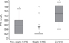
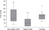
Significantly different clinical and clinicopathological results between septic SIRS and non-septic SIRS dogs are summarized in Table 2. The septic SIRS dogs had a significantly higher APPLEfast score and significantly lower concentrations of serum albumin, TT3, and TT4 than those in non-septic SIRS and control dogs (Figs. 1 and 2). Non-survivors (n = 4) had significantly lower serum albumin (median 15.6 g/L, range 11.3–23.0 g/L) and TT4 concentrations (median 1.5 nmol/L, range 1.5–6.0 nmol/L) compared to survivors (n = 37; median 26.2 g/L, range 18.1–42.9 g/L; median 16 nmol/L, range 1.5–66.0 nmol/L, respectively). Among the survivors, there were no significant correlations between duration of hospital stay and serum thyroid hormone concentrations. Both serum TT4 and TT3 concentrations were negatively correlated with APPLEfast scores (r = −0.4, p < 0.01 and r = −0.3; p < 0.05, respectively). Serum TT4 and albumin concentrations were positively correlated (r = 0.56; p = 0.0001), while the correlation between TT3 and albumin was not significant (r = 0.3; p = 0.05).
Discussion
The presence of NTI has been documented in different human and veterinary critical conditions including systemic inflammation [120]. In the current study, a panel of serum thyroid hormones was assayed in specific canine diseases: acute pancreatitis, parvoviral enteritis, and septic peritonitis. These diseases were included as they are representative and homogeneous spontaneous models of canine infectious and non-infectious SIRS. The high serum CRP concentration in the SIRS dogs confirmed the presence of systemic inflammation in our study population [5]. In order to stratify SIRS patients according to disease severity and mortality risk, as has been previously done [920], the dogs' APPLEfast scores were calculated. There was a significantly higher value APPLEfast score in the septic SIRS group than in the non-septic SIRS and control dogs. The reduction in serum TT4 value in the septic SIRS group is in agreement with previous veterinary reports investigating NTI in similar settings [202425], and is a common finding in clinical studies on canine NTI [1315].
Changes in serum TT3, fT3, and rT3 concentrations have been widely documented in critically ill human patients, but are less reported in veterinary literature [7131518]. Low levels of TT3 are the most common finding and had the strongest correlation with outcome in a retrospective evaluation of thyroid hormones among heterogeneous non-thyroidal diseases in dogs [15]. Similar results were obtained in a retrospective evaluation of thyroid hormones in critically ill dogs requiring intensive care therapy; low TT3 levels were frequently detected and associated with mortality [7]. In our study, a significant reduction in serum TT3 concentration in canine SIRS was observed, indicating its potential as a sensitive marker of NTI, as has been described in humans [27].
An increase in serum rT3 has been reported during NTI in humans [27]. A similar increase has been reported in healthy euthyroid dogs during general anesthesia and surgery [28], and in a small population of healthy dogs following endotoxin administration [18]; however, no similar results in canine species during spontaneous SIRS have been reported. In the present study, the median concentration of rT3 was not significantly different between SIRS and control dogs. This may indicate that rT3 variations may be less susceptible to NTI in spontaneous severe canine disease, at least with respect to TT3 abnormalities. It is also possible that rT3 variations are somehow influenced by the onset of the disease, and that serial monitoring of that hormone may reveal different changes in its concentrations. The relevance of the measurement of serum rT3 during canine NTI was apparently limited in this population of SIRS dogs, and that limited role needs to be examined in further studies.
Regarding serum fT3 concentrations, the difference between SIRS and control dogs was not significant, although lower values were detected in the SIRS group. This may indicate that, as observed in humans [27], serum fT3 values do not parallel changes in serum TT3 concentrations and are little affected by the presence of NTI in dogs, at least under acute inflammatory conditions. However, the performance and accuracy of the assay used in this study should be considered when interpreting our result.
The pathogenesis of NTI is incompletely described but is assumed to be multifactorial. The binding of thyroid hormones to circulating proteins and their metabolism at the tissue level are possibly involved. Circulating thyroid hormones are tightly bound to thyroid-binding proteins, including albumin. Such molecules are negative acute phase proteins and may decrease in acute critical illness. The high prevalence of hypoalbuminemia in SIRS dogs, particularly in septic SIRS, may account for the decreased thyroid hormones concentrations observed in our study population. This observation is partially supported by the moderate correlation between TT4 and albumin concentrations. However, other mechanisms in the fall of serum thyroid hormones, particularly for TT3, should be considered. However, such investigations were beyond the scope of the present study.
Dogs with septic SIRS had significantly lower serum thyroid hormones (TT3 and TT4) and higher APPLEfast scores than those in non-septic SIRS and control dogs. These results may suggest that the prevalence and the degree of NTI is strictly related to severity of illness. The negative correlation observed between thyroid hormones (TT3 and TT4) and the APPLEfast score may further support this statement. Derangement in serum thyroid hormone levels have been previously demonstrated in a cohort of dogs with SIRS; however, no relationship with survival or with SIRS origin (infectious versus non-infectious) was reported [20]. Different analytical methods, case series compositions, and disease categories may have accounted for the different results observed in our study. The presence of NTI has been associated with a negative outcome in different canine diseases [15242526], and low TT3 levels were correlated with mortality in critically ill dogs and in canine heterogeneous non-thyroidal diseases [715]. In addition, low TT4 concentrations were significantly associated with a negative outcome in puppies with parvoviral enteritis at 24 and 48 hours after admission [24].
In our study, significantly lower TT4 values were found in non-survivor dogs with SIRS. In contrast, there was no difference detected between survivors and non-survivors among the other serum thyroid hormones assessed in this study. However, our survival analysis was limited by the low number of non-survivors in our population; the prognostic significance of thyroid hormones in terms of outcome prediction in canine SIRS should be addressed by further studies.
There are some limitations to be considered before interpreting the results of the current study. The retrospective nature of the study limited the measurement of thyroid hormones in multiple standardized time points, and partially restricted the availability of serum samples for evaluation of a more extended thyroid panel (e.g., to also include fT4 and TSH). However, only dogs diagnosed with selected causes of SIRS and with complete clinical and clinicopathological data were included in the study, allowing improved completeness of data available for analysis upon admission. Concerning the method of subgrouping our patients, we decided to include both dogs with parvoviral enteritis and septic peritonitis in the septic SIRS category. Despite both diseases being considered reproducible models of abdominal sepsis [316], a potential age-related difference in clinical and clinicopathological variables among disease groups, including controls, could be a major concern. Specifically, younger dogs with parvoviral enteritis may have partially influenced concentrations of some of the variables investigated (e.g., serum albumin). However, statistical tests performed to compare the different groups divided according to final diagnosis (acute pancreatitis, parvoviral enteritis, septic peritonitis, and controls) produced similar results without adding any other significant information (data not shown). The predominance of variant CPV-2c in our population did not allow comparative analysis of variants in dogs affected by CPV. In addition, breed and sex-related differences have been reported to affect thyroid hormone concentrations in healthy dogs [1221]. Although sex distribution was homogeneous in our population, and only medium-large breed dogs were included, no breed- or sex-matched controls were considered, which may have partially biased the results. It is theoretically possible that some of the SIRS dogs included in the study may have had concurrent hypothyroidism despite the low prevalence of this disease and the lack of historical and clinical features consistent with its presence. Although the authors consider the occurrence of hypothyroidism unlikely in this population, the additional measurement of TSH and fT4 would have better ruled out this hypothesis and completed the thyroidal evaluation in our SIRS dogs. Finally, the data generated from the current study refer to specific categories of canine SIRS and should not be overinterpreted or extended to different diseases or more chronic situations.
In conclusion, our study confirms a wide frequency of serum thyroid hormones alterations can indicate the presence of NTI in a cohort of dogs with SIRS. Serum concentrations of TT3 and TT4 might be considered useful and reproducible markers of NTI during acute inflammatory states in dogs. Thyroid hormones abnormalities were more severe in septic than in non-septic SIRS dogs, and they were positively correlated with APPLEfast scores. The results suggest the presence of extensive thyroid axis impairment in SIRS dogs with severe illness. Whether the presence of NTI should be considered as an adaptive response to a critical disease or the consequence of endocrine system dysfunction and failure remains a topic of debate; as well, there is uncertainty about the need for therapeutic strategies with hormone supplementation. Further prospective, large-scale studies investigating the pathogenesis and the prognostic role of NTI in canine SIRS are warranted.


 XML Download