

 PDF
PDF Citation
 Print
Print


Introduction
Holstein is one of the most preferred cattle breeds in dairy farming. Elite sires have been used extensively all around the world via artificial insemination (AI) to achieve genetic improvement in Holstein cattle breeding. However, as an outcome of inbreeding, the frequency of undesired recessive alleles has increased in Holstein cattle populations. Complex vertebral malformation (CVM) is the most frequent autosomal recessive inherited disorder of Holstein cattle [2]. The solute carrier family 35 member A3 (SLC35A3) gene encodes the golgi-resident nucleotide sugar transporter protein, which has an essential role in a mechanism controlling the formation of vertebrae. A point mutation at the 559th nucleotide that substitutes guanine to thymine (G>T) in SLC35A3 gene is the cause of a critical amino acid divergence that abolishes the function of the nucleotide sugar transporter protein and results in vertebral malformations [24]. Affected calves, which are usually aborted or stillborn, carry two copies of the mutant allele. Affected but live-born calves exhibit low birth-weight, cervical and thoracic vertebral anomalies, scoliosis, and malformations in carpal and tarsal joints, as well as cardiac anomalisms, and they generally die within a few days [117]. The degree of vertebral malformation among affected calves can vary [4]. Cattle that carry one copy of both the mutant and wild-type alleles, are CVM carriers and can transfer the cause of the disorder to the next generation. CVM was first identified in a Danish Holstein population in 1999 [3]. The USA Holstein sire Penstate Ivanhoe Star (US1441440) and his son Carlin-M Ivanhoe Bell (US1667366), which were used intensively in AI, were declared to be the worldwide distributors of the undesired mutant allele [24]. Thereafter, sporadic CVM cases were reported from the USA [9] and the United Kingdom [18]. Subsequently, studies to identify CVM carriers were conducted in Japan [1116], Germany [14], Sweden [5], Czech Republic [8], Poland [20], Turkey [15], China [7232627], Slovakia [10], and Iran [1219]. Allele-specific polymerase chain reaction (AS-PCR) [11], created restriction-site PCR (CRS-PCR) [25], PCR with primer-introduced restriction analysis (PCR-PIRA) [13], and DNA sequencing [15] methods have been reported as capable of detecting the mutant allele that causes the CVM disorder. The aim of this study was to compare the sensitivity, specificity, positive and negative predictive values, accuracy, and rapidity of the AS-PCR, PCR-PIRA, and CRS-PCR methods and to determine the most accurate one for screening the mutant allele in SLC35A3 gene in a Holstein cattle population.
Materials and Methods
Animal Samples and DNA isolation
Holstein cattle (n = 311) were chosen randomly from four different provinces within the Thrace region of Turkey: Edirne (n = 58), Istanbul (n = 54), Kırklareli (n = 91) and Tekirdağ (n = 108). Blood samples from vena jugularis and vena coccygea were taken by using sterile vacuumed EDTA tubes and transferred to our laboratory under appropriate cold chain conditions (between ice packs). Isolation of DNA was performed by using an automated DNA extraction system (Exiprep 16 Plus; Bioneer, Korea).
DNA sequencing and alignment
Amplification of SLC35A3 gene, in order to perform DNA sequencing, was carried out with forward (F): 5′CAGATT CTCAAGAGCTTAATTCTA3′ and reverse (R): 5′TATTTGC AACAACAAGCAGTT3′ primer pairs [15] in a 50 µL reaction mixture including lyophilized PCR PreMix (K-2013 AccuPower PCR PreMix; Bioneer), 1 µL of each primer (50 pM), 5 µL of genomic DNA, and 43 µL of sterile ddH2O (AccuGENE; Lonza, Belgium) under the following conditions: 94° for 5 min and 30 cycles of 94℃ for 45 sec, 52℃ for 45 sec, 72℃ for 1 min, and 72℃ for 10 min. Amplicons were sequenced in both directions on an ABI 3730XL DNA Analyzer by the DNA Sequencing Service, Bioneer, Korea. In order to validate mutant and wild-type alleles, sequences were aligned with the reference sequence of the bovine SLC35A3 gene from the National Center for Biotechnology Information (GenBank No. HM183012.1) and screened for G>T mutation by using the BioEdit sequence alignment editor software (ver. 7.2.5; Ibis Therapeutics, USA).
Amplification of SLC35A3 gene
The related region of SLC35A3 gene was amplified by applying AS-PCR, CRS-PCR, and PCR-PIRA methods with a common PCR mixture in a 20 µL volume including lyophilized PCR PreMix (K-2012 AccuPower PCR PreMix; Bioneer), 0.5 µL 10 pM of each primer, 3 µL of genomic DNA and 16 µL of sterile ddH2O (AccuGENE Water; Lonza).
AS-PCR
In order to identify wild-type and mutant alleles of SLC35A3 gene, two PCR reactions were carried out for each sample by using two allele-specific forward primers for mutant (CACAATTTGTAGGTCTCATGGCAG) and wild-type alleles (CACAATTTGTAGGTCTCATGGCAT), and a common reverse primer (GTTATACTACAGGAGTCACCTCT) [11]. Amplification was performed under the following conditions: denaturing at 95℃ for 2 min and 30 cycles of 95℃ for 30 sec, 62℃ for 30 sec, 72℃ for 1 min, and final extension at 72℃ for 5 min.
CRS-PCR
Amplification of CRS-PCR was performed with F: 5′-GC TCTCCTCTGTAATCCCCA-3′ and R: 5′-CCACTGGAAA AACTAGCTGTGAGTA-3′ primer pairs [25] under the following conditions: denaturing at 95℃ for 2 min and 35 cycles of 95℃ for 30 sec, 60℃ for 30 sec, 72℃C for 30 sec, and final extension at 72℃ for 10 min. Amplicons were restricted by using the RsaI fast digest enzyme (Thermo Scientific, USA) at 37℃ for 15 min.
PCR-PIRA
Amplification of SLC35A3 gene was performed by applying PCR-PIRA with CACAATTTGTAGGTCTCACTGCA and CGATGAAAAAGGAACCAAAAGGG primer pairs [13] under the following conditions: denaturing at 94℃ for 2 min and 35 cycles of 94℃ for 30 sec, 56℃ for 30 sec, 72℃ for 60 sec, and final extension at 72℃ for 10 min. Amplicons were digested by using the PstI fast digest enzyme (Thermo Scientific) at 37℃ for 15 min.
The difference between the CRS-PCR and PCR-PIRA methods may be briefly summarized as follows: in CRS-PCR a restriction site is created for the RsaI enzyme (CATG) with the reverse primer in the wild-type allele, whereas in PCR-PIRA a restriction site is introduced for the PstI enzyme (CTGCAG) with the forward primer in the wild-type allele.
Results
DNA sequencing
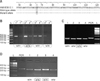
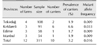
Discrimination of wild-type and mutant alleles among 311 Holstein cattle was performed by monitoring for the G>T mutation. Ten of the sampled cattle were identified as carriers based on the DNA sequencing results (panel A in Fig. 1). The prevalence of carriers and the frequency of the mutant allele were 3.2% and 0.016, respectively. The distribution of CVM carriers and the frequencies of mutant allele in cattle sampled in the four Thrace region provinces are summarized in Table 1.
Amplification of SLC35A3 gene
AS-PCR: Two separate amplification reactions were performed for each sample, each with allele-specific primer pairs for mutant and wild-type alleles and a common reverse primer. The expected results were the presence of DNA bands after separate amplification with both mutant and wild-type allele-specific primers and presence of a DNA band with only the wild-type allele-specific primer in carrier and non-carrier samples, respectively. Amplicons of 395 bp were visualized on 2% agarose gel (panel B in Fig. 1). Two carrier (true positive [TP]) and 241 non-carrier (true negative [TN]) cattle were identified accurately by using the AS-PCR method; however, it misidentified 60 non-carriers as carriers (false positive [FP]) and 8 carriers as non-carriers (false negative [FN]).
CRS-PCR: The amplicons obtained via CRS-PCR comprised 225 bp. After digesting the samples with the RsaI enzyme, the mutant allele remained undigested at 225 bp, but the wild-type allele was digested into two DNA fragments (201 bp and 14 bp). Therefore, non-carriers consisted of 201 bp, while carriers included both 225 bp and 201 bp on 3% agarose gel (panel C in Fig. 1). Although all non-carrier cattle were identified accurately by CRS-PCR, one carrier was misidentified as a non-carrier (FN = 1).
PCR-PIRA: Amplification of SLC35A3 gene via PCR-PIRA resulted in amplicons of 287 bp. After performing digestion of the PCR-PIRA products with the PstI enzyme, non-carrier amplicons with the wild-type allele were cut into 264 bp and 23 bp DNA fragments, but only the 264 bp was visualized on 3% agarose gel. After PstI digestion, carrier amplicons showed two DNA fragments (264 bp and 287 bp) on 3% agarose gel electrophoresis. In addition to using the DNA ladder (100 bp), the PCR-PIRA amplicons (287 bp) were also used as markers for evaluating the band pattern of the mutant allele (panel D in Fig. 1). All PCR-PIRA samples gave the same results as those obtained from DNA sequencing (TP = 301; TN = 10).
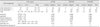
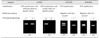
Sensitivity, specificity, negative and positive predictive values, accuracy, and inaccuracy values obtained from the AS-PCR, CRS-PCR, PCR-PIRA methods were compared by applying 2 × 2 table analysis of the TP, FP, FN, and TN values for each method. DNA sequencing provided the gold standard test results. All formulae and results, including estimates of the duration of each method, are summarized in Table 2. Brief protocols of the AS-PCR, CRS-PCR, and PCR-PIRA methods are displayed in Table 3.
Discussion
Thrace is an animal health and transport link between Europe and Asia. In the present study, prevalence of CVM carriers in the Thrace region was identified via DNA sequencing. Of the 10 CVM-carrier cattle, 6 were identified in Kırklareli province, which is a border province between Turkey and Bulgaria. The prevalence of CVM carriers in Kırklareli was higher (6.6%) than that in Tekirdağ (1.9%), Istanbul (1.9%), and Edirne (1.7%) provinces, and the mutant allele frequency was also the highest in Kırklareli (0.033). Total prevalence of CVM carriers and total frequency of the mutant allele were 3.2% and 0.016, respectively, in the sampled population of Holstein cattle raised in the Thrace region of Turkey. Meydan et al. [15] reported that the prevalence of CVM carrier and frequency of the associated mutant allele were 3.4% and 0.017, respectively, in Ankara and Şanlıurfa, which are two provinces in the Asian part of Turkey. Although that study and the present one were conducted in two different continental regions of Turkey, the results were very similar. On that basis, it can be concluded that the prevalence of CVM carriers remains approximately the same within the borders of Turkey.
The prevalence of CVM carriers in Thrace region of Turkey (3.2%) was lower than those for Holstein cattle reared in Japan (32.5% and 13.0%) [1116], Denmark (31.0%) [24], Poland (24.8%) [20], Sweden (23.0%) [5], Germany (13.2%) [14], China (15.6%, 14.7%, 9.54%, and 7.7%) [7232627], and Slovakia (8.5%) [10], but it was higher than that in Iran [1219] (0%). The instructions on procedures and principles associated with sperm imports to Turkey was revised for CVM-carrier bull sperm by the Ministry of Food Agriculture and Livestock in 2007. According to this regulation, a tested free/CVM free indicator should be included in the pedigree of Holstein bulls in order to identify them as CVM disease free. If this indicator is not present in the bull's pedigree, then a certificate approved by the competent authority must be submitted in order to prove that the bull is not a CVM carrier. The lower prevalence of CVM carriers in Turkey compared to prevalences in other countries might be maintained via this regulation. The widespread use of untested sperm before the 2007 revision of procedures and principles for sperm import and the continued usage of untested bulls in breeding might be the main reasons why Turkey still has CVM-carrier cattle.
The frequency of CVM carriers decreased in the German Holstein sire population from 16.6% to 4.6% between the years 2002 and 2007 [21] through elimination efforts. The main challenging factor in eradication strategies for autosomal recessive inherited disorders is detecting the carrier animals accurately. Therefore, we compared AS-PCR, CRS-PCR and PCR-PIRA methods according to their sensitivity, specificity, positive and negative predictivity, accuracy, and inaccuracy in a Holstein cattle population. Among three methods, AS-PCR was the most rapid one (≤ 2 h). However, it showed the lowest specificity (80%), sensitivity (20%), positive predictivity (3%), negative predictivity (97%), and accuracy (78%) but the highest inaccuracy (22%). The time required to perform CRS-PCR and PCR-PIRA methods was approximately equal (≤ 3 h). Sensitivity of CRS-PCR (90%) was less than PCR-PIRA (100%), but specificity and positive predictive value of CRS-PCR and PCR-PIRA were equal (100%). Accuracy and negative predictive value of PCR-PIRA (100%) was higher than that of CRS-PCR (99.7%), and CRS-PCR showed higher inaccuracy (0.3%) than that of PCR-PIRA (0%).
In conclusion, the presence of the CVM mutant allele among the Holstein cattle population in the Thrace region of Turkey was identified for the first time in this study. We assume that, before implementation of the 2007 regulation on the import of CVM-carrier sperm, untested bulls and/or untested sperm were used more extensively in Kırklareli province than in other provinces in the Thrace region. In order to prevent the spread of the CVM mutant allele, further studies should be carried out to identify CVM carriers among breeding sires and imported Holstein heifers. In addition, all semen samples used in AI should be tested. The results of this study may encourage researchers to select the right methodology to use in studies into the prevention of CVM disorder and might constitute a step toward CVM mutant allele elimination strategies in the Thrace region. Considering specificity, sensitivity, positive and negative predictive values, accuracy and inaccuracy values, and rapidity of the three methods, PCR-PIRA is suggested to be the most efficient approach to screening for CVM carriers in a Holstein cattle population.
 XML Download