

 PDF
PDF ePub
ePub Citation
 Print
Print


Introduction
Canine elbow dysplasia (ED) is abnormal development of the elbow joint [11,12]. This condition was defined by the International Elbow Working Group (IEWG) in 1993 to include fragmented medial coronoid process (FMCP), osteochondrosis of the humerus (OC), ununited anconeal process (UAP), articular cartilage injury, and incongruity of the elbow joint. These disorders are associated with varying degrees of joint instability, inflammation, and loose fragments within the joint that result in lameness and osteoarthrosis (OA) [6,8,11,12,16,17]. Several epidemiological studies have examined the genetic basis of ED, a condition that appears to be inherited differently among different breeds [8,16,18]. Most cases of ED first present at 6-12 months of age as forelimb lameness although some dogs present later in life (<6 years old) [11,12,16].
Under normal circumstances, dogs should be radiologically evaluated at 12 months of age. Radiography has been the standard-of-care imaging modality for the diagnosis, grading, and registry of ED. Because radiography is widely available, efficient, and cost-effective, comprehensive radiographic assessment will likely continue to be a valuable component for diagnosing ED [6,11,12,17]. Classification of ED cases according to the IEWG protocol is based on the existence and severity of arthritic changes on the joint surfaces as well as the presence of one or more of the following changes: UAP, OC, FMCP, and joint malformation or incongruity [6,8,15].
Treatment of ED should ideally involve correcting the underlying causes before significant joint damage has occurred. During the early stages of the disease different non-surgical therapeutic measures such as analgesic therapy (NSAIDs), weight loss, exercise restriction, functional food consumption, nutritional supplements, physiotherapy, and other complementary modalities can be considered [8,10,11]. In addition, numerous surgical procedures and chondroprotective formulations for managing established cases and the accompanying symptoms have been developed [8,11,16,17]. However, little is known about preventive treatments for ED.
Taking into account the data presented above, the current investigation focused on two different objectives. First, an oral supplement as a preventative measure was evaluated by comparing the number of dogs that developed ED in treated versus untreated groups. Secondly, effect of the treatment on symptoms of ED once the illness has been diagnosed was assessed.
Materials and Methods
This randomised, controlled, prospective, phase IV pilot study was approved by the Animal Welfare Committee of the Organización Nacional de Ciegos Españoles (ONCE) Guide Dog Foundation. The investigation was conducted at the ONCE Guide Dog Foundation training centre (Spain). All animals included in the study were from this facility. Animal care was conducted according to the protocols of the centre. Clinical evaluation and continuous monitoring of the animals were also performed.
Case selection criteria
Several factors were considered when selecting animals for the study. The dogs had to be healthy, at least 3 months old, purebred Labrador retriever or Golden retriever mix, and from internal litters (these litters were born at the animal facility where the study was conducted). Animals of both genders were included without distinction and were neutered during the study. All the dogs were vaccinated (Eurican Pneumo and Eurican MHPPi2-L; Merial, France). Canines were excluded from the study if they had any disease that could confound or interfere with the evaluation, had sustained a previous bone fracture or trauma, were taking concomitant chondroprotective medication, or had been selected for breeding. A total of 105 Labrador dogs met the selection criteria.
Clinical study design
All the dogs were divided into two groups using random number generator software before the start of the study. The control group was fed a specific diet (Puppy; Eukanuba, Canada) without supplementation while the treatment group received the same diet in addition to oral supplement tablets (Hyaloral; Pharmadiet, Spain) during all the study. The supplement contained 20 mg of hyaluronic acid, 2.2 g of enzymatically hydrolysed collagen, 312.5 mg of crystallised glucosamine, 200 mg of chondroitin sulphate, and 100 mg of gamma oryzanol per tablet. Dosage was based on body weight (dose: ½ tablet/day for each 10 kg of body weight, 1 tablet/day for each 20 kg of weight, 1.5 tablets/day for each 30 kg of weight, and 2 tablets/day for each 40 kg of weight).
Clinical evaluation was performed during four follow-up visits when the animals were 3, 6, 12 and 20 months old. The following outcomes were evaluated by two different veterinarians (SMA and ADR): physical examination results (medical history and vital signs), orthopaedic evaluation of the elbow joints (lameness, range of motion, and swelling), radiographs, serology and blood analysis data, and subjective veterinarian assessment of clinical signs and symptoms of dysplasia for each joint (palpation, pain, and gait). A qualitative analysis was performed for the orthopaedic evaluation. Scoring was based on a scale from 0 to 3 for the following characteristics:
We have used this modified orthopaedic scale as a tool at the ONCE Guide Dog Foundation for routine clinical evaluation without anaesthesia or sedation. Based on our experience, this scale can be used to identify positive correlations between radiograph findings and clinical signs of the disease.
For the radiological examination, we evaluated two different X-ray images for the elbow joint: one taken in the medio-lateral position with forced flexion (ML) and the other in a cranio-caudal position with the animals under general anaesthesia or deep sedation. The current IEWG elbow screening protocol includes submission of these specific quality flexed ML radiographs of both elbows for osteophyte evaluation [18]. Next, we classified the degree of dysplasia according to the IEWG protocol [8]: normal (grade 0), mild (grade 1, mild joint incongruity, osteophytes less than 2 mm high), moderate (grade 2, clear incongruity, osteophytes 2-5 mm high), or severe (grade 3, evidence of primary pathology, osteophytes higher than 5 mm) dysplasia.
Current, three-dimensional imaging techniques such as computed tomography (CT) and magnetic resonance imaging (MRI) are the most reliable diagnostic methods. Due to financial considerations, radiographs remain the most cost-effective method of diagnosing ED. Optimal radiographic detail is essential to accurately evaluate elbow pathology, and we used the same technique and independent radiologist (JPS) to perform and evaluate all radiographs. However, we only evaluated radiographic changes and their evolution given that the imaging modality used relies on the progression of OA signs for ED to be detected.
The study was divided in two phases. During the first phase (when the animals were 3 to 12 months old), we evaluated the oral supplement as a preventative measure by comparing the number of dogs that developed ED in each group. After the animals were old enough (12 months) to confirm a diagnosis of ED, radiological control was performed. Then the second phase of the study was initiated (until the animals were 20 months old). Animals in both groups with radiological signs of dysplasia were considered to be unsuitable and were withdrawn from being guide dogs, but remained in the study until the last follow-up visit to evaluate the effect of the supplement on ED signs and symptoms. All the animals were also closely monitored for the possible appearance of undesirable side effects.
Statistical analysis
Statistical analysis was carried out using SPSS for Windows (ver. 19.0; IBM, USA). All outcome measures were summarised using descriptive statistics. Baseline characteristics were compared to verify that both groups were similar before starting the study. Normal distribution was monitored for all parameters in order to perform the correct inferential statistical analysis. Fisher's exact test was used to compare differences in the incidence of dysplasia and symptoms between groups. Mann-Whitney U and Friedman and Wilcoxon tests were performed to compare values between or within groups, respectively, for the orthopaedic controls and supplement efficacy. An intention-to-treat analysis was conducted. If a dog dropped out in both groups the latest values were carried forward. The confidence interval (1-α) was set at 95% with a significance cut-off value of 0.05 and power of 90%.
Results
We included 105 dogs in our investigation. Three left the study for reasons unrelated to the experiments, two left due to behaviour not compatible with training, and one died due to a clotting disorder. Since most of the animals were affected bilaterally by ED, the most affected joint was always considered when assessing signs and symptoms.
The animals were randomly divided into two groups. At the start of the study, the groups were homogeneous in terms of weight and data for all physical examinations performed (this homogeneity was maintained throughout the study). No differences were observed between the groups for any of the findings from the physiological or joint evaluations. None of the animals included in either group presented signs or symptoms of dysplasia at 3 months of age (baseline).
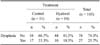
For the primary endpoint of prevention, we analysed outcomes relating to the incidence of dysplasia (radiologically confirmed) at 12 months and found that 33.3% of dogs in the control group had dysplasia compared to 18.5% in the treatment group (Table 1). All cases of ED were classified as grade 2 (moderate) with 100% (n = 13) in the control group being OC while 75% (n = 6) of the cases in the treatment group were OC and 25% (n = 2) were FMCP.
To evaluate the therapeutic efficacy (the secondary endpoint) of the supplement, all the analyses were performed for animals with radiologically confirmed diagnoses of ED both from the treatment and control groups. The rest of the animals (ones without ED) were followed. None of these dogs developed signs or symptoms of dysplasia throughout the study.
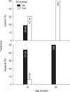
When analysing the symptoms of dysplasia at 12 months of age (Fig. 1), differences were found between the treatment group (12.5%) and control group (61.5%; p = 0.067). These differences were found to be significant at the last visit (p < 0.05). When the animals were 20 months old, none of the treated dogs had joint symptoms associated with joint dysplasia while these symptoms persisted in the control group (Fig. 1).
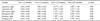
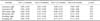
Changes in orthopaedic evaluation findings (lameness, range of motion, and swelling) over time were significantly different only in the control group for which symptom severity increased throughout the study (Table 2). In the treatment group, symptoms occurred to a lesser extent or were not observed, and there were no significant differences over time since the symptoms improved (Table 3). Differences in orthopaedic evaluation data for the most affected joints were found when comparing the groups (p < 0.05) at 12 months. The control group had mainly left-sided lameness as well as a lesser range of motion and swelling both on the right and left. At the last follow-up visit, differences between the two groups increased and were significant (p < 0.05) for all the parameters evaluated: lameness, range of motion, and swelling in the right and left elbows (Table 4). However, radiographic signs of dysplasia were still observed in animals from both groups at 20 months of age.
For the veterinarian clinical evaluation during which the general state of the elbow joints was analyzed, we observed that there was a statistically significant difference within the treatment group between visits 4 and 3 for the right joint in which symptoms significantly improved. The left joint also improved but not significantly. In the control group, statistically significant differences between visits 3 and 4 compared to visit 2 were identified. In both cases, symptoms for both the right and left joints worsened. When we compared the veterinarian assessment data between groups for each visit, we observed statistically significant differences for both joints at visits 3 and 4. No differences were found between the study groups for the additional control parameters (blood and serology analyses). Finally, no adverse events were observed for either of the study groups.
Discussion
ED is a joint development disorder associated with visible clinical signs that present in the animal between 6 and 12 months of age [9,11,14,16]. This is a serious problem in large breed dogs. The present study was performed at a guide dog training school. This is important because dogs that are most appropriate for guide work are nowadays golden retrievers, Labradors, and German shepherds that have a high genetic disposition for ED.
The use of oral chondroprotective agents for treating joint diseases such as osteoarthritis (OA) in humans and animals has been widely studied, and the synergistic effects of different nutraceuticals is a step forward in the management of OA [19]. However, a therapeutic effect has not been clearly proven. Some research has been done in animals to study the effects of chondroprotective agents for OA [1,2,4,5,8], but few studies have investigated the use of these reagents as prophylactics or for treating ED [3,7,13,14].
Considering that joints with dysplasia usually show signs of OA and administration of a chondroprotector may ameliorate the progression of clinical osteoarthritis symptoms, this study was designed to evaluate an oral supplement. As one of the objectives, efficacy of the supplement was analyzed by comparing the development of ED in each group (treated versus control). According to the protocols for animal use and management established by the ONCE Guide Dog Foundation training school, the dogs included in our study were followed up at 3, 6, 12, and 20 months of age. Mild signs appeared in some animals and cases of dysplasia were radiologically confirmed in both groups at 12 months as described in several previous studies [11,12,14].
We observed that the number of dogs developing radiographic evidence of ED decreased in the treatment group to 18.5% compared to 33.3% in the control group. In Europe, the prevalence of elbow dysplasia in Labrador dogs is 20~30% [12,15,16]. At our training centre, however, litters selected from parents chosen according to behavioural criteria had a somewhat higher incidence rate. Thus, our results suggest that the treatment modality we developed represents a probable way to prevent the progression of osteoarthritic changes associated with joint dysplasia.
Once joint dysplasia has been detected, treatment modalities should be designed to prevent the progression of the disorder (whenever possible) or minimise symptoms. Most studies conducted to date have evaluated the efficacy of different surgical treatments for a middle stage of elbow dysplasia when there is articular damage [8,11,16]. In contrast, we evaluated the effects of a daily supplement on symptoms and signs of ED over a 17-month period. The results demonstrated that the treatment group showed significant improvement in terms of lameness and range of motion compared to the control animals. All symptoms of dysplasia had improved by 20 months of age in the treatment group compared to the control dogs although radiological signs persisted in both groups. These results suggest that using a daily supplement containing hyaluronate, collagen, and other glycans could be an option for treating the signs and symptoms of joint dysplasia as an alternative to surgery.
Our data coincide with those obtained from a study on the efficacy of pharmacological treatment compared to traditional surgical techniques for FMCP and OC of the elbow [3]. Dogs assigned to the medical treatment group received 3 mg/kg pentosan polysulfate once a week for 4 weeks. The other group underwent medial arthrotomy and partial collateral desmotomy. At the end of the study, lameness and pain had decreased in both groups with no significant differences observed. These results suggest that treatment with pentosan polysulfate is a valid alternative to surgery. Additionally, another study investigated the effect of different doses of glycosaminoglycans for treating various signs of hip dysplasia [7]. The dogs that received 4.4 mg/kg glycosaminoglycans showed the greatest effects in terms of improved orthopaedic scores as opposed to the placebo group that showed the least improvement.
Our findings have identified many questions in need of further investigation. Long-term and blinded clinical trials are required to confirm whether the results of our study could be replicated in other training schools and regular veterinarian practice. This is because most breeders do not currently evaluate the elbows of all of their dogs. Furthermore, reductions of clinical signs associated with ED are judged based on subjective veterinarian analyses.
A number of important limitations also need to be considered. Limitations of the present study included restrictions associated with radiography versus CT and MRI as a method of diagnosing ED, a lack of blinding of the investigators (only the radiologist was independent and blinded), and the inclusion of breeds that have an unusual prevalence of OC compared to FCP. These limitations, especially interpretation of the radiographic data, could have influenced the results of this investigation.
More research and clinical consensus is needed to more reliably verify the use of chondroprotective substances for preventing and treating ED. However, results obtained in our study enable us to conclude that administration of hyaluronic acid, enzymatically hydrolysed collagen, glucosamine, chondroitin sulfate, and gamma oryzanol (Hyaloral) to animals diagnosed with ED significantly reduces clinical signs and symptoms. Moreover, we concluded that the results of this study indicate that giving this dietary supplement to Labradors starting at the age of 3 months may have a potential cumulative action that confers protection against the progression of radiographic osteoarthritic changes associated with ED. This is particularly important considering that the breeds used as guide dogs are most often Labrador retrievers whose main obstacle for being accepted for training is the prevalence of ED. Finally, we were able to confirm the safety and tolerability of the Hyaloral supplement.
 XML Download