Journal List > J Vet Sci > v.11(2) > 1041029

Pacheco and Mason: Evaluation of infectivity and transmission of different Asian foot-and-mouth disease viruses in swine

Abstract

Most isolates of foot-and-mouth disease virus (FMDV) display a broad host range. Since the late 1990s, the genetic lineage of PanAsia topotype FMDV serotype O has caused epidemics in the Far East, Africa, the United Kingdom, France, the Netherlands, and numerous other countries throughout Europe and Asia. In contrast, there are several FMDV isolates that exhibit a more restricted host range. A Cathay topotype isolate of FMDV serotype O from the 1997 epizootic in Taiwan (O/TAW/97) demonstrated restricted host specificity, only infecting swine. Methods used to evaluate infectivity and pathogenicity of FMDV isolates in cattle are well-documented, but there has been less progress studying transmission and pathogenicity of FMDV isolates in pigs. In previous studies designed to examine pathogenicity, various chimeric viruses derived from O/TAW/97 were intradermally inoculated in the heel bulb of pigs. Subsequent quantitative scoring of disease and evaluation of virus released into nasal secretions and blood was assessed. Here we prove the usefulness of this method in direct and contact inoculated pigs to evaluate infectivity, pathogenicity and transmission of different Asian FMDV isolates. Virus strains within the Cathay topotype were highly virulent in swine producing a synchronous disease in inoculated animals and were efficiently spread to in-contact naive pigs, while virus strains from the PanAsia topotype displayed more heterogeneous properties.

Introduction

Foot-and-mouth disease (FMD) is one of the most dreaded ailments of livestock due to its broad host range and high rate of contagious spread. Efforts to control FMD outbreaks have resulted in regional and international quarantine laws and regulations. Recent outbreaks in historically FMD-free countries (e.g., reintroductions into Argentina, Brazil, Uruguay [25,27] and Western Europe in the 2000s, in Japan in 2000 after 90 years [36], and in Korea in 2000 and 2002 after 66 years [30,31]) suggest a need for more information about how FMD spreads. Among the seven serotypes of FMD virus (FMDV), serotype O has the broadest distribution, occurring worldwide [22]. FMDV can infect all even-toed ungulates, although some FMDV isolates exhibit a restricted host range. One of these, the virus responsible for the devastating epizootic in Taiwan in 1997 (O/TAW/97), affects swine but does not cause disease in cattle [10]. We have previously reported that O/TAW/97 has a shortened form of the non-structural viral protein 3A, which is associated with the inability of this virus to replicate or grow in bovine cells in culture [5,21,29]. We have also reported that this deletion is associated with attenuation in cattle but not in pigs [5,33]. This shortened form of 3A is characteristic of one topotype of Asian viruses [21], designated the Cathay topotype [37]. Interestingly, the earliest available member of the Cathay topotype examined, a 1970 virus recovered from a pig in Hong Kong (O/HKN/21/70), grows well in bovine cells, despite expressing a truncated form of 3A [21]. Other members of this topotype were recently isolated in Hong Kong in the years 2001 and 2002, named O/HKN/2001 and O/HKN/2002, respectively [7,11,28]. Following the multibillion-dollar outbreak in Taiwan caused by O/TAW/97 [39] a second FMDV strain was isolated in 1999 in the Kinmen Island prefecture of Taiwan, several kilometers from the coast of mainland China. The Kinmen Island virus is a member of the PanAsia topotype of FMDV [22,24] and contains a full-length 3A-coding region [21]. Interestingly, the 1999 Kinmen Island isolate (represented in this study by O/TAW/2/99) did not cause disease in cattle (the presence of virus was only detected serologically and in probang samples collected from disease-free animals [21]). FMDV was subsequently discovered on the main part of Taiwan, where it caused clinically apparent disease in cattle and goats, but not pigs (although some isolates could induce disease in experimentally infected pigs) [20]. In 2000, PanAsia topotype FMD viruses closely related to O/TAW/2/99 were detected in Japan, South Korea, Far Eastern Russia, and Mongolia, where they affected various species of even-toed ungulates [12,23]. PanAsia topotype viruses eventually spread out of Asia, and were responsible for an outbreak in the South African Republic in 2000 and the catastrophic European epidemic in 2001 [23]. In 2002, a PanAsian virus was isolated again in South Korea, designated as O/SKR/02 [30]. More recently, outbreaks in Turkey caused by this topotype were associated with myocarditis in lambs [15].
The efficiency and speed of dissemination of FMD depends on the FMDV strain involved, the quantities of virus shed, the rate of contact and the susceptibility of the recipient animals. As mentioned by Alexandersen et al. [3], there is an urgent need for additional quantitative information on excretion and transmission of FMDV and on disease parameters. This information will help to improve models used to predict the spread of the disease, especially if such predictions are to be used in FMD control (i.e., vaccination or treatment with biotherapeuthics). This report illustrates findings of FMDV excretion and transmission in pigs infected by direct or contact inoculation with isolates representative of the two topotypes described above (Cathay and PanAsia). When studied in cattle, FMDV infectivity can be readily accomplished by inoculating graded dilutions of virus intradermally into sites in the tongue, with scoring of lesions forming 24 to 72 h later [16,17]. However, the epidermis of the porcine tongue is thinner and more fragile than the bovine tongue, preventing application of this method to pigs [6]. Here we describe the adaptation of an intradermal heel-bulb inoculation method described by Burrows in 1966 [6] for the determination of porcine infectious dose of several FMDV isolates. This method was previously successfully applied for the comparison of swine infectivity with different chimeric viruses derived from O/TAW/97 [33]. Because the primary route of infection in pigs is by direct contact [1,2], we decided to evaluate transmission from directly inoculated pigs to pigs in direct contact for a limited period of 4 h. Preliminary application of these methods to the same strains evaluated in vitro demonstrates their utility in comparison of virulence, and suggests that the PanAsian viruses are more heterogeneous than the Cathay topotype viruses in terms of their ability to cause disease in pigs.

Materials and Methods

Virus strains and cell cultures

Table 1 describes the sources of viruses as well as the precise passage history of the viruses at the time of receipt and further passages made in our lab. Viruses prepared as described in this table were stored frozen at -70℃ until needed. Baby hamster kidney cell monolayers (BHK, strain 21, clone 13, ATCC CL10, passage 62 to 66) were used to determine virus titers in terms of plaque forming units (PFU) [18]. The ability of these viruses to replicate in BHK, primary fetal bovine kidney cells (FBK) and primary fetal porcine kidney cells (FPK) [13,14] was determined as described previously [33]. All samples were run simultaneously to avoid inter-assay variability.

Evaluation of the disease after pig inoculation, and determination of porcine infectivity and pathogenicity

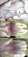
All animal manipulations were performed following protocols approved by the Plum Island Animal Disease Center (PIADC) Animal Care and Use Committee. Determination of virus infectious dose in pigs was performed as previously described [33]. Briefly, four co-housed 20~40 kg out-bred white pigs were sedated and inoculated intradermally in the heel bulb of each major digit of each foot with 102, 103, 104 or 105 PFU of virus/5 µL (estimated volume retained in the inoculation site as described by Burrows [6]) achieved by inserting a 23G needle 1 cm along the superficial layer of the epidermis (Fig. 1A). Immediately following injection, the titer of the virus stock was confirmed by plaque assay in BHK cells. Formation of vesicles at the inoculation sites was scored 24 h after inoculation (Figs. 1B and C), and data from all four animals was used to determine the number of PFUs of virus capable of producing a 50% pig heel infectious dose (PHID50) [34]. To demonstrate ability of transmission by direct contact between pigs, two days after inoculation the four directly inoculated pigs were combined with four similar-sized naive pigs in a fenced 4 m2 area within an animal isolation room, with water but without food during the exposure time. Four h later, the exposed pigs were placed into four rooms with separate HEPA-filtered ventilation systems containing hermetically sealed doors facing clean hallways. Heparinized blood and nasal swabs were collected and processed for virus isolation or IgM detection as previously described [33]. Records of the sites containing vesicles were prepared each day as previously described [33]. A maximum lesion score of 12 was possible in directly inoculated pigs (the eight injected digits were not counted in the former determinations) and a score of 20 was possible for direct contact pigs. The same team of investigators visited the 8 pigs for the indicated number of days, showering and changing clothing between each animal room. In all cases directly inoculated animals were examined after the direct contact animals. Pigs were humanely euthanized 5~7 days after they produced lesions or, in absence of lesions, when virus-specific IgM could be detected by ELISA or on day 20 after exposure. In some cases, once secondary vesicles appeared, animals were sedated and euthanized to collect samples that were used for other experiments (animals # 48, 97, 103, 292, 293 and 296). No animals died as a result of FMDV infection.

Results

Different topotypes show different species specificity when analyzed in vitro

To determine species specificity in vitro, an assay was developed to measure the minimum infectious dose of virus able to propagate an infection on various cell types (Fig. 2). For these experiments, multi-well plates with BHK, FBK, and FPK cells were infected with 10-fold dilutions of virus [starting with 107 PFU/mL (as measured previously on BHK cells)] and examined to determine the lowest dose of virus able to cause complete cytopathic effect (CPE) at 48 h. These results were expressed as TCID50/mL. All six strains and a positive control virus (vCRM8, a genetically engineered virus which is highly virulent in bovine and swine [4,35]) showed similar ability to cause CPE in hamster- and swine-derived cells (values close to 107 TCID50/mL). In bovine-derived cells, all viral isolates except O/TAW/97 were able to replicate at similar titers.

Different topotypes demonstrate different infectivity when inoculated in swine

To study differences of infectivity (PFU/PHID50), each one of the six strains were inoculated intradermally in the heel bulb of four pigs as described above. At 24 h post-inoculation the inoculation sites were scored as negative or positive (Fig. 1) to determine the PHID50/mL present in the inoculums. Table 2 shows an example with the results obtained for O/SKR/00. Table 3 shows the PHID50/mL and the infectivity obtained for each strain. Although we started the experiment with all viruses at similar PFU/mL values (ranging from 9.5 × 10⁶ to 2.8 × 10⁷), the PHID50/mL values were broadly different. With O/TAW/97 we obtained a value of 105 PHID50/mL; with O/HKN/21/70, O/SKR/00 and O/UKG/35/01 we obtained values of 103; and with O/TAW/2/99 and O/SAR/19/00 we obtained values less than 102. The five strains also showed different relative infectivities when compared to O/TAW/97. Twenty to 62 times more virus would be needed to produce a vesicle at the inoculation site with O/HKN/21/70, O/SKR/00 and O/UKG/35/01 while greater than 5,000 times more virus would be needed for the remaining two strains, O/TAW/2/99 and O/SAR/19/00.

Cathay topotype FMDVs are highly infectious and produce severe disease in pigs

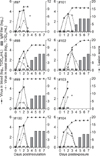
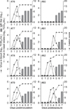
When we studied the various parameters of infection of O/TAW/97 (Fig. 3), the direct inoculation of 105 PFU (103 PHID50, Table 3) produced an acute and synchronous disease in the inoculated animals. In the O/TAW/97-inoculated animals, virus in blood was detected as early as 1 day post-inoculation (dpi) and virus was isolated from nasal swabs at 1 to 3 dpi. At 2 dpi, all the animals expressed peak amounts of virus in blood, nasal swabs and vesicles at secondary sites of replication. The highest value of lesion score was equal to or higher than 10 (out of a maximum of 12) and was reached at 2 to 4 dpi. In all O/TAW/97-infected pigs, blood IgM was detected following clearance of detectable virus (a similar result was obtained in all six experiments in all pigs showing detectable viremia regardless of the strain they were inoculated with, Figs. 3-8). After direct contact at 2 dpi with O/TAW/97 direct inoculated pigs for a limited period of 4 h, FMDV readily spread to direct contact animals that also showed a rapid, acute and synchronous disease indistinguishable from directly inoculated animals.
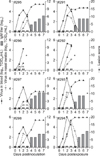
When we studied the various parameters of infection with O/HKN/21/70 (Fig. 4), the direct inoculation of 105 PFU (38 PHID50, Table 3) produced an acute and synchronous disease in the inoculated animals, similar to O/TAW/97-inoculated pigs. FMDV readily spread to direct contact animals that also displayed a rapid, acute and synchronous disease indistinguishable from directly inoculated animals. In conclusion, independent of previous history of amplification in cells or differences in infectivity, the disease in these 16 animals inoculated with strains from the Cathay topotype was synchronous, acute and severe.

Some PanAsia topotype viruses are as virulent as the Cathay topotype, whereas others are less virulent in pigs

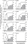
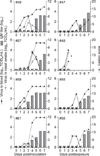
When we studied the various parameters of infection of O/SKR/00 (Fig. 5) or O/UKG/35/01 (Fig. 6), the direct inoculation of 105 PFU (47 to 82 PHID50, Table 3) produced an acute and synchronous disease in the inoculated animals, indistinguishable from the results obtained in pigs directly inoculated with the Cathay topotypes. However, differences between the Cathay topotype and these two PanAsian strains were found after limited direct contact. Specifically, for both strains, O/SKR/00 or O/UKG/35/01 (Figs. 5 and 6, respectively) one out of four direct contact animals never displayed clinical disease; only subclinical disease was detected by means of detection of IgM antibodies at 5 dpi for pig #80 (Fig. 6) and 13 dpi for pig #815 (results not shown). Additionally, in O/UKG/35/01 direct contact animals, 2 pigs produced a delayed disease (pigs # 81 and 83, Fig. 6).
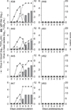
For the two remaining strains, O/TAW/2/99 and O/SAR/19/00 (Figs. 7 and 8), the various parameters of disease differed drastically from the Cathay topotype. Even though the pigs received a direct inoculation of 105 PFU, the PHID50 was below 1 for both strains (Table 3). Accordingly, these two strains produced a less acute and less synchronized disease in the inoculated animals when compared to the Cathay topotype. After direct contact FMDV did not readily spread to direct contact animals, probably due to the low shedding of FMDV by the donor animals. In conclusion, disease in animals inoculated with the PanAsia topotype strains was much more heterogeneous than the disease in animals inoculated with the Cathay topotype.

Discussion

Experimental infection of livestock with FMDV can be achieved by a variety of methods. These include contact with infected animals, contact with aerosols produced by infected animals, contact with men who have been in contact with infected animals, and parenteral administration by intradermal, intravenous, intramuscular, and subcutaneous inoculation. Using these methods, virulence can be quantified by evaluating the dose of virus capable of causing disease, severity and kinetics of development of disease, amount of virus present in blood, amount of virus shed by infected animals, and ability of virus to spread to other animals [38]. Taken together, these properties can be used to predict the pathogenic potential of different FMDV isolates.
To learn more about the Cathay topotype of FMDV, we attempted to develop a model for evaluation of transmission by aerosol. These initial experiments showed that O/TAW/97 as well as a South American virus, O1 Campos, were highly virulent in directly inoculated pigs and were readily transmitted by contact, but clinical disease could not be transmitted to naive pigs across a 1.3 m gap between open fencing in an animal isolation room. Preliminary experiments demonstrated that 3 of 19 exposed animals displayed a subclinical infection as measured by specific IgM responses to FMDV detected 10 or more days following exposure (results not shown). These results are consistent with the relatively low susceptibility of pigs to infection by the aerosol route, as reviewed by Alexandersen et al. [1,2].
Based on these findings, we selected a different experimental design to evaluate virulence and spread of six different FMDV isolates in pigs. First, we started determining the infectivity (PFU/PHID50) of each strain by means of inoculating intradermally in the heel bulb [6] with graded dilutions of virus and scored for the appearance of lesions at the inoculation sites 24 h later, to determine the concentration of virus expressed as PHID50/mL of each of the individual isolates. An example of the reproducible nature of the heel-bulb inoculation method in pigs and its usefulness to calculate a PHID50/mL value are shown in this study. Second, a quantitative evaluation of disease in all direct-inoculated animals was accomplished using a variety of criteria. These included a quantitative lesion score (based on a daily thorough examination of each animal, which included a close examination of the mouth, nose, feet and all four digits) as well as evaluation of virus titers in the blood, nasal swab samples, and titer of IgM in blood. Third, a direct contact transmission was accomplished for the six strains involved. For this, we selected day 2 post direct inoculation to place four naive animals for 4 h in close contact with the four injected animals. This time point was chosen based on previous experience with different strains of FMDV serotype O in our lab (results not shown). Quantitative evaluation of disease in contact inoculated animals was accomplished as described for direct inoculated animals.
The most substantial differences among the strains we have evaluated, namely the ratio of tissue culture infectious doses to animal infectious dose, could be interpreted as resulting from adaptation of some of the strains to grow well in BHK cells, rather than inherent differences in their infectivity in pigs. Although there is some evidence to indicate that that cell lines may not be as sensitive as primary cell systems for assaying animal-derived FMDV [19], we have found that carefully maintained low-passage cultures of BHK cells are just as sensitive as other methods utilized for evaluation of pig-derived strains. Specifically, in our hands, BHK cells (passage 62~66), IBRS2 cells (passage 117~122) [8], and FPK cells gave nearly identical titers in vitro with all six strains used, including the isolates that had never been in cell culture like O/TAW/97 (results not shown). Another widely used primary cell culture system, BTY cells, has been reported to be more sensitive than other systems [32]. However, this cell type does not support the growth of O/TAW/97 [10], thus, we did not employ BTY cells in our studies. It is also described that BTY and BHK cells were equally sensitive in titrations of PanAsia isolates derived from pig and cow [9]. These small differences, readily explainable in terms of batch-to-batch variation in virus production, emphasize that BHK cells display similar sensitivity to infection by all six strains used in our studies, irrespective of the fact that several of these strains were propagated in cell culture. Thus, based on all of these analyses, it appears certain that the differences we have reported in PFU/PHID50 in Table 3 are due to differences in porcine infectivity, and not to differences in tissue culture infectivity of these six FDMV strains.
Using our virulence evaluation systems, we found that the Cathay topotype virus from the 1997 outbreak in Taiwan, which has never been amplified in cells, was highly virulent in swine, producing a synchronous disease in inoculated pigs and efficient spread to direct contact animals. These results are consistent with reports of rapid spread of disease in the 1997 porcine outbreak in Taiwan [39]. Interestingly, the second Cathay virus we examined, O/HKN/21/70, showed similar infectivity, pathogenicity and spread, despite a substantially different passage history (including multiple passages in cell culture at WRL prior to our acquisition.). Additionally, we found that O/HKN/21/70 was not able to replicate at the intradermal lingual inoculation site in a single cow (results not shown) even though it grew well in bovine-derived cells, as described here and previously [21]. Thus, these results suggest that O/TAW/97 does not display any significant new properties in vivo; rather, it appears to be similar to a virus isolated almost 30 years earlier. Furthermore, in our hands, animals directly inoculated with other FMDV serotype O strains (O1 Campos and O1 Manisa) in an identical method of inoculation have shown the same pattern of disease as those animals inoculated with the Cathay topotype (results not shown).
The results obtained from the PanAsia topotype viruses showed much more heterogeneity. Two strains in this topotype, O/SKR/00 and O/UKG/35/01, produced a porcine disease pattern similar to the two Cathay topotype viruses in direct inoculated animals and their PFU/PHID50 values were similar to O/HKN/21/70. Interestingly, this similar infectivity among the three isolates (O/HKN/21/70, O/SKR/00 and O/UKG/35/01) is independent of the fact that these three viruses have undergone different numbers of cell amplification cycles. However, pigs exposed to O/SKR/00- or O/UKG/35/01-inoculated pigs showed a pattern of disease different from that obtained with the Cathay topotype viruses. Specifically, these PanAsian virus-exposed animals had lower virus titers in blood and nasal secretions, and a delayed appearance of clinical signs. In addition, one pig in each of these two contact exposure groups only displayed evidence of a subclinical infection, without any detectable virus recovered from blood or nasal secretions. The two remaining PanAsia topotype viruses (O/TAW/2/99 and O/SAR/19/00) produced milder disease in directly inoculated animals, consistent with a much lower infectious dose inoculated into these animals (in terms of PHID50). In addition, these two strains transmitted very poorly, especially O/SAR/19/00, which failed to produce a clinical disease in any contact exposed pig (one pig, #52 displayed a subclinical disease based on the detection of IgM at 8 dpi, results not shown). This poor transmission is consistent with the fact that on the day of exposure (day 2) we were unable to detect virus in any nasal swab samples obtained from the four SAR/19/00-inoculated animals, and only from one-of-four O/TAW/2/99-inoculated animals. Thus, transmission of these strains cannot be readily compared to the other four strains we tested. Since these last two strains had been amplified extensively in tissue culture under conditions that we cannot readily confirm, we cannot be sure that their dramatically reduced virulence in animals is not due to some type of selection in cell culture, or related to the fact that the virus was isolated from a probang sample, in the case of O/TAW/2/99. A complete sequence analysis of the four PanAsian strains identified here reveals only a small number of differences [26], which are not readily reconcilable with the observed differences in pathogenicity in livestock. The disease patterns induced by PanAsia topotype viruses were much more heterogeneous than those produced by O/TAW/97 and O/HKN/21/70, even though PanAsian viruses are much more closely related to each other [23] than this pair of Cathay topotype viruses [21].
In conclusion, we have described a method to evaluate the pathogenicity and transmission of different FMDV strains that allow us to demonstrate diverse FMD pathogenesis outcomes after direct inoculation of swine. Additionally, differences in FMD pathogenesis were also found after limited direct contact of naive pigs with severely sick donor animals. Future projects include development of similar experiments in swine with FMDV strains A24 Cruzeiro and O1 Manisa to be used for FMD challenge and pathogenesis studies, as well as in vaccine efficacy and preventive biotherapeutic trials.

Figures and Tables

Fig. 1
(A) Intradermal inoculation in the heel bulb. The space between the two arrows marks the portion of the needle that lays within the dermis, approximately 1.2 cm. Inoculums were released while slowly removing the needle. (B and C) Replication of O/UKG/35/01 at the inoculation site 24 h after intradermal inoculation with 700 PFU/5 µL (1 and 2) and 70,000 PFU/5 µL (3 and 4). The presence of a vesicle (POS) indicates a positive result. The absence of a vesicle (NEG) indicates a negative result.
jvs-11-133-g001
Fig. 2
Species specificity in primary bovine and porcine kidney cells (FBK and FPK, respectively) compared with BHK cells. TCID50/mL was estimated for each virus starting with 107 PFU/mL, as previously determined in BHK cells. The vertical bar and the extended bar illustrate the results from two independent experiments. The name of each virus and species of origin are described in Table 1. vCRM8 (a chimeric virus experimentally shown to be infectious in cattle and swine) was added as an internal positive control.
jvs-11-133-g002
Fig. 3
Viremia, virus in nasal secretions, vesicular lesions and IgM titers in pigs directly inoculated with O/TAW/97 (left panels) or exposed to O/TAW/97 (right panels). *Indicates the day animals were euthanized to obtain samples for other studies.
jvs-11-133-g003
Fig. 4
Viremia, virus in nasal secretions, vesicular lesions and IgM titers in pigs directly inoculated with O/HKN/21/70 (left panels) or exposed to O/HKN/21/70 (right panels). *Indicates the day animals were euthanized to obtain samples for other studies.
jvs-11-133-g004
Fig. 5
Viremia, virus in nasal secretions, vesicular lesions and IgM titers in pigs directly inoculated with O/SKR/00 (left panels) or exposed to O/SKR/00 (right panels).
jvs-11-133-g005
Fig. 6
Viremia, virus in nasal secretions, vesicular lesions and IgM titers in pigs directly inoculated with O/UKG/35/01 (left panels) or exposed to O/UKG/35/01 (right panels).
jvs-11-133-g006
Fig. 7
Viremia, virus in nasal secretions, vesicular lesions and IgM titers in pigs directly inoculated with O/TAW/2/99 (left panels) or exposed to O/TAW/2/99 (right panels). *Indicates the day this animal was euthanized to obtain samples for other studies.
jvs-11-133-g007
Fig. 8
Viremia, virus in nasal secretions, vesicular lesions and IgM titers in pigs directly inoculated with O/SAR/19/00 (left panels) or exposed to O/SAR/19/00 (right panels).
jvs-11-133-g008
Table 1
Origin and passage history of foot-and-mouth disease virus (FMDV) strains used in this study
jvs-11-133-i001

*Nomenclature for all viruses is as follows: serotype/three-letter location code for country or place of origin/accession number (if applicable)/year of isolation. TAW: Taiwan, HKN: Hong Kong, SKR: South Korean Republic, SAR: South African Republic, UKG: United Kingdom. Livestock species of origin. Institute of origin (FADDL: Foreign Animal Disease Diagnostic Laboratory, APHIS, VS, Plum Island, NY, USA; WRL: World Reference Laboratory for FMD, Pirbright, Woking, Surrey, UK; OVI: Onderstepoort Veterinary Institute, Onderstepoort, SAR). Passages (p) were performed in pigs (PIG), bovine thyroid cells (BTY), IBRS2 porcine kidney cell line (RS), baby hamster kidney cell line 21 clone 13 (BHK), or porcine kidney cells (PK) prior to receipt. §Amplifications made in our lab for inoculation studies. The sample received was vesicular fluid that was inoculated in a pig and vesicular fluid from this pig was harvested and aliquoted for inoculations in vivo and in vitro. The virus was isolated from an esophageal/pharyngeal sample obtained from an animal exhibiting a subclinical infection. **Original sample from the outbreak was directly inoculated in one pig, and vesicular fluid was harvested from another pig placed in direct contact with the inoculated one. Aliquots from this vesicular fluid were used for all inoculations in vivo and in vitro.

Table 2
Results obtained after direct inoculation with FMDV O/SKR/00 in porcine heel bulb for determination of concentration of the inoculum as 50% pig heel infectious doses per mL (PHID50/mL)
jvs-11-133-i002

*Animal number. Amount of virus in each inoculation site. Each sign represents one main digit. "+" indicates a macroscopic lesion was formed at the site of inoculation. "-" indicates that no lesion was visible.

Table 3
Infectivity results obtained with different Asian FMDV strains in comparison with O/TAW/97
jvs-11-133-i003

*Viral concentration of inoculums expressed as plaque forming units per mL (PFU/mL) obtained in BHK cells. Determination of 50% pig heel infectious doses (PHID50/mL) in inoculums after intradermal inoculation in the heel bulb. PFU of virus calculated to be one PHID50. §Each pig received values of 105 PFU of virus, projected by multiplying the value of PFU/mL by 11.11 µL the estimated volume of inoculums (5 µL × 2 of first dilution plus three more dilutions of a volume of 1/10 of the immediate previous dilution). Determination of PHID50 that each animal received, based on volume estimated above. Relative infectivity of each virus compared with porcine infectivity of O/TAW/97. This number indicates how many more viral particles than O/TAW/97 are needed to obtain 1 PHID50.

Acknowledgments

We thank Dr. Juan Lubroth, FADDL, PIADC, Greenport, NY, USA for supplying O/TAW/97, O/SKR/00, O/UKG/35/01; Dr. Wilna Vosloo, OVI, Onderstepoort, SAR, for supplying O/SAR/19/00, and Dr. Nick Knowles, WRL, Pirbright, UK for supplying O/HKN/21/70 and O/TAW/2/99. We thank the PIADC Animal Caretakers for assistance with animal experiments. This work was partially supported by the Agricultural Research Service of the USDA (CRIS Project #1940-32000-035-00D) and by a grant from the National Research Initiative Competitive Grants program of USDA/CSREES (Grant #99-35204-7949). We also thank Ms. Melanie Prarat for editing the manuscript.

References

1. Alexandersen S, Brotherhood I, Donaldson AI. Natural aerosol transmission of foot-and-mouth disease virus to pigs: minimal infectious dose for strain O1 Lausanne. Epidemiol Infect. 2002. 128:301–312.
crossref
2. Alexandersen S, Donaldson AI. Further studies to quantify the dose of natural aerosols of foot-and-mouth disease virus for pigs. Epidemiol Infect. 2002. 128:313–323.
crossref
3. Alexandersen S, Zhang Z, Donaldson AI, Garland AJ. The pathogenesis and diagnosis of foot-and-mouth disease. J Comp Pathol. 2003. 129:1–36.
crossref
4. Almeida MR, Rieder E, Chinsangaram J, Ward G, Beard C, Grubman MJ, Mason PW. Construction and evaluation of an attenuated vaccine for foot-and-mouth disease: difficulty adapting the leader proteinase-deleted strategy to the serotype O1 virus. Virus Res. 1998. 55:49–60.
crossref
5. Beard CW, Mason PW. Genetic determinants of altered virulence of Taiwanese foot-and-mouth disease virus. J Virol. 2000. 74:987–991.
crossref
6. Burrows R. The infectivity assay of foot-and-mouth disease virus in pigs. J Hyg (Lond). 1966. 64:419–429.
crossref
7. Chen X, Feng Q, Wu Z, Liu Y, Huang K, Shi R, Chen S, Lu W, Ding M, Collins RA, Fung YW, Lau LT, Yu AC, Chen J. RNA-dependent RNA polymerase gene sequence from foot-and-mouth disease virus in Hong Kong. Biochem Biophys Res Commun. 2003. 308:899–905.
crossref
8. de Castro MP. Behaviour of the foot-and-mouth disease virus in cell cultures: Susceptibility of the IB-RS-2 cell line. Arq Inst Biol (Sao Paulo). 1964. 31:63–78.
9. Donaldson AI, Sellers RF. Transmission of FMD by people. Vet Rec. 2003. 153:279–280.
10. Dunn CS, Donaldson AI. Natural adaption to pigs of a Taiwanese isolate of foot-and-mouth disease virus. Vet Rec. 1997. 141:174–175.
crossref
11. Feng Q, Chen X, Ma O, Liu Y, Ding M, Collins RA, Ko LS, Xing J, Lau LT, Yu AC, Chen J. Serotype and VP1 gene sequence of a foot-and-mouth disease virus from Hong Kong (2002). Biochem Biophys Res Commun. 2003. 302:715–721.
crossref
12. Ferguson NM, Donnelly CA, Anderson RM. Transmission intensity and impact of control policies on the foot and mouth epidemic in Great Britain. Nature. 2001. 413:542–548.
crossref
13. Freshney RI. Culture of Animal Cells: A Manual of Basic Technique. 1987. New York: Liss;144.
14. George VG, Hierholzer JC, Ades EW. Mahy BWJ, Kangro HO, editors. Cell culture. Virology Methods Manual. 1996. London: Academic Press;3–23.
crossref
15. Gulbahar MY, Davis WC, Guvenc T, Yarim M, Parlak U, Kabak YB. Myocarditis associated with foot-and-mouth disease virus type O in lambs. Vet Pathol. 2007. 44:589–599.
crossref
16. Henderson WM. The Quantitative Study of Foot-and-Mouth Disease Virus. Report Series of Agricultural Research Council. No. 8. 1949. Her Majesty's Stationery Office: London;1–50.
17. Henderson WM. A comparison of different routes of inoculation of cattle for detection of the virus of foot-andmouth disease. J Hyg (Lond). 1952. 50:182–194.
crossref
18. Hierholzer JC, Killington RA. Mahy BW, Kangro HO, editors. Virus isolation and quantitation. Virology Methods Manual. 1996. London: Academic Press;25–46.
crossref
19. House C, House JA. Evaluation of techniques to demonstrate foot-and-mouth disease virus in bovine tongue epithelium: comparison of the sensitivity of cattle, mice, primary cell cultures, cryopreserved cell cultures and established cell lines. Vet Microbiol. 1989. 20:99–109.
crossref
20. Huang CC, Lin YL, Huang TS, Tu WJ, Lee SH, Jong MH, Lin SY. Molecular characterization of foot-and-mouth disease virus isolated from ruminants in Taiwan in 1999-2000. Vet Microbiol. 2001. 81:193–205.
crossref
21. Knowles NJ, Davies PR, Henry T, O'Donnell V, Pacheco JM, Mason PW. Emergence in Asia of foot-and-mouth disease viruses with altered host range: characterization of alterations in the 3A protein. J Virol. 2001. 75:1551–1556.
crossref
22. Knowles NJ, Samuel AR. Molecular epidemiology of foot-and-mouth disease virus. Virus Res. 2003. 91:65–80.
crossref
23. Knowles NJ, Samuel AR, Davies PR, Kitching RP, Donaldson AI. Outbreak of foot-and-mouth disease virus serotype O in the UK caused by a pandemic strain. Vet Rec. 2001. 148:258–259.
24. Knowles NJ, Samuel AR, Davies PR, Midgley RJ, Valarcher JF. Pandemic strain of foot-and-mouth disease virus serotype O. Emerg Infect Dis. 2005. 11:1887–1893.
crossref
25. Malirat V, de Barros JJ, Bergmann IE, Campos Rde M, Neitzert E, da Costa EV, da Silva EE, Falczuk AJ, Pinheiro DS, de Vergara N, Cirvera JL, Maradei E, Di Landro R. Phylogenetic analysis of foot-and-mouth disease virus type O re-emerging in free areas of South America. Virus Res. 2007. 124:22–28.
crossref
26. Mason PW, Pacheco JM, Zhao QZ, Knowles NJ. Comparisons of the complete genomes of Asian, African and European isolates of a recent foot-and-mouth disease virus type O pandemic strain (PanAsia). J Gen Virol. 2003. 84:1583–1593.
crossref
27. Mattion N, König G, Seki C, Smitsaart E, Maradei E, Robiolo B, Duffy S, León E, Piccone M, Sadir A, Bottini R, Cosentino B, Falczuk A, Maresca R, Periolo O, Bellinzoni R, Espinoza A, Torre JL, Palma EL. Reintroduction of foot-and-mouth disease in Argentina: characterisation of the isolates and development of tools for the control and eradication of the disease. Vaccine. 2004. 22:4149–4162.
crossref
28. Mingqiu Z, Qingli S, Jinding C, Lijun C, Yanfang X. Sequence analysis of the protein-coding regions of foot-and-mouth disease virus O/HK/2001. Vet Microbiol. 2008. 130:238–246.
crossref
29. O'Donnell VK, Pacheco JM, Henry TM, Mason PW. Subcellular distribution of the foot-and-mouth disease virus 3A protein in cells infected with viruses encoding wild-type and bovine-attenuated forms of 3A. Virology. 2001. 287:151–162.
30. Oem JK, Lee KN, Cho IS, Kye SJ, Park JH, Joo YS. Comparison and analysis of the complete nucleotide sequence of foot-and-mouth disease viruses from animals in Korea and other PanAsia strains. Virus Genes. 2004. 29:63–71.
crossref
31. Oem JK, Lee KN, Cho IS, Kye SJ, Park JY, Park JH, Kim YJ, Joo YS, Song HJ. Identification and antigenic site analysis of foot-and-mouth disease virus from pigs and cattle in Korea. J Vet Sci. 2005. 6:117–124.
crossref
32. OIE. Manual of Standards for Diagnostic Tests and Vaccines. Chapter 2.1.1. Foot and Mouth Disease. 2000. Paris: OIE;77–92.
33. Pacheco JM, Henry TM, O'Donnell VK, Gregory JB, Mason PW. Role of nonstructural proteins 3A and 3B in host range and pathogenicity of foot-and-mouth disease virus. J Virol. 2003. 77:13017–13027.
crossref
34. Reed LJ, Muench H. A simple method of estimating fifty percent endpoints. Am J Hyg. 1938. 27:493–497.
35. Sa-Carvalho D, Rieder E, Baxt B, Rodarte R, Tanuri A, Mason PW. Tissue culture adaptation of foot-and-mouth disease virus selects viruses that bind to heparin and are attenuated in cattle. J Virol. 1997. 71:5115–5123.
crossref
36. Sakamoto K, Kanno T, Yamakawa M, Yoshida K, Yamazoe R, Murakami Y. Isolation of foot-and-mouth disease virus from Japanese black cattle in Miyazaki Prefecture, Japan, 2000. J Vet Med Sci. 2002. 64:91–94.
crossref
37. Samuel AR, Knowles NJ. Foot-and-mouth disease type O viruses exhibit genetically and geographically distinct evolutionary lineages (topotypes). J Gen Virol. 2001. 82:609–621.
crossref
38. Sellers RF. Quantitative aspects of the spread of foot and mouth disease. Vet Bull. 1971. 41:431–439.
39. Yang PC, Chu RM, Chung WB, Sung HT. Epidemiological characteristics and financial costs of the 1997 foot-and-mouth disease epidemic in Taiwan. Vet Rec. 1999. 145:731–734.
crossref
TOOLS
Similar articles