

 PDF
PDF ePub
ePub Citation
 Print
Print


Introduction
Mycobacterium avium subsp. paratuberculosis (MAP) is the causative agent of Johne's disease (JD), a chronic intestinal granulamatous infection affecting domestic and wild ruminants [7,11,15,32]. Although cattle are usually infected early in life, clinical signs do not develop until 2-4 years of age, which makes early diagnosis of this infection a difficult task. JD is considered to be an economically important disease and accounts for an annual loss of $220 million to the US dairy industry [25]. The proposed, but poorly defined association of MAP with Crohn's disease in human beings, is also of concern [13,18,23,24]. The in vivo diagnosis of MAP infections is quite challenging and difficult in the pre-clinical stages since the majority of infected animals do not show symptoms of the disease. Although the isolation and identification of MAP is the most definitive test for diagnosis, it is time-consuming and labor-intensive, requiring 8-12 weeks. Contamination is an added problem when MAP is cultured from fecal samples. Although, PCR for IS900 sequences is of diagnostic value, at times PCR leads to false positive amplification due to the presence of environmental bacteria with similar sequences [10]. Novel sequences recently identified in the genome of MAP appear specific and may also be used in nucleic acid-based diagnostic tests [6,16]. Real time PCR-based assays, which involve high equipment costs and trained personnel, can be used only under well-established laboratory conditions and serological tests may lack sensitivity [8].
Most diagnostic laboratories continue to use traditional culture methods; few laboratories use molecular methods along with culture methods [14,21,26,30,31]. Development of bioanalytical systems, such as biosensors coupled with a reverse transcriptase PCR to achieve low limits of detection, will be useful in the rapid and accurate detection of MAP.
Biosensors based on nucleic acid hybridization and liposome signal amplification have been shown to be very useful in developing rapid, inexpensive, and easy-to-handle systems for the detection and quantification of RNA molecules [1,2,4,5]. A biosensor is a lateral flow assay that provides visual or reflectance data within about 20 min of overall assay time [3]. A biosensor uses a membrane flow-through system with an immobilized DNA probe that hybridizes with the target. Signal amplification is provided when the target sequence hybridizes to a second DNA probe coupled to liposomes encapsulating the dye, sulforhodamine B (SRB). The amount of liposomes captured in the detection zone can be either read visually or quantified with a handheld reflectometer.
For MAP diagnosis, the IS900 gene, with 15-20 copies [27], has been routinely used in PCR-based detection systems. However, in the past, IS900 primers have also amplified IS900-like PCR products, probably from environmental mycobacteria, and resulting in false positive results [10]. Despite this possibility, IS900 gene amplification should still serve as a good indicator when coupled to a high-specificity hybridization reaction, as proposed here. Apart from IS900, other novel sequences, such as ISMav2 [26] and ISMap02 [20,27], could also be potential candidates in PCR-based assays. In the current study, the development of a rapid biosensor assay for the detection of live MAP organisms employing IS900 gene sequences is described. This is the first time the biosensor assay for MAP has been demonstrated.
Materials and Methods
Bacterial strain and growth
Mycobacterium avium subsp. paratuberculosis-66115-98, a clinical isolate available from the Department of Population Medicine and Diagnostic Sciences at Cornell University, was grown in 7H9 medium, supplemented with 10% oleic acid-albumin-dextrose-catalase (Becton, Dickinson and Company, USA) and Mycobactin J (Allied Monitor, USA). The cultures were grown at 37℃ for 8 weeks and used in this study.
RNA extraction
MAP cultures were centrifuged at 12,000 rpm for 10 min. One ml of Trizol was added to the pellet, and the mixture was passed through the syringe and needle (22 gauge) several times. The mixture was kept at room temperature for 5 min. Two hundred µl of chloroform was added and mixed vigorously for 15 sec and incubated at room temperature for 3 min. The mixture was spun in a microcentrifuge at 12,000 rpm for 15 min at 4℃. The supernatant was transferred to a fresh microcentrifuge tube and an equal volume of 70% alcohol was added at room temperature. The mixture was transferred to the minispin column of a RNeasy kit (Qiagen, USA) and RNA was isolated following the manufacturer's protocol. The isolated RNA samples were treated with 10 U/µl of RNase-free DNase I (Qiagen, USA) at 37℃ for 10 min, followed by heat inactivation at 95℃ for 5 min, and then chilled on ice.
Estimation of cell quantity by optical density
MAP organisms were quantified by measuring the optical density at 550 nm as described earlier [17]. An optical density of 0.25 at 550 nm was equivalent to approximately 108 organisms per ml.
Quantitation of cell number
The organisms were harvested by centrifugation, diluted in phosphate buffered saline (PBS; NaCl, 0.8%; KCl, 0.02%; Na2HPO4, 0.115%; and KH2PO4, 0.02% [pH 7.2]) containing 0.05% Tween-80, loaded on the platform of an improved Neubauer haemocytometer chamber, and visually counted.
Preparation of spiked fecal samples
Fecal samples were collected from healthy animals for initial standardization. Ten-fold serial dilutions of viable MAP organisms were prepared from a stock suspension of 108 organisms. Aliquots of each bacterial dilution (900 µl) were added to 100 mg of feces to yield bacterial numbers between 101 and 106. For samples from infected animals, 25-50 gm of fecal samples were collected from 8 calves challenged with 107 MAP cells/animal in milk replacer for 7 consecutive days. One hundred mg of fecal samples collected 2, 4, 6, 8, and 10 days after challenge were used for RNA isolation.
RNA extraction from spiked fecal samples
RNA was extracted from spiked fecal samples containing 101 to 106 organisms using Trizol (Invitrogen, USA) and the extracted RNA was resuspended in 10 µl RNase-free water. The isolated RNA samples were treated with 10 U/µl of RNase-free DNase I (Qiagen, USA) at 37℃ for 10 min, followed by heat inactivation at 95℃ for 5 min, and then chilled on ice.
Reverse-Transcriptase PCR
RNA isolated from spiked fecal samples was amplified using a one-step RT-PCR kit (Qiagen, USA). IS900 primers were used for amplification. The RT-PCR products were electrophoresed and checked on a 1% agarose gel containing 5 µg of ethidium bromide. The amplified products were used in the biosensor assay.
Preparation of membranes
Polyethersulfone membranes (Pall, USA) were cut into 4.5 mm × 7.5 cm strips. Streptavidin was diluted in 0.4 M NaHCO3/Na2CO3 buffer (pH 9.0) containing 5% methanol in a final concentration of 20 pmol/µl. Streptavidin was spotted on the membrane strips using a Camag Linomat IV TLC sample applicator (Camag Scientific, USA) and incubated for 20 min at room temperature. The membranes were dried for an additional 1.5 h in a vacuum oven (-15 psi) at 55℃. Subsequently the membranes were incubated in a blocking solution of 0.5% polyvinylpyrrolidone, 0.015% casein in Tris-buffered saline (TBS, 20 mmol/l Tris; 150 mmol/l NaCl; and 0.01% NaN3 [pH 7.5]) for 30 min. Following this, the membranes were dried in a vacuum oven (-15 psi) at 30℃ for 3 h, and stored in vacuum-sealed bags at 4℃ until used.
Preparation of liposomes
A slightly modified protocol [3] of the reverse phase evaporation method [28] was used for the preparation of liposomes. Briefly, 40.3 µmol dipalmitoyl phosphatidyl choline, 21 µmol dipalmitoyl phosphatidyl glycerol, and 51.7 µmol cholesterol were dissolved in a mixture of chloroform, methanol, and isopropyl ether (30 ml : 5 ml : 30 ml) by sonication using a round bottom flask in a water bath at 45℃. Subsequently, 50 µl of cholesterol-tagged reporter probe (corresponding to 0.013 mol%) was added to the mixture and sonicated in a 45℃ water bath. To the lipid mixture, a total of 4 ml of 150 mM SRB in 0.02 mol/l phosphate buffer (pH 7.5; 516 mmol/kg) was added and sonicated for 5 min. The organic solvents were evaporated in a rotary evaporator so that the liposomes formed spontaneously, entrapping SRB. The liposomes were extruded 11 times through 2 µm and 0.6 µm filters using a mini- extruder and polycarbonate filters (Avanti Polar Lipids, USA) to obtain a uniform particle size. Liposomes were purified from the free dye by gel filtration using a Sephadex G50 column, followed by dialysis against 0.01 mol/l PBS (pH 7.0) containing sucrose to increase the osmolarity to 590 mmol/l. Purified liposomes were stored at 4℃ until used.
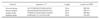
Primers and probes
The details of primers and probes used in this study are presented in Table 1. The capture and reporter probes used in this study were prepared synthetically. A synthetic target with the following sequence was used to optimize the assay conditions, which has been found to be useful in previous RNA biosensor assay developments. This sequence is essentially made up of sequences antisense to the capture (bold and italics) and reporter (bold and underlined) probes plus additional sequences at the 5' and 3' ends homologous to the IS900 sequence, as follows: (5'CGATCAGCAACGCGGCGCCGCCGGCGTTGAGGTCGATCGCCCACGTGACCTCGCCTCCATCGGCCAACGTCGTCACCGCCGCAATCA 3').
Lateral flow biosensor assay
The assay was performed by mixing 1.5 µl (1.5 µg) of the target sequence (RT-PCR product), 0.5 µl of forward primer (1 µM), 0.5 µl of reverse primer (1 µM), 1 µl of capture probe (1 pmol), 1 µl of reporter probe (2 pmol), and 4 µl of master mix (20% formamide, 4× sodium saline citrate [SSC], 0.4% Ficoll type 400, and 0.4 M sucrose) in a microcentrifuge tube. The mixture was denatured at 95℃ for 5 min, annealed at 60℃ for 1 min, and transferred to a glass tube. To this mixture, 2 µl of liposomes (tagged with the reporter probe) was added and incubated at 60℃ for 20 min. After incubation, the membrane strip (with 20 pmol of streptavidin) was inserted into the glass tube, and the hybridization mixture was allowed to migrate up the strip. Subsequently, 35 µl of running buffer (40% formamide, ×8 SSC [1.35 M sodium chloride, 0.135 M sodium citrate, and 0.01% sodium azide {pH 7.0}], 0.2% Ficoll, and 0.2 M sucrose) was added to the glass tube to flush the solution up the membrane. After 8-10 min, when all of the running buffer had run the length of the strip, the signal at the capture zone was analyzed with the BR-10 reflectometer (ESECO Speedmaster, USA). The reflectometer measures the reflectance of light at a wavelength of 560 nm, which is close to the maximum absorbance of the SRB that is encapsulated within the liposomes.
Microtiter assay
Reacti-Bind Neutravidin-linked microtiter plates were obtained from Pierce Biotechnology (USA). The plates were washed twice with 200 µl of wash buffer (PBS containing 0.05% [v/v] Tween-20 and 0.01% bovine serum albumin), and once with 200 µl of PBS. To each well, 100 µl of biotinylated capture probe (0.1 µM in 50 mM potassium phosphate buffer [pH 7.5] containing 1 mM EDTA) was added and incubated for 30 min at room temperature. Unbound capture probe was removed and the wells were washed thoroughly with 200 µl of wash buffer, followed by 200 µl of hybridization buffer (4× SSC, 20% formamide, 0.2% Ficoll, and 0.2 M sucrose). The target (RT-PCR product) and the reporter probe (0.2 µM in 50 mM potassium phosphate buffer [pH 7.5] containing 1 mM EDTA) were diluted in hybridization buffer, and denatured at 95℃ for 5 min. To this mixture, 3 µl of liposomes for each well was added and incubated at 60℃ for 20 min. One hundred µl of this mixture was added to each well and incubated at 60℃ for 30 min. The plates were washed twice with 200 µl PBS-sucrose buffer and 50 µl of 30 mM OG was added. After a 5 min incubation period, the fluorescence of the bound liposomes was measured at λex = 540/35 nm and λem = 590/25 nm.
Results
Optimization and development of a lateral-flow biosensor assay based on a synthetic IS900 sequence
The lateral-flow biosensor assay was developed and optimized using universal membranes, liposomes, and specific capture and reporter probes for IS900.
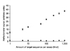
Initially, a synthetic DNA target was used to optimize the assay to assess the signal-to-noise ratios with a relatively large dynamic range and the highest signal obtainable. The standard lateral-flow biosensor assay was run in triplicate with 1 µl of synthetic DNA with 8 different concentrations ranging from 1-1,000 fmol µ/l. The limit of detection was determined using the signal obtained for the negative control plus three times the standard deviation at that point. The data showed that the limit of detection was as low as 1 fmol of the synthetic target sequence per assay with a dynamic range from 1-1,000 fmol (Fig. 1). The negative control contained water instead of target sequence and had a value of 1.04 ± 3.
Lateral-flow biosensor assay with the RT-PCR product of the IS900 gene
After optimization the lateral-flow biosensor assay with the synthetic IS900 sequence, the assay was performed with the RT-PCR product of the IS900 gene sequence. The RT-PCR product of RNA isolated from cultured MAP was used for optimization of the assay. In order to allow the capture and reporter probes to hybridize with the double-stranded DNA target sequence, denaturing, and hybridization conditions were optimized. For final assays, the target (RT-PCR product), probes, and primers were denatured at 95℃ for 5 min, annealed at 60℃ for 1 min, and hybridized with liposomes. Annealing at 60℃ was done to prevent the re-association of thermally-denatured double-stranded DNA strands.
Lateral-flow biosensor assay with the RT-PCR product of the RNA extracted from spiked fecal samples
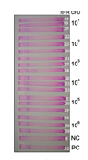
Positive signals were noticed at the capture zone, even with the RT-PCR product of RNA extracted from fecal samples containing only 10 organisms of MAP per 100 mg of feces (Fig. 2). We also performed the assay with a limited number of fecal samples collected from 2, 4, 6, 8, and 10 days from calves orally challenged with MAP. Fecal samples collected 2 and 4 days after challenge gave positive signals in the biosensor assay, which concurred with the MAP culture results. The MAP culture results of the positive fecal samples had 8 and 5 colony forming units (CFU), respectively, 2 and 4 days post-challenge. However, fecal samples collected 6, 8, and 10 days after challenge were found to be negative by both the biosensor assay and MAP culture studies. The coefficient of variance ranged from 0.59-3.7 for the different levels of MAP organisms in the spiked fecal samples tested by the biosensor assay.
Microtiter plate assay with the RT-PCR product of RNA extracted from spiked fecal samples
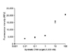
For optimization, the biotinylated capture probe was immobilized to the microtiter plates coated with neutravidin and the synthetic single-stranded DNA target for IS900 was allowed to hybridize prior to the addition of the reporter probe and the SRB encapsulating liposomes. The assay was run in triplicate with 1 µl of synthetic DNA target at 7 different concentrations ranging from 0.001 - 1,000 nM. In order to decrease the limit of detection, liposomes were lysed with a detergent, releasing the otherwise selfquenched SRB dye and detected using fluorescence (Fig. 3). A limit of detection of 0.1 nM was obtained calculating the lowest concentration detected that is above a value of the negative control plus three times the standard deviation of the negative control.
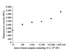
The lateral-flow biosensor assay was compared with the microtiter plate assay employing the same probe and target sequences for the detection of RNA extracted from fecal samples. The microtiter plate assay was performed with the RT-PCR product of the IS900 gene after denaturation at 95℃ for 5 min and hybridized at 60℃. Positive signals were obtained when 1.5 µl of target was used in the assay. The detection limit was found to be as low as 10 CFU when RT-PCR product of RNA extracted from fecal samples spiked with 101 to 106 organisms (Fig. 4). This was the same limit of detection obtained for the simple LF assay.
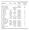
Specificity of the assay
The specificity of the lateral-flow biosensor assay was evaluated with samples from closely related mycobacteria for false positive reactions. These mycobacteria were cultured under optimal conditions and the RNA extracted was used in the RT-PCR reactions. No false positive signals were detected for any of the mycobacteria tested (Table 2).
Discussion
The majority of the diagnostic tests available for MAP detection is based upon the amplification of insertion sequences (IS elements). In this study, we used the IS900 gene because of its uniqueness in the MAP genome [9,22,29] and its comparably high copy number. Diagnostic tests based on IS900 elements have a high level of sensitivity because of the copy number [27]. In this study, we developed lateral flow and a microtiter assays. In the microtiter assay, the detection of the amplified target sequence is achieved through surfactant-induced liposome lysis and release of encapsulated dye molecules with subsequent fluorescent detection [12]. Although the hybridization of the probes with the target is usually done at 41℃ [3] in the case of single-stranded RNA sequences, the hybridization was optimized at 60℃ to suit the high G + C content of the MAP genome.
Generally, milk and feces are considered to be the most suitable clinical specimens for the diagnosis of JD. However, because of the presence of large amounts of fat and calcium ions, milk is regarded as a difficult specimen for the detection of MAP organisms [19]. Hence, we used fecal samples in this assay. The lateral flow biosensor assay was performed with the RT-PCR product of RNA extracted from spiked fecal samples containing 101 to 106 organisms in order to assess the sensitivity of the assay.
The results of our study indicated that the lateral flow biosensor assay was effective, even in the detection of 10 MAP organisms in the spiked fecal samples. Apart from the spiked fecal samples, we also tested fecal samples from experimentally infected animals, wherein fecal samples collected 2 and 4 days post-challenge gave positive results by the lateral flow biosensor assay. Shedding of MAP in feces has been reported to be inconsistent after challenge, at least during early stages. Moreover, there could be colonization of the organisms in the intestines which could have resulted in the non-detection of MAP at 6, 8, and 10 days post-challenge. However, 2 and 4 days post-challenge samples were also positive by MAP culture results with 8 and 5 CFU, respectively, which in turn indicated the ability of this method in detecting low levels of MAP organisms. Moreover, with the use of the RT-PCR product, in general only viable organisms present in the feces will be detected which provides an excellent tool for diagnosis. The existing cultural and serological methods accurately predict MAP infections during clinical stages when most animals shed large numbers of organisms, compared to subclinical stages when fecal shedding occurs at low levels with lesser frequencies. The present study with detection limits as low as 10 organisms is well-suited for the present day diagnostic requirements of JD. These results indicated that this assay is highly sensitive and could be used to detect animals in the early stage of infection with very low MAP shedding.
In addition to the rapid lateral flow assay that is suitable for low-sample numbers, a microtiter plate assay was developed for the detection of the RT-PCR product of RNA extracted from spiked fecal samples. Comparison of the lateral flow biosensor assay with the microtiter plate assay indicated that the detection limit of both assays were similar (10 CFU). With no false positive signals with the closely related mycobacteria tested in this study, this assay was considered to have excellent specificity.
In conclusion, the results of our study indicated that the IS900 gene sequence-based lateral flow biosensor assay developed is sensitive and specific for the detection MAP organisms in fecal samples. The assay was found to be effective in detecting as few as 10 organisms per 100 mg of feces. This assay will be useful in identifying animals in their early clinical stage, shedding low numbers of MAP in their feces, which can allow their quick removal from the rest of the herd, thereby avoiding further environmental contamination. Although one would expect a perfect dose response in the results between 10 and 106 organisms, the results were not as expected, which could possibly be due to the presence of PCR inhibitors in the fecal samples. This assay is comparatively cheaper and does not require costly equipments in comparison to real-time PCR or PCR coupled with Southern blotting. In this assay, reverse transcription PCR is being used instead of regular PCR which will help in detecting live MAP organisms. Moreover, the results can be obtained in a shorter time, in contrast to MAP culture techniques which take at least 6-8 weeks. Therefore, the present work was carried out with an idea of developing bioanalytical systems that are simple and yet highly sensitive. With the availability of small, easy-to-carry thermal cyclers, this assay could be developed as a portable assay which may cater to the needs of first responder emergency teams and clinicians in the field.
 XML Download