

 PDF
PDF ePub
ePub Citation
 Print
Print


INTRODUCTION
Epidermal cysts are uncommon and can arise from any part of the body [1]. Penile epidermal cysts in children are usually congenital and are caused by abnormal embryologic closure of the median raphe; these cysts are termed median raphe cysts [2-4]. Penile epidermal cysts in adults commonly develop after trauma or surgery. During wound healing, when shedding of the epithelium occurs within a closed space, cysts are formed by squamous epithelium undergoing keratinization [5-7]. According to this theory, an augmentation penoplasty using a dermal fat graft can cause a penile epidermal cyst. Here we report a case of a penile epidermal cyst that developed after augmentation penoplasty.
CASE REPORT
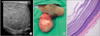
A 44-year-old male patient who was diagnosed with liver cirrhosis was referred to the urologic department because of a slowly growing penoscrotal swelling. The patient had a history of augmentation penoplasty with a dermal fat graft 20 years previously. The mass was recognized 4 years ago, but it was very small and asymptomatic at that time. As the swelling slowly increased in size, however, the patient had difficulty with sexual intercourse. He had no associated urinary symptoms. On physical examination, the swelling was located at the lateral surface of the penile proximal shaft. It was round, firm, and nontender and the penile skin overlying the lesion was intact and mobile. A scrotal ultrasonography to rule out any scrotal disorder showed a 5 cm×3 cm×5 cm heterogeneous echoic extratesticular mass with an internal linear hypoechoic lesion with no communication with the spermatic cord (Fig. 1A). Excision of the mass was performed under spinal anesthesia. After incision of the skin, we performed meticulous dissection around the mass. The capsule of the mass was preserved and enucleated from the surrounding tissues (Fig. 1B). Histologic findings showed the mass to be a cyst lined by stratified squamous epithelium (Fig. 1C). There were no adnexal structures of skin or mesenchymal structures. The final diagnosis was a penile epidermal cyst.
DISCUSSION
Penile epidermal cysts are broadly classified into congenital cysts and secondary cysts caused by trauma or surgery. Congenital cysts, such as median raphe cysts, can arise from an abnormal embryological closure of the median raphe [2-4]. Secondary cysts can occur because of occluded hair follicles, mechanical implantation involving the injection of epidermal fragments, and obstructed eccrine ducts [5-7]. Because the patient in this case had undergone an augmentation penoplasty with a dermal fat graft, we suspect that the cyst originated from the dermal fat graft.
Amid controversies regarding the indications, the demand for and supply of augmentation penoplasty techniques in Korea has increased for the improvement of personal satisfaction and self-confidence [8,9]. These techniques include subcutaneous injection of fat, dermal fat grafts, autologous tissue engineering, and a fasciocutaneous flap. Dermal fat grafts have commonly been used recently for augmentation penoplasty in Korea [9].
Penile epidermal cysts can be diagnosed by careful examination and radiological evaluation, including ultrasonography and computed tomography [5-7]. Magnetic resonance imaging may be useful in cases of suspected extension into the pelvis, although such cases are rare [6]. If a urethral diverticula or urethrocutaneous fistula is suspected, retrograde urethrography or voiding cystourethrography should be carried out [5-7]. In addition, the differential diagnosis of an epidermal cyst includes teratoma and dermoid cyst. However, skin and its appendages are present in a dermoid cyst, and derivatives of other germ cells are present in a teratoma [7]. In the present case, no appendages of skin or derivatives of other germ cells were found in the surgical specimen. The patient was misdiagnosed with a hydrocele at the time of referral. Physical examination and scrotal ultrasonography were performed to rule out any scrotal disorder. Surprisingly, the mass was not cystic on ultrasound, but was heterogeneous in character with an internal hypoechoic lesion.
Surgical excision of penile epidermal cysts is the treatment of choice [4-7]. The resection should be completed without leaving any epithelium to prevent a recurrence. Although no cases of malignancy arising in the wall of an epidermal cyst of the penis have previously been reported, patients with epidermal cysts should be followed up after complete cyst removal.
To our knowledge, there is little literature regarding penile epidermal cysts after augmentation penoplasty, and the incidence of cyst development related to augmentation penoplasty is also unknown. We believe the present study may be helpful to further our understanding of penile epidermal cysts after augmentation penoplasty.
 XML Download