

 PDF
PDF ePub
ePub Citation
 Print
Print


INTRODUCTION
Platelet-derived therapies are a growing trend across multiple medical and surgical specialties [12345]. Evidence suggests that platelets play an important role in tissue repair, vascular remodeling and inflammatory and immune responses through secretion of growth factors, cytokines and chemokines [67]. These biologically active proteins include transforming growth factor-β, platelet-derived growth factor, platelet-derived epithelial growth factor, insulin-like growth factor, vascular endothelial growth factor, basic fibroblast growth factor, as well as many others [8]. These growth factors are implicated in many aspects of natural wound healing, including chemotaxis, cell proliferation, cell differentiation and angiogenesis. They also control and conduct synthesis, modification and degeneration of extracellular matrix proteins. Coordination of these cellular and molecular processes is integral to proper wound healing and tissue regeneration [9]. The key role of platelets in these processes makes them an attractive candidate for therapies aimed at accelerating natural healing.
One of the most well described platelet-based therapies is autologous platelet-rich plasma (PRP) [10]. PRP is derived from the centrifugation of whole blood with a separator gel to remove the red and white blood cells. The resulting supernatant has a greater than four-fold increase in platelets and other plasma proteins [11]. This concentrate is then administered via injection. Newer strategies to prolong the anti-inflammatory and wound healing properties of platelets have focused on creating a fibrin matrix (platelet rich fibrin matrix, PRFM) to bind the platelets and prevent extravasation from the site of injection, thereby addressing the concern of early washout with PRP [12]. In addition, PRFM offers a potential scaffold for tissue ingrowth and may allow continued release of platelet-related factors for a longer duration.
Autologous blood-based biomaterials are promising therapeutic options for varied pathology. Rapid generation of therapeutic material following collection allows for point-of-care therapy [13]. Furthermore, an autologous therapy avoids the need for immunosuppression and eliminates concern of rejection. Within urology, as with many other specialties, there are numerous conditions where tissue regeneration is desirable. In a prior rodent model, Wu et al. [1415] performed intracavernosal injection of PRP after cavernous nerve crush injury and noted increased myelinated axons and improved recovery of erectile function. Currently, there are no reports of PRP or PRFM for the treatment of urologic conditions in humans, and thus, no assessment of safety. The aim of this study was to evaluate the safety and feasibility of PRFM injections in a subset of patients treated for erectile dysfunction (ED), Peyronie's disease (PD), or stress urinary incontinence (SUI).
MATERIALS AND METHODS
The study was approved by the Institutional Review Board of Wake Forest School of Medicine (approval number: IRB00042919). Data was prospectively collected and retrospectively reviewed for patients treated with PRFM for ED, PD, or SUI by a single surgeon from November 2012 to July 2017 as part of our novel therapeutics program. Informed consent was obtained and patients were aware of off-label use. Demographic data, clinical pathology, procedural details, outcomes data, and pre- and postprocedural International Index of Erectile Function (IIEF-5) questionnaires (for male patients) were collected. Each participant was injected with autologous PRFM using a proprietary system (Selphyl, Aesthetic Factors Inc., Wayne, NJ, USA).
1. Preparation and injection process
Venipuncture was performed in the clinic. Two separate collection tubes were filled with 9 mL of whole blood. The samples were centrifuged at 6,000 RPMS for six minutes, and the supernatant was separated from the remaining blood sample using a proprietary system. Ten percent calcium chloride solution was then added to the PRP in a 1:10 ratio, converting fibrinogen to fibrin. This process would generally yield approximately 5.5 mL of injectable PRFM per tube with patients receiving either 1 or 2 tubes. PRFM, referred to some as ‘activated PRP’ was chosen so as to allow better local retention of product and thus avoid early washout. Administration was performed within ten minutes of final preparation.
Injections were performed based on the targeted genitourinary pathology. Between 4 and 9 mL of PRFM was injected per treatment session. Intracavernosal injection was performed for ED. For patients with PD, an artificial erection was induced with 20 µg of alprostadil to assess curvature, and injections were placed directly into tunical plaques under ultrasound guidance. After a thorough discussion of potential risks and benefits, three patients elected needle fracture of plaque(s) with concomitant 10 mL saline injection prior to PRFM injections. For SUI, a pediatric cystoscope and transurethral injection needle were used to inject PRFM into the urethral submucosa, distal to the bladder neck.
Patients were observed in the clinic for 20–30 minutes post-procedurally for potential complications or side effects. Clinical information, safety related questions, survey data, and IIEF-5 questionnaires were collected at the time of clinical follow-up and telephone calls were used to evaluate for possible adverse events for which no medical attention was sought.
RESULTS
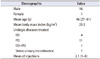
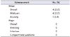
Seventeen patients underwent injections for the treatment of organic ED (4), PD (11), coexisting ED with PD (1), and female SUI (1) (Table 1). Cited reasons for ED included vasculogenic, penile fracture, medication-related and electrical injury to the genitalia. Mean patient age at time of first injection was 46 years (range, 27–61 years). Patients received an average of 2.1 (range, 1–8) injection procedures during the study period. Additional injections were provided upon patient request. Injections were well tolerated in all cases. Three patients reported mild pain at the injection site, one of whom also noted mild penile bruising after the injection (Table 2). All patients who noted bruising were PD patients who were given intracavernosal alprostadil were also given planned injection of 250 µg of phenylephrine at the conclusion of the procedure to detumescence. No systemic complications were noted initially or during follow-up. Mean follow-up was 15.5 months.
Among ED and/or PD patients queried with IIEF-5 (7), no patient reported a worsening of overall score or of any individual domain score. IIEF-5 scores improved by an average of 4.14 points after PRFM therapy. In patients with PD with subsequent follow-up, 80% (4/5) initially reported subjective improvement in their degree of curvature. One female patient underwent transurethral injection for SUI with 50% reduction in pad usage. When asked whether they would be likely to undergo further PRFM injections, 80% of patients answered affirmatively.
DISCUSSION
Platelet based therapies are being increasingly utilized in multiple medical settings, including dermatology, ophthalmology, cardiology, colorectal surgery, and plastic surgery [111]. PRP has been frequently used for orthopedic conditions such as bone and soft tissue trauma, inflammatory conditions, and chronic pain syndromes [1710]. Across multiple disciplines, PRP has been used both as a primary treatment modality and as a supplement to other therapies in hopes of supplementing wound healing, tissue regeneration, and angiogenesis. Although most of the studies focusing on PRP injections have been relatively small and heterogenous, they largely support safety and efficacy. Additionally, the concept of autologous therapy may be particularly attractive to some patients [16].
ED affects as many as 1 in 4 men, and evidence indicates the incidence is rising [1718]. The pathophysiology is multifactorial, but a significant proportion results from endothelial dysfunction secondary to inflammation [19]. The most common treatments for ED aim to improve endothelial function through augmentation of the nitric oxide pathway [20]. To date, there are no treatments that address the underlying cause of endothelial dysfunction. Platelet-derived therapies targeting inflammation and promoting tissue regeneration may represent a potential treatment option.
PD, while less common than ED, affects roughly 1%–8% of men [21]. The pathophysiology appears to involve increased inflammation from tissue disruption, followed by aberrant wound healing resulting in fibrotic plaques [22]. Current treatment regimens include plaque injection, plication, grafting, or insertion of penile prosthesis to restore appropriate form and function. Currently there are no therapies targeting either the inflammatory processes or the aberrant wound healing that causes PD. Furthermore, therapies focusing on disrupting the fibrotic plaques through mechanical manipulation, or more recently, collagenase injection, do not address appropriate wound healing or regeneration of the damaged tissue [23]. Theoretically, injection of PRFM could combine mechanical disruption of the plaque, via needle fracture, while simultaneously neutralizing destructive inflammatory processes in an effort to promote a better wound-healing response and stabilize the disrupted plaque.
Biologic materials have been used for decades in the treatment of SUI. Multiple products have been used as bulking agents to supplement urethral coaptation. While generally less efficacious than surgical repairs, injectable agents remain attractive given their relative ease of administration and lack of need for implantable mesh-based materials. When it was previously available, glutaraldehyde cross-linked bovine collagen was the most commonly injected biomaterial used to treat female SUI and was associated with a cure rate of 53% [24]. Theoretically, injection of autologous PRFM could provide both urethral bulking and potential regenerative effects to a damaged female urethra.
Investigations of PRFM for the urologic conditions noted in this report have not been previously reported. Wu et al. [15] investigated the effects of several different preparations of PRP injections in rat models with bilateral cavernous nerve crush injuries. Their data suggest that an “optimized” PRP formulation with a high level of growth factors was more stable than other preparations of PRP. Rats receiving this formulation showed significantly greater increases in intracavernosal pressure, higher mean arterial pressure, higher levels of nitric oxide synthase, and greater recovery of erectile function than those receiving saline injections or other formulations of PRP. Tang et al. [25] also showed that PRP injections at the site of cavernous nerve crush injuries helped facilitate nerve regeneration and erectile function in a rat model. More recently, Shirvan et al. [26] described injection of PRP and interposition platelet rich fibrin glue into the fistulous tracts of 12 patients with vesicovaginal fistulas (most <5 mm). All patients showed significant improvement with 11 patients cured at six-month follow-up, both subjectively and by examination.
We recognize that a variety of preparations, delivery modalities, and dosing schedules are available for PRP/PRFM therapies. A mean of 2.1 injection procedures per patient were performed during the study period. In our study, the PRP was added to a calcium chloride preparation to create PRFM. This was done to theoretically prevent rapid washout of the PRP from the corpora. One potential safety concern about using a colloid/hydrogel type of material in the corpora was the possibility of interrupting corporal blood flow, creating the possibility of a ‘penile compartment syndrome,’ akin to priapism. This did not occur in any of the ED or PD patients in our study, as each of these injections was well tolerated.
Data from this report regarding functional assessments must be interpreted with caution. This was not a prospective study, and we believe a significant placebo effect exists for research involving male sexual health. Objective improvements in the IIEF-5 score (4.14 points, 9.1%) were seen in patients receiving PRFM therapy for ED and PD. This level of improvement was similar to the average IIEF score increase (4.45 points, [3.42, 5.29]) seen in patients using PDE5Is after nerve sparing prostatectomy in a recent meta-analysis [20]. At follow-up interviews, patients expressed specific improvements in the rigidity of erections and improvements in satisfaction due to increased confidence. Of PD patients available for follow-up, 80% noticed an initial subjective improvement in their degree of curvature. Additionally, the one patient who received PRFM injections for SUI noted a 50% decrement in pad usage. Patients injected with silicone polymers (Macroplastique, Cogentix, Minnetonka, MN, USA) reported a 77% subjective cure rate but only a 9% objective cure rate on urodynamic testing [27]. No conclusions can be drawn from a single patient, but a 50% objective improvement from a transurethral injection procedure using an autologous product seems promising. With regards to feasibility of the procedure, there were no concerns related to the preparation of the PRFM or the injection process itself into the corpora cavernosa, tunical plaques, or urethral submucosa for patients with ED, PD, or SUI, respectively.
While this study attests to safety in this selected population, it has multiple limitations. This was a retrospective review of a small cohort of patients with a spectrum of pathology that may not be representative of the general population. As an autologous product, we expect that reabsorption rates are high, such that repetitive therapy will be required. This raises the possibility of treatment-related fibrosis from injection site trauma. As mentioned, although there was no detriment in IIEF score, the lack of a placebo arm prevents a detailed context. Future work will involve placebo control, with structured assessments for efficacy.
CONCLUSIONS
Our initial experience suggests that PRFM injections for ED, PD, and female SUI are feasible and safe. Although the limited data is suggestive of efficacy, a placebo control will be required in subsequent efforts for confirmation. Future studies evaluating efficacy of PRFM injections for genitourinary pathology appear warranted.
 XML Download