

 PDF
PDF ePub
ePub Citation
 Print
Print


INTRODUCTION
Vitamin C is a well-known anti-tumor agent as well as essential nutrients. According to the report by Kang et al., relatively high concentration of vitamin C (10 mM) induces apoptosis of B16 murine melanoma cells via the destruction of mitochondrial membrane potential and release of cytochrome C (1). In addition, vitamin C effectively suppresses the translocation of transferring receptor from cytosol to membrane. And it interfere the uptake iron into tumor cells, which is essential process for maintenance of the proliferation of tumor cells (2). There are other reports regarding anti-tumor effect of relatively low concentration vitamin C, less than 1.0 mM. Most of the cases, low concentration of vitamin C could not induce extensive apoptosis, but shows the suppression of tumor proliferation and inhibition of the growth factor production (3). Definite growth arrest of B16 melanoma cells at G1 stage by the treatment of 0.2 mM of vitamin C treatment was recently reported. It was closely related with the increase of p53-p21Waf1/Cip1 and inhibition of CDK2 activity (3,4). In the case of SK-MEL-2, human melanoma cell line, it is reported that its proliferation is suppressed by 1 mM of vitamin C and it is mediated by the decrease of insulin-like growth factor (IGF)-II and its receptor expression (5). In addition, vitamin C could also suppress the production of endogenous molecules, which is essential for their growth and metastasis (6-8).
There are several kinds of immune escape mechanisms in tumor. The most well-known mechanism is the down-regulation of major histocompatibility complex (MHC) I expression on the surface of tumor cells for the evasion from killing by tumor specific cytolytic T lymphocytes (CTLs) (9). Down-regulation of Fas (CD95) expression is also known as one of the immune escape mechanisms of tumor (10). Taken together, CTLs first recognize tumor cells through the MHC I on the surface of tumor cells, and then induce the apoptosis on tumor cells through the transduction of death signals by the interaction between Fas on tumor and Fas ligand (FasL; CD154) on CTLs. It is generally known that there are two ways for the induction of apoptosis (11,12). One is mediated by tumor necrosis factor (TNF) receptor family such as Fas and TRAIL. The other is mediated by the destruction of membrane potential in mitochondria, which is essential for ATP generation. The former is called as Type I and the latter is Type II. However, Type I and II pathways are not totally independent process. Caspase-8 is activated by the death signals through TNF receptor family and activated Caspase-8 triggers the activation of Type II pathway through the cleavage of Bid into truncated form of Bid. Even though it is already discovered that vitamin C induces Type II apoptotic pathway in tumor cells in a Caspase-8 independent manner, but the enhanced apoptosis by vitamin C via the increase of Fas (CD95) also should be considered.
Therefore, we investigated whether vitamin C could increase the susceptibility of tumor cells to anti-Fas mAbs and the expression of Fas (CD95) and MHC class I on stomach cancer cell line, SNU1.
MATERIALS AND METHODS
Cells
Human stomach cancer cell line, SNU1 was obtained from American Type Culture Collection (Manassas, VA, USA). Cells were cultured in RPMI 1640 supplemented with 2 mM L-glutamine, 100 units/ml penicillin, 100µg/ml streptomycin, and 10% heat-inactivated fetal bovine serum. This cell line was used for experiments while in the log phase of growth.
Vitamin C treatment and measurement of apoptosis
Human stomach cancer cell line, SNU1 (1×10⁶ cells) was cultured in the presence or absence of vitamin C (1 and 2 mM) for 36 hrs. The cells were collected and washed twice with cold PBS, and then resuspended in 1× binding buffer at a concentration of 1×10⁶ cells/ml. 100µl of the solution (1×10⁵ cells) was then transferred to a 5 ml culture tube. After the addition of 5µl of Annexin V-FITC, cells were incubated at room temperature for 15 min in the dark with gentle vortexing. 400µl of 1x binding buffer was then added to each tube. One microliter of propidium of iodide was added, prior to analyze with FACSCaliber (BD Pharmingen, San Diego, CA, USA). The Annexin V-FITC apoptosis detection kit was purchased from BD Pharmingen (San Diego, CA, USA).
Trypan blue dye exclusion assay
Human stomach cancer cell line, SNU1 (1×10⁶ cells) was cultured in the presence or absence of vitamin C (1 and 2 mM) for 24hrs, and then 200 and 400 ng/ml of anti-Fas mAbs (CH-11, MBL International, Woburn, MA, USA) were added. After further incubation for 18 hrs, cells were harvested and washed once with phosphate-buffered saline (PBS). Cell viability was examined by the trypan blue dye exclusion assay. Quadruplicate wells were run for each group.
Flow cytometry analysis
The changing of Fas and MHC I expression on human stomach cancer cell line, SNU1 by the treatment of vitamin C was assessed by flow cytometry using the PE conjugated rat anti-human Fas and MHC I mAbs (BD Pharmingen, San Diego, CA, USA). Briefly, SNU1 (1×10⁶ cells) was cultured in the presence or absence of vitamin C (2 mM) for 3, 6, 12, and 24 hrs. And then Cells were washed twice with phosphate buffered saline (PBS) and then stained with 1µg of anti-human Fas and MHC I mAbs for 30 min on ice. After washing twice with PBS, and cells were analyzed by FACSCaliber (BD Pharmingen, San Diego, CA, USA).
RESULTS
Vitamin C increases the expression of Fas and MHC I on the surface of stomach cancer cell line, SNU1
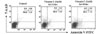
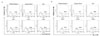
Since we have found that the susceptibility of tumor cells to vitamin C is quite diverse in our previous reports (1-8), we first investigated the optimal concentration of vitamin C, which does not show cytotoxic effect during the experiment. As shown in Fig. 1, we could not observe the extensive apoptosis on stomach cancer cell line, SNU1, when it was in cubated for 36 hrs in the presence of 1 and 2 mM of vitamin C. And then we next investigated the changing on Fas and MHC I expression on SNU1 after treatment of 2 mM of vitamin C for indicated time. Interestingly, Fas expression was increased at 3 hrs after treatment of vitamin C (Fig. 2A). In addition, the expression of MHC I on the surface of SNU1 was increased at 3 hrs after treatment of vitamin C and it lasted untill 24 hrs after treatment of vitamin C (Fig. 2B).
Fas mediated apoptosis on stomach cancer cell line, SNU1 is increased by the ligation of anti-Fas mAbs
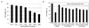
As we showed that Fas expression on SNU1 was increased by the treatment of vitamin C in Fig. 2A, the changing on cell viability by the ligation of Fas with anti-Fas mAb was examined. To determine the optimal amount of anti-Fas mAb, which cannot induce the extensive apoptosis, the viability of cells was examined by trypan blue dye exclusion assay, after ligation of cells with anti-Fas mAbs (200~2,000 ng/ml) for 18 hrs. We found that cell viability was more than 90% by the treatment of 200 and 400 ng/ml of anti-Fas mAbs (Fig. 3A). And then, we examined the synergistic effect of vitamin C and anti-Fas mAb on the changing on viability of SNU1. As shown in Fig. 3B, when the cells were pre-treated with 1 and 2 mM of vitamin C for 24 hrs, prior to ligate with anti-Fas mAb for 18 hrs, the viability of cells was decreased, when it compared with the control and the cells treated with 400 ng/ml of anti-Fas mAb only.
DISCUSSION
Surgical excision, chemotherapy and radiotherapy are conventional approaches for the treatment of cancer. Surgical excision is used to get rid of tumor burden. However, it is useful only for the early stage of cancer, not for metastatic cancer. For this reason, chemotherapy or radiotherapy is usually performed for the elimination of metastatic or residual tumors. However, severe side effects and high costs are major problems, which should be overcome. Recently, immunotherapy through the enhancing anti-tumor activity of immune responses is also considered as approach for the treatment of cancer (13,14). The basic principle of immunotherapy for the eradication of cancer is the activation of dendritic cells (DCs), natural killer (NK) cells and tumor infiltrating lymphocytes (TILs) (15,16). Therefore, it is thought that the investigation of the appropriate substances, which is able to induce the activation of anti-tumor immune cell subsets, is the best way for immunotherapy.
From this point of view, vitamin C is considered as one of the best candidates, since it is already reported that NK cells show increased anti-tumor activity through the activation by the treatment of vitamin C (17). In addition, we found that Vitamin C-treated murine bone marrow-derived dendritic cells preferentially drive naïve T cells into Th1 cells by increased IL-12 secretions (18). Even though it is cleared the role of vitamin C on the enhancement of anti-tumor immune responses, the investigation regarding the increase of immune susceptibility of tumor cells is also needed to maximize anti-tumor immune responses by vitamin C. So, we did this experiment whether vitamin C can modulate immune susceptibility of tumor cells. Among various kinds of molecule involved in immune susceptibility of tumor cells, Fas is one of the most well-known execute molecules (10). Briefly, cytolytic T lymphocytes (CTLs) first recognize tumor cells through the MHC I on the surface of tumor cells, and then induce the apoptosis on tumor cells through the transduction of death signals by the interaction between Fas on tumor and Fas ligand (FasL; CD154) on CTLs. Thus, down-regulation of Fas and MHC I expression is also known as the general characteristics of tumors for their escape from immune system (10). Therefore, our findings shown here suggest the utility of vitamin C as an adjuvant in immunotherapy, since vitamin C can act dual function, which are enhancing anti-tumor immune responses and increasing immune susceptibility of tumor cell.
The physiological vitamin C concentration in human serum is known as 70~85µM (19,20). Serum concentration of vitamin C is determined by the route of administration. According to the report by Sebastian J et al, when 3 grams of vitamin C is administered via intravenous injection, vitamin C concentration in serum is reached at 1,700µM, but when it was administered via oral route, it is reached at 220µM (21). Even though vitamin C used in our experiment (1 and 2 mM) is higher than its physiological concentration under normal diet condition, it is possible to increase serum vitamin C concentration to 1 and 2 mM by the administration via intravenous injection of vitamin C. When we consider that the recent trials of anti-cancer therapy by using of vitamin C is done by intravenous injection, it seems that the combination approach of vitamin C and immunotherapy or chemo-/radiotherapy will give us the new insights of cancer therapy.
 XML Download